 Movie
Movie Controller


+ Open data

- Basic information
| Entry |  | |||||||||||||||
|---|---|---|---|---|---|---|---|---|---|---|---|---|---|---|---|---|
| Title | Cryo-EM structure of the SARS-CoV-2 Spike bound to TMEM106B | |||||||||||||||
 Map data Map data | Cryo-EM map filtered by local resolution | |||||||||||||||
 Sample Sample |
| |||||||||||||||
 Keywords Keywords |  SARS-CoV-2 / Spike / ectodomain / TMEM106B / luminal domain / VIRAL PROTEIN SARS-CoV-2 / Spike / ectodomain / TMEM106B / luminal domain / VIRAL PROTEIN | |||||||||||||||
| Function / homology |  Function and homology information Function and homology informationpositive regulation of hydrolase activity / lysosomal protein catabolic process / regulation of lysosome organization / lysosomal lumen acidification / lysosome localization / positive regulation of dendrite development / dendrite morphogenesis / lysosomal transport / lysosome organization / neuron cellular homeostasis ...positive regulation of hydrolase activity / lysosomal protein catabolic process / regulation of lysosome organization / lysosomal lumen acidification / lysosome localization / positive regulation of dendrite development / dendrite morphogenesis / lysosomal transport / lysosome organization / neuron cellular homeostasis / late endosome membrane / ATPase binding / lysosome / endosome / lysosomal membraneSimilarity search - Function | |||||||||||||||
| Biological species |   Severe acute respiratory syndrome coronavirus 2 / Severe acute respiratory syndrome coronavirus 2 /  Homo sapiens (human) Homo sapiens (human) | |||||||||||||||
| Method | single particle reconstruction / cryo EM / Resolution: 3.52 Å | |||||||||||||||
 Authors Authors | CHEREPANOV P | |||||||||||||||
| Funding support |  United Kingdom, 4 items United Kingdom, 4 items
| |||||||||||||||
 Citation Citation | Journal: Cell / Year: 2023 Title: TMEM106B is a receptor mediating ACE2-independent SARS-CoV-2 cell entry. Authors: Jim Baggen / Maarten Jacquemyn / Leentje Persoons / Els Vanstreels / Valerie E Pye / Antoni G Wrobel / Valeria Calvaresi / Stephen R Martin / Chloë Roustan / Nora B Cronin / Eamonn Reading ...Authors: Jim Baggen / Maarten Jacquemyn / Leentje Persoons / Els Vanstreels / Valerie E Pye / Antoni G Wrobel / Valeria Calvaresi / Stephen R Martin / Chloë Roustan / Nora B Cronin / Eamonn Reading / Hendrik Jan Thibaut / Thomas Vercruysse / Piet Maes / Frederik De Smet / Angie Yee / Toey Nivitchanyong / Marina Roell / Natalia Franco-Hernandez / Herve Rhinn / Alusha Andre Mamchak / Maxime Ah Young-Chapon / Eric Brown / Peter Cherepanov / Dirk Daelemans /   Abstract: SARS-CoV-2 is associated with broad tissue tropism, a characteristic often determined by the availability of entry receptors on host cells. Here, we show that TMEM106B, a lysosomal transmembrane ...SARS-CoV-2 is associated with broad tissue tropism, a characteristic often determined by the availability of entry receptors on host cells. Here, we show that TMEM106B, a lysosomal transmembrane protein, can serve as an alternative receptor for SARS-CoV-2 entry into angiotensin-converting enzyme 2 (ACE2)-negative cells. Spike substitution E484D increased TMEM106B binding, thereby enhancing TMEM106B-mediated entry. TMEM106B-specific monoclonal antibodies blocked SARS-CoV-2 infection, demonstrating a role of TMEM106B in viral entry. Using X-ray crystallography, cryogenic electron microscopy (cryo-EM), and hydrogen-deuterium exchange mass spectrometry (HDX-MS), we show that the luminal domain (LD) of TMEM106B engages the receptor-binding motif of SARS-CoV-2 spike. Finally, we show that TMEM106B promotes spike-mediated syncytium formation, suggesting a role of TMEM106B in viral fusion. Together, our findings identify an ACE2-independent SARS-CoV-2 infection mechanism that involves cooperative interactions with the receptors heparan sulfate and TMEM106B. | |||||||||||||||
| History |
|
- Structure visualization
| Supplemental images |
|---|

- Downloads & links
-EMDB archive
| Map data | emd_17169.map.gz | 12.6 MB | EMDB map data format | |
|---|---|---|---|---|
| Header (meta data) | emd-17169-v30.xmlemd-17169.xml | 21.4 KB 21.4 KB | Display Display | EMDB header |
| FSC (resolution estimation) | emd_17169_fsc.xml | 13.9 KB | Display | FSC data file |
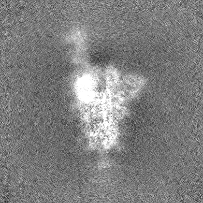
| Images | 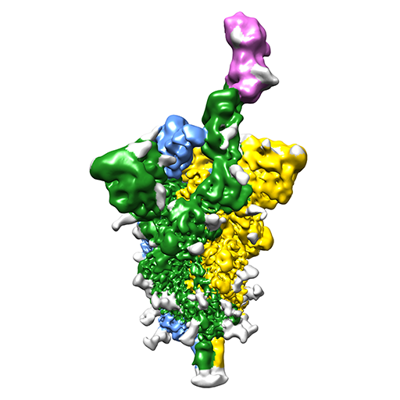 emd_17169.png emd_17169.png | 89.7 KB | ||
| Others | emd_17169_half_map_1.map.gzemd_17169_half_map_2.map.gz | 262.6 MB 262.6 MB | ||
| Archive directory |  http://ftp.pdbj.org/pub/emdb/structures/EMD-17169ftp://ftp.pdbj.org/pub/emdb/structures/EMD-17169 http://ftp.pdbj.org/pub/emdb/structures/EMD-17169ftp://ftp.pdbj.org/pub/emdb/structures/EMD-17169 | HTTPS FTP |
-Related structure data
-Links
| EMDB pages | EMDB (EBI/PDBe) / EMDataResource |
|---|
-Map
| File | Download / File: emd_17169.map.gz / Format: CCP4 / Size: 282.6 MB / Type: IMAGE STORED AS FLOATING POINT NUMBER (4 BYTES) | ||||||||||||||||||||
|---|---|---|---|---|---|---|---|---|---|---|---|---|---|---|---|---|---|---|---|---|---|
| Annotation | Cryo-EM map filtered by local resolution | ||||||||||||||||||||
| Voxel size | X=Y=Z: 0.85 Å | ||||||||||||||||||||
| Density |
| ||||||||||||||||||||
| Symmetry | Space group: 1 | ||||||||||||||||||||
| Details | EMDB XML:
|
-Supplemental data
-Half map: Half map 2
| File | emd_17169_half_map_1.map | ||||||||||||
|---|---|---|---|---|---|---|---|---|---|---|---|---|---|
| Annotation | Half map 2 | ||||||||||||
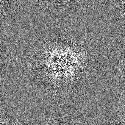
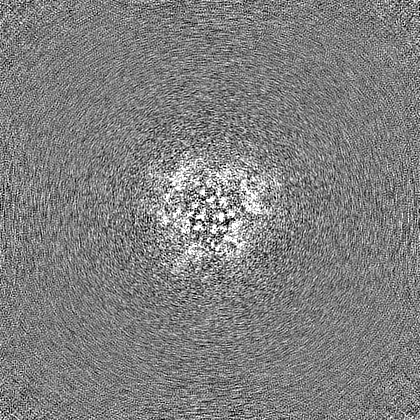
| Projections & Slices |
| ||||||||||||
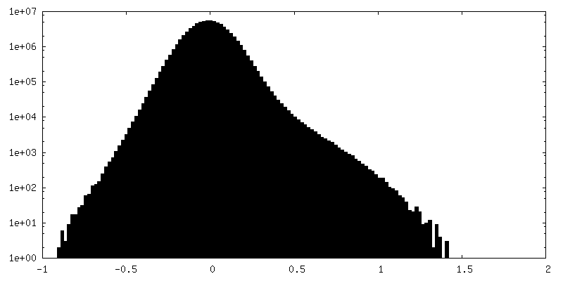
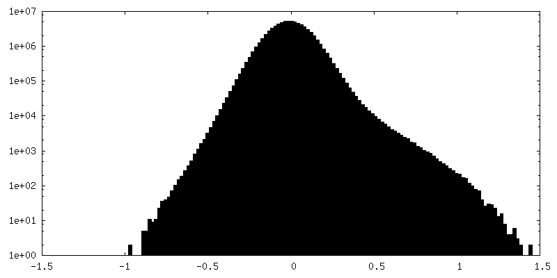
| Density Histograms |
 Z
Z Y
Y X
X


-Half map: Half map 1
| File | emd_17169_half_map_2.map | ||||||||||||
|---|---|---|---|---|---|---|---|---|---|---|---|---|---|
| Annotation | Half map 1 | ||||||||||||
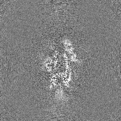
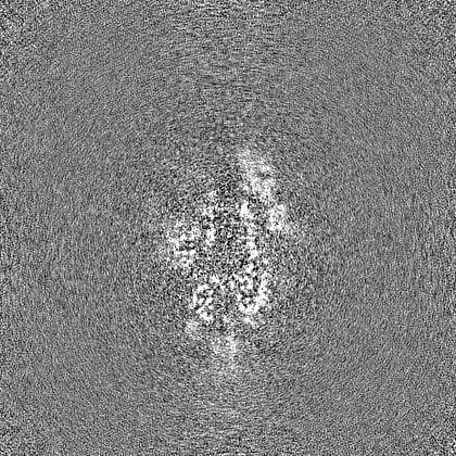
| Projections & Slices |
| ||||||||||||
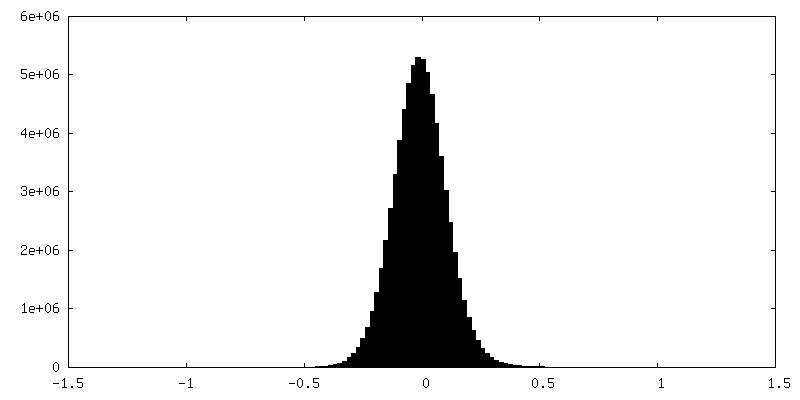
| Density Histograms |


- Sample components
-Entire : Trimeric SARS-CoV-2 Spike bound to one molecule of TMEM106B
| Entire | Name: Trimeric SARS-CoV-2 Spike bound to one molecule of TMEM106B |
|---|---|
| Components |
|
-Supramolecule #1: Trimeric SARS-CoV-2 Spike bound to one molecule of TMEM106B
| Supramolecule | Name: Trimeric SARS-CoV-2 Spike bound to one molecule of TMEM106B type: complex / ID: 1 / Parent: 0 / Macromolecule list: all |
|---|---|
| Molecular weight | Theoretical: 450 KDa |
-Supramolecule #2: SARS-CoV-2 Spike Ectodomain, hexa-Pro stabilized; Belgium/GHB-030...
| Supramolecule | Name: SARS-CoV-2 Spike Ectodomain, hexa-Pro stabilized; Belgium/GHB-03021 variant (Asp-484) type: complex / ID: 2 / Parent: 1 / Macromolecule list: #1 |
|---|---|
| Source (natural) | Organism: Severe acute respiratory syndrome coronavirus 2 |
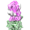
-Supramolecule #3: TMEM106B lumenal domain
| Supramolecule | Name: TMEM106B lumenal domain / type: complex / ID: 3 / Parent: 1 / Macromolecule list: #2 |
|---|---|
| Source (natural) | Organism: Homo sapiens (human) |
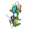
-Macromolecule #1: SARS-CoV-2 Spike Ectodomain, hexa-Pro stabilized; Belgium/GHB-030...
| Macromolecule | Name: SARS-CoV-2 Spike Ectodomain, hexa-Pro stabilized; Belgium/GHB-03021 variant (Asp-484) type: protein_or_peptide / ID: 1 Details: SARS-CoV-2 Spike ectodomain, stabilized in trimeric prefusion conformation, twin strep-tagged. Enantiomer: LEVO |
|---|---|
| Source (natural) | Organism: Severe acute respiratory syndrome coronavirus 2 / Strain: Belgium/GHB-03021 |
| Recombinant expression | Organism: Homo sapiens (human) |
| Sequence | String: MFVFLVLLPL VSSQCVNLTT RTQLPPAYTN SFTRGVYYPD KVFRSSVLHS TQDLFLPFFS NVTWFHAKRF DNPVLPFNDG VYFASTEKSN IIRGWIFGTT LDSKTQSLLI VNNATNVVIK VCEFQFCNDP FLGVYYHKNN KSWMESEFRV YSSANNCTFE YVSQPFLMDL ...String: MFVFLVLLPL VSSQCVNLTT RTQLPPAYTN SFTRGVYYPD KVFRSSVLHS TQDLFLPFFS NVTWFHAKRF DNPVLPFNDG VYFASTEKSN IIRGWIFGTT LDSKTQSLLI VNNATNVVIK VCEFQFCNDP FLGVYYHKNN KSWMESEFRV YSSANNCTFE YVSQPFLMDL EGKQGNFKNL REFVFKNIDG YFKIYSKHTP INLVRDLPQG FSALEPLVDL PIGINITRFQ TLLALHRSYL TPGDSSSGWT AGAAAYYVGY LQPRTFLLKY NENGTITDAV DCALDPLSET KCTLKSFTVE KGIYQTSNFR VQPTESIVRF PNITNLCPFG EVFNATRFAS VYAWNRKRIS NCVADYSVLY NSASFSTFKC YGVSPTKLND LCFTNVYADS FVIRGDEVRQ IAPGQTGKIA DYNYKLPDDF TGCVIAWNSN NLDSKVGGNY NYLYRLFRKS NLKPFERDIS TEIYQAGSTP CNGVDGFNCY FPLQSYGFQP TNGVGYQPYR VVVLSFELLH APATVCGPKK STNLVKNKCV NFNFNGLTGT GVLTESNKKF LPFQQFGRDI ADTTDAVRDP QTLEILDITP CSFGGVSVIT PGTNTSNQVA VLYQDVNCTE VPVAIHADQL TPTWRVYSTG SNVFQTRAGC LIGAEHVNNS YECDIPIGAG ICASYQPRRA RSVASQSIIA YTMSLGAENS VAYSNNSIAI PTNFTISVTT EILPVSMTKT SVDCTMYICG DSTECSNLLL QYGSFCTQLN RALTGIAVEQ DKNTQEVFAQ VKQIYKTPPI KDFGGFNFSQ ILPDPSKPIK RSPIEDLLFN KVTLADAGFI KQYGDCLGDI AARDLICAQK FNGLTVLPPL LTDEMIAQYT SALLAGTITS GWTFGAGPAL QIPFPMQMAY RFNGIGVTQN VLYENQKLIA NQFNSAIGKI QDSLSSTPSA LGKLQDVVNQ NAQALNTLVK QLSSNFGAIS SVLNDILSRL DPPEAEVQID RLITGRLQSL QTYVTQQLIR AAEIRASANL AATKMSECVL GQSKRVDFCG KGYHLMSFPQ SAPHGVVFLH VTYVPAQEKN FTTAPAICHD GKAHFPREGV FVSNGTHWFV TQRNFYEPQI ITTDNTFVSG NCDVVIGIVN NTVYDPLQPE LDSFKEELDK YFKNHTSPDV DLGDISGINA SVVNIQKEID RLNEVAKNLN ESLIDLQELG KYEQGSGYIP EAPRDGQAYV RKDGEWVLLS TFLGRSLEVL FQGPGSAWSH PQFEKGGGSG GGGSGGSAWS HPQFEK |
-Macromolecule #2: TMEM106B lumenal domain
| Macromolecule | Name: TMEM106B lumenal domain / type: protein_or_peptide / ID: 2 Details: TMEM106B lumenal domain expressed with an N-terminal signal peptide and a C-terminal His6 tag Enantiomer: LEVO |
|---|---|
| Source (natural) | Organism: Homo sapiens (human) |
| Recombinant expression | Organism: Homo sapiens (human) |
| Sequence | String: METDTLLLWV LLLWVPGSTG DAAQPRSIDV KYIGVKSAYV SYDVQKRTIY LNITNTLNIT NNNYYSVEVE NITAQVQFSK TVIGKARLNN ITIIGPLDMK QIDYTVPTVI AEEMSYMYDF CTLISIKVHN IVLMMQVTVT TTYFGHSEQI SQERYQYVDC GRNTTYQLGQ ...String: METDTLLLWV LLLWVPGSTG DAAQPRSIDV KYIGVKSAYV SYDVQKRTIY LNITNTLNIT NNNYYSVEVE NITAQVQFSK TVIGKARLNN ITIIGPLDMK QIDYTVPTVI AEEMSYMYDF CTLISIKVHN IVLMMQVTVT TTYFGHSEQI SQERYQYVDC GRNTTYQLGQ SEYLNVLQPQ QGSGENLYFQ SAGHHHHHH UniProtKB: Transmembrane protein 106B |
-Experimental details
-Structure determination
| Method | cryo EM |
|---|---|
 Processing Processing | single particle reconstruction |
| Aggregation state | particle |
-Sample preparation
| Concentration | 1 mg/mL | |||||||||||||||
|---|---|---|---|---|---|---|---|---|---|---|---|---|---|---|---|---|
| Buffer | pH: 8 Component:
Details: 150 mM NaCl, 1 mM EDTA, and 25 mM Tris-HCl, pH 8.0 | |||||||||||||||
| Grid | Model: Quantifoil R1.2/1.3 / Mesh: 400 / Pretreatment - Type: GLOW DISCHARGE | |||||||||||||||
| Vitrification | Cryogen name: ETHANE-PROPANE / Chamber humidity: 100 % / Chamber temperature: 293 K / Instrument: FEI VITROBOT MARK IV |
- Electron microscopy
| Microscope | FEI TITAN KRIOS |
|---|---|
| Electron beam | Acceleration voltage: 300 kV / Electron source: FIELD EMISSION GUN |
| Electron optics | Illumination mode: FLOOD BEAM / Imaging mode: BRIGHT FIELDBright-field microscopy / Nominal defocus max: 2.1 µm / Nominal defocus min: 0.5 µm |
| Specialist optics | Energy filter - Name: GIF Bioquantum / Energy filter - Slit width: 20 eV |
| Sample stage | Cooling holder cryogen: NITROGEN |
| Image recording | Film or detector model: GATAN K3 (6k x 4k) / Number grids imaged: 2 / Number real images: 37079 / Average electron dose: 30.0 e/Å2 |
| Experimental equipment |  Model: Titan Krios / Image courtesy: FEI Company |
-Image processing
| Particle selection | Number selected: 1799554 |
|---|---|
| Startup model | Type of model: OTHER |
| Initial angle assignment | Type: MAXIMUM LIKELIHOOD / Software - Name: cryoSPARC (ver. 4.1) |
| Final 3D classification | Number classes: 4 / Avg.num./class: 50000 / Software - Name: RELION (ver. 4.0) |
| Final angle assignment | Type: MAXIMUM LIKELIHOOD / Software - Name: cryoSPARC (ver. 4.1) |
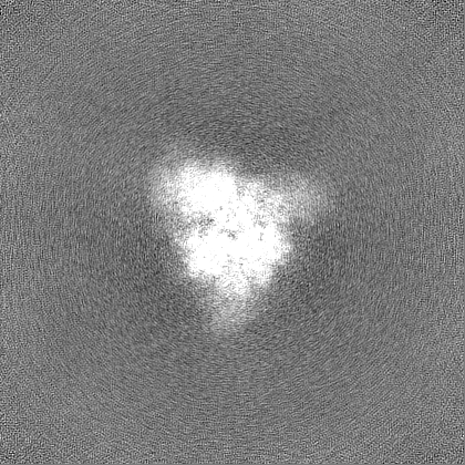
| Final reconstruction | Number classes used: 1 / Applied symmetry - Point group: C1 (asymmetric) / Resolution.type: BY AUTHOR / Resolution: 3.52 Å / Resolution method: FSC 0.143 CUT-OFF / Software - Name: cryoSPARC (ver. 4.1) / Details: Non-uniform refinement as implemented in cryoSPARC / Number images used: 25781 |
| FSC plot (resolution estimation) | 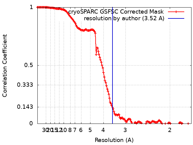 |
