 Movie
Movie Controller


+ Open data

- Basic information
| Entry |  | |||||||||
|---|---|---|---|---|---|---|---|---|---|---|
| Title | Vimentin intermediate filament structure | |||||||||
 Map data Map data | VIF 3D structure | |||||||||
 Sample Sample |
| |||||||||
 Keywords Keywords |  vimentin / intermediate filament / cytoskeleton / STRUCTURAL PROTEIN vimentin / intermediate filament / cytoskeleton / STRUCTURAL PROTEIN | |||||||||
| Function / homology |  Function and homology information Function and homology informationkeratin filament binding / lens fiber cell development / intermediate filament organization / cellular response to muramyl dipeptide / structural constituent of eye lens / astrocyte development / Striated Muscle Contraction / intermediate filament / RHOBTB1 GTPase cycle / intermediate filament cytoskeleton ...keratin filament binding / lens fiber cell development / intermediate filament organization / cellular response to muramyl dipeptide / structural constituent of eye lens / astrocyte development / Striated Muscle Contraction / intermediate filament / RHOBTB1 GTPase cycle / intermediate filament cytoskeleton / microtubule organizing center / cell leading edge / Bergmann glial cell differentiation / positive regulation of collagen biosynthetic process / Caspase-mediated cleavage of cytoskeletal proteins / phagocytic vesicle / regulation of mRNA stability / Late endosomal microautophagy / structural constituent of cytoskeleton / nuclear matrix / Aggrephagy / cellular response to type II interferon / Chaperone Mediated Autophagy / peroxisome / neuron projection development / negative regulation of neuron projection development / double-stranded RNA binding / scaffold protein binding / Interleukin-4 and Interleukin-13 signaling / cellular response to lipopolysaccharide / molecular adaptor activity / cytoskeleton / axon / protein domain specific binding / focal adhesion / positive regulation of gene expression / extracellular exosome / identical protein binding / plasma membrane / cytosol / cytoplasmSimilarity search - Function | |||||||||
| Biological species |  Homo sapiens (human) Homo sapiens (human) | |||||||||
| Method | helical reconstruction / cryo EM / Resolution: 7.2 Å | |||||||||
 Authors Authors | Eibauer M / Medalia O | |||||||||
| Funding support |  Switzerland, 1 items Switzerland, 1 items
| |||||||||
 Citation Citation | Journal: Nat.Struct.Mol.Biol. / Year: 2024 Title: Vimentin filaments integrate low-complexity domains in a complex helical structure Authors: Eibauer M / Weber MS / Kronenberg-Tenga R / Beales CT / Boujemaa-Paterski R / Turgay Y / Sivagurunathan S / Kraxner J / Koster S / Goldman RD / Medalia O | |||||||||
| History |
|
- Structure visualization
| Supplemental images |
|---|

- Downloads & links
-EMDB archive
| Map data | emd_16844.map.gz | 195.4 MB | EMDB map data format | |
|---|---|---|---|---|
| Header (meta data) | emd-16844-v30.xmlemd-16844.xml | 15.3 KB 15.3 KB | Display Display | EMDB header |
| FSC (resolution estimation) | emd_16844_fsc.xml | 13.6 KB | Display | FSC data file |
| Images | 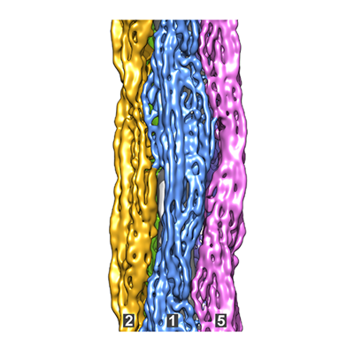 emd_16844.png emd_16844.png | 117.2 KB | ||
| Masks | emd_16844_msk_1.map | 209.3 MB | Mask map | |
| Filedesc metadata | emd-16844.cif.gz | 4.4 KB | ||
| Others | emd_16844_additional_1.map.gzemd_16844_half_map_1.map.gzemd_16844_half_map_2.map.gz | 3.8 GB 164 MB 164.1 MB | ||
| Archive directory |  http://ftp.pdbj.org/pub/emdb/structures/EMD-16844ftp://ftp.pdbj.org/pub/emdb/structures/EMD-16844 http://ftp.pdbj.org/pub/emdb/structures/EMD-16844ftp://ftp.pdbj.org/pub/emdb/structures/EMD-16844 | HTTPS FTP |
-Related structure data
| Related structure data |  8rveMC M: atomic model generated by this map C: citing same article ( |
|---|---|
| Similar structure data |


-Links
| EMDB pages | EMDB (EBI/PDBe) / EMDataResource |
|---|---|
| Related items in Molecule of the Month |


-Map
| File | Download / File: emd_16844.map.gz / Format: CCP4 / Size: 209.3 MB / Type: IMAGE STORED AS FLOATING POINT NUMBER (4 BYTES) | ||||||||||||||||||||||||||||||||||||
|---|---|---|---|---|---|---|---|---|---|---|---|---|---|---|---|---|---|---|---|---|---|---|---|---|---|---|---|---|---|---|---|---|---|---|---|---|---|
| Annotation | VIF 3D structure | ||||||||||||||||||||||||||||||||||||
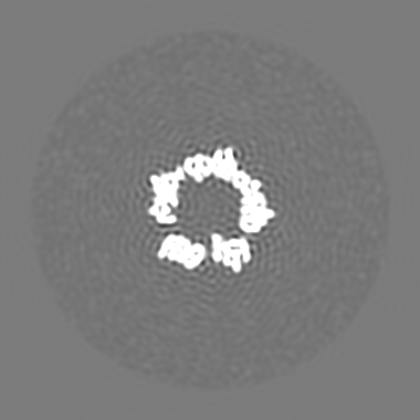
| Projections & slices | Image control
Images are generated by Spider. | ||||||||||||||||||||||||||||||||||||
| Voxel size | X=Y=Z: 1.0021 Å | ||||||||||||||||||||||||||||||||||||
| Density |
| ||||||||||||||||||||||||||||||||||||
| Symmetry | Space group: 1 | ||||||||||||||||||||||||||||||||||||
| Details | EMDB XML:
|
 Z (Sec.)
 Y (Row.)
 X (Col.)


-Supplemental data
-Mask #1
| File | emd_16844_msk_1.map | ||||||||||||
|---|---|---|---|---|---|---|---|---|---|---|---|---|---|
| Projections & Slices |
| ||||||||||||
| Density Histograms |


-Additional map: VIF helical symmetry applied to the VIF 3D structure
| File | emd_16844_additional_1.map | ||||||||||||
|---|---|---|---|---|---|---|---|---|---|---|---|---|---|
| Annotation | VIF helical symmetry applied to the VIF 3D structure | ||||||||||||
| Projections & Slices |
| ||||||||||||
| Density Histograms |


-Half map: VIF 3D structure / half map 1
| File | emd_16844_half_map_1.map | ||||||||||||
|---|---|---|---|---|---|---|---|---|---|---|---|---|---|
| Annotation | VIF 3D structure / half map 1 | ||||||||||||
| Projections & Slices |
| ||||||||||||
| Density Histograms |


-Half map: VIF 3D structure / half map 2
| File | emd_16844_half_map_2.map | ||||||||||||
|---|---|---|---|---|---|---|---|---|---|---|---|---|---|
| Annotation | VIF 3D structure / half map 2 | ||||||||||||
| Projections & Slices |
| ||||||||||||
| Density Histograms |


- Sample components
-Entire : Vimentin intermediate filament
| Entire | Name: Vimentin intermediate filament |
|---|---|
| Components |
|
-Supramolecule #1: Vimentin intermediate filament
| Supramolecule | Name: Vimentin intermediate filament / type: complex / ID: 1 / Parent: 0 |
|---|---|
| Source (natural) | Organism: Homo sapiens (human) |
| Molecular weight | Theoretical: 56 kDa/nm |
-Experimental details
-Structure determination
| Method | cryo EM |
|---|---|
 Processing Processing | helical reconstruction |
| Aggregation state | filament |
-Sample preparation
| Buffer | pH: 7.5 |
|---|---|
| Grid | Model: Quantifoil R2/1 / Material: COPPER / Mesh: 200 / Support film - Material: CARBON / Support film - topology: HOLEY |
| Vitrification | Cryogen name: ETHANE / Instrument: HOMEMADE PLUNGER |
- Electron microscopy
| Microscope | FEI TITAN KRIOS |
|---|---|
| Electron beam | Acceleration voltage: 300 kV / Electron source: FIELD EMISSION GUN |
| Electron optics | Illumination mode: FLOOD BEAM / Imaging mode: BRIGHT FIELDBright-field microscopy / Nominal defocus max: 2.8000000000000003 µm / Nominal defocus min: 0.8 µm |
| Image recording | Film or detector model: GATAN K3 BIOQUANTUM (6k x 4k) / Average electron dose: 62.0 e/Å2 |
| Experimental equipment |  Model: Titan Krios / Image courtesy: FEI Company |
-Image processing
| Segment selection | Number selected: 1462717 / Software - Name: crYOLO |
|---|---|
| Startup model | Type of model: OTHER |
| Final angle assignment | Type: NOT APPLICABLE |
| Final reconstruction | Applied symmetry - Helical parameters - Δz: 42.461 Å Applied symmetry - Helical parameters - Δ&Phi: 73.7308 ° Applied symmetry - Helical parameters - Axial symmetry: C1 (asymmetric) Algorithm: FOURIER SPACE / Resolution.type: BY AUTHOR / Resolution: 7.2 Å / Resolution method: FSC 0.143 CUT-OFF / Software - Name: RELION / Number images used: 236920 |
| FSC plot (resolution estimation) |  |
