 Movie
Movie Controller

[English] 日本語
 Yorodumi
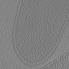
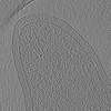
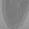
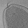
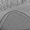
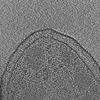
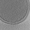
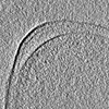
- EMDB-16479: Cryo-electron tomogram of a Roseburia intestinalis DSM 14610 cell. -
+ Open data

- Basic information
| Entry | 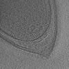 | |||||||||
|---|---|---|---|---|---|---|---|---|---|---|
| Title | Cryo-electron tomogram of a Roseburia intestinalis DSM 14610 cell. | |||||||||
 Map data Map data | A cryo-electron tomogram of an intact Roseburia intestinalis DSM 14610 cell. | |||||||||
 Sample Sample |
| |||||||||
 Keywords Keywords |  Bacteria / Roseburia / UNKNOWN FUNCTION Bacteria / Roseburia / UNKNOWN FUNCTION | |||||||||
| Biological species |  Roseburia intestinalis L1-82 (bacteria) Roseburia intestinalis L1-82 (bacteria) | |||||||||
| Method | electron tomography / cryo EM | |||||||||
 Authors Authors | Wimmer BH / Medalia O | |||||||||
| Funding support | 1 items
| |||||||||
 Citation Citation | Journal: Microlife / Year: 2023 Title: Phylogenetic diversity of core rumen microbiota as described by cryo-ET. Authors: Benedikt H Wimmer / Sarah Moraïs / Ran Zalk / Itzhak Mizrahi / Ohad Medalia /   Abstract: Microbial taxonomy is critical for describing ecosystem composition, yet the link between taxonomy and properties of microbes, such as their cellular architecture, remains poorly defined. We ...Microbial taxonomy is critical for describing ecosystem composition, yet the link between taxonomy and properties of microbes, such as their cellular architecture, remains poorly defined. We hypothesized that the cellular architecture represents microbial niche adaptation. We used cryo-electron microscopy and tomography to analyze microbial morphology in order to associate cellular architecture with phylogeny and genomic contents. As a model system, we chose the core rumen microbiome and imaged a large isolate collection covering 90% of its richness at the order level. Based on quantifications of several morphological features, we found that the visual similarity of microbiota is significantly related to their phylogenetic distance. Up to the level, closely related microbes have similar cellular architectures, which are highly correlated with genome similarity. However, in more distantly related bacteria, the correlation both with taxonomy and genome similarity is lost. This is the first comprehensive study of microbial cellular architecture and our results highlight that structure remains an important parameter in classification of microorganisms, along with functional parameters such as metabolomics. Furthermore, the high-quality images presented in this study represent a reference database for the identification of bacteria in anaerobic ecosystems. | |||||||||
| History |
|
- Structure visualization
| Supplemental images |
|---|

- Downloads & links
-EMDB archive
| Map data | emd_16479.map.gz | 204.2 MB |  EMDB map data format EMDB map data format | |
|---|---|---|---|---|
| Header (meta data) | emd-16479-v30.xmlemd-16479.xml | 10.5 KB 10.5 KB | Display Display | EMDB header |
| Images |  emd_16479.png emd_16479.png | 216.3 KB | ||
| Filedesc metadata | emd-16479.cif.gz | 3.7 KB | ||
| Archive directory |  http://ftp.pdbj.org/pub/emdb/structures/EMD-16479ftp://ftp.pdbj.org/pub/emdb/structures/EMD-16479 http://ftp.pdbj.org/pub/emdb/structures/EMD-16479ftp://ftp.pdbj.org/pub/emdb/structures/EMD-16479 | HTTPS FTP |
-Related structure data
-Links
| EMDB pages | EMDB (EBI/PDBe) / EMDataResource |
|---|
-Map
| File | Download / File: emd_16479.map.gz / Format: CCP4 / Size: 401 MB / Type: IMAGE STORED AS SIGNED BYTE | ||||||||||||||||||||
|---|---|---|---|---|---|---|---|---|---|---|---|---|---|---|---|---|---|---|---|---|---|
| Annotation | A cryo-electron tomogram of an intact Roseburia intestinalis DSM 14610 cell. | ||||||||||||||||||||
| Voxel size | X=Y=Z: 8.839 Å | ||||||||||||||||||||
| Density |
| ||||||||||||||||||||
| Symmetry | Space group: 1 | ||||||||||||||||||||
| Details | EMDB XML:
|
-Supplemental data
- Sample components
-Entire : Roseburia intestinalis DSM 14610
| Entire | Name: Roseburia intestinalis DSM 14610 |
|---|---|
| Components |
|
-Supramolecule #1: Roseburia intestinalis DSM 14610
| Supramolecule | Name: Roseburia intestinalis DSM 14610 / type: cell / ID: 1 / Parent: 0 |
|---|---|
| Source (natural) | Organism: Roseburia intestinalis L1-82 (bacteria) |
-Experimental details
-Structure determination
| Method | cryo EM |
|---|---|
 Processing Processing | electron tomography |
| Aggregation state | cell |
-Sample preparation
| Buffer | pH: 7.4 / Details: TBS |
|---|---|
| Grid | Model: Quantifoil R2/1 / Material: COPPER / Mesh: 200 / Support film - Material: CARBON / Support film - topology: HOLEY / Pretreatment - Type: PLASMA CLEANING / Pretreatment - Time: 25 sec. |
| Vitrification | Cryogen name: ETHANE / Instrument: HOMEMADE PLUNGER / Details: blotting 3 - 4s. |
| Details | diluted to OD = 0.15, grown in YCFA + 1% Glucose |
| Sectioning | Other: NO SECTIONING |
| Fiducial marker | Manufacturer: Aurion / Diameter: 10 nm |
- Electron microscopy
| Microscope | FEI TITAN KRIOS |
|---|---|
| Electron beam | Acceleration voltage: 300 kV / Electron source: FIELD EMISSION GUN |
| Electron optics | C2 aperture diameter: 50.0 µm / Illumination mode: FLOOD BEAM / Imaging mode: BRIGHT FIELDBright-field microscopy / Cs: 2.7 mm / Nominal defocus max: 6.0 µm / Nominal defocus min: 6.0 µm / Nominal magnification: 64000 |
| Sample stage | Specimen holder model: FEI TITAN KRIOS AUTOGRID HOLDER / Cooling holder cryogen: NITROGEN |
| Image recording | Film or detector model: GATAN K2 SUMMIT (4k x 4k) / Detector mode: COUNTING / Number real images: 41 / Average electron dose: 3.4 e/Å2 |
| Experimental equipment |  Model: Titan Krios / Image courtesy: FEI Company |
-Image processing
| Final reconstruction | Algorithm: BACK PROJECTION / Software - Name: IMOD / Number images used: 41 |
|---|---|
| Details | IMOD reconstruction |
