 Movie
Movie Controller

[English] 日本語
 Yorodumi
- EMDB-1638: Proteome organization in a genome-reduced bacterium -Gyrase of My... -
+ Open data

- Basic information
| Entry | Database: EMDB / ID: EMD-1638 | |||||||||
|---|---|---|---|---|---|---|---|---|---|---|
| Title | Proteome organization in a genome-reduced bacterium -Gyrase of Mycoplasma pneumoniae - | |||||||||
 Map data Map data | Gyrase of mycoplasma pneumoniae | |||||||||
 Sample Sample |
| |||||||||
 Keywords Keywords |  Gyrase / Mycoplasma pneumoniae / single particle Gyrase / Mycoplasma pneumoniae / single particle | |||||||||
| Biological species |  Mycoplasma pneumoniae (Filterable agent of primary atypical pneumonia) Mycoplasma pneumoniae (Filterable agent of primary atypical pneumonia) | |||||||||
| Method | single particle reconstruction / negative staining / Resolution: 25.0 Å | |||||||||
 Authors Authors | Kuhner S / vanNoort V / Betts MJ / Leo-Macias A / Batisse C / Rode M / Yamada T / Maier T / Bader S / Beltran-Alvarez P ...Kuhner S / vanNoort V / Betts MJ / Leo-Macias A / Batisse C / Rode M / Yamada T / Maier T / Bader S / Beltran-Alvarez P / Castano-Diez D / Chen W-H / Devos D / Guell Cargol M / Norambuena T / Racke I / Rybin V / Schmidt A / Yus E / Aebersold R / Herrmann R / Bottcher B / Frangakis AS / Russell RB / Serrano L / Bork P / Gavin A-C | |||||||||
 Citation Citation | Journal: Science / Year: 2009 Title: Proteome organization in a genome-reduced bacterium. Authors: Sebastian Kühner / Vera van Noort / Matthew J Betts / Alejandra Leo-Macias / Claire Batisse / Michaela Rode / Takuji Yamada / Tobias Maier / Samuel Bader / Pedro Beltran-Alvarez / Daniel ...Authors: Sebastian Kühner / Vera van Noort / Matthew J Betts / Alejandra Leo-Macias / Claire Batisse / Michaela Rode / Takuji Yamada / Tobias Maier / Samuel Bader / Pedro Beltran-Alvarez / Daniel Castaño-Diez / Wei-Hua Chen / Damien Devos / Marc Güell / Tomas Norambuena / Ines Racke / Vladimir Rybin / Alexander Schmidt / Eva Yus / Ruedi Aebersold / Richard Herrmann / Bettina Böttcher / Achilleas S Frangakis / Robert B Russell / Luis Serrano / Peer Bork / Anne-Claude Gavin /  Abstract: The genome of Mycoplasma pneumoniae is among the smallest found in self-replicating organisms. To study the basic principles of bacterial proteome organization, we used tandem affinity purification- ...The genome of Mycoplasma pneumoniae is among the smallest found in self-replicating organisms. To study the basic principles of bacterial proteome organization, we used tandem affinity purification-mass spectrometry (TAP-MS) in a proteome-wide screen. The analysis revealed 62 homomultimeric and 116 heteromultimeric soluble protein complexes, of which the majority are novel. About a third of the heteromultimeric complexes show higher levels of proteome organization, including assembly into larger, multiprotein complex entities, suggesting sequential steps in biological processes, and extensive sharing of components, implying protein multifunctionality. Incorporation of structural models for 484 proteins, single-particle electron microscopy, and cellular electron tomograms provided supporting structural details for this proteome organization. The data set provides a blueprint of the minimal cellular machinery required for life. | |||||||||
| History |
|
- Structure visualization
| Movie |
 Movie viewer Movie viewer |
|---|---|
| Structure viewer | EM map: SurfViewMolmilJmol/JSmol |
| Supplemental images |

- Downloads & links
-EMDB archive
| Map data | emd_1638.map.gz | 14.6 KB | EMDB map data format | |
|---|---|---|---|---|
| Header (meta data) | emd-1638-v30.xmlemd-1638.xml | 10.3 KB 10.3 KB | Display Display | EMDB header |
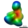
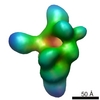
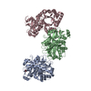
| Images | 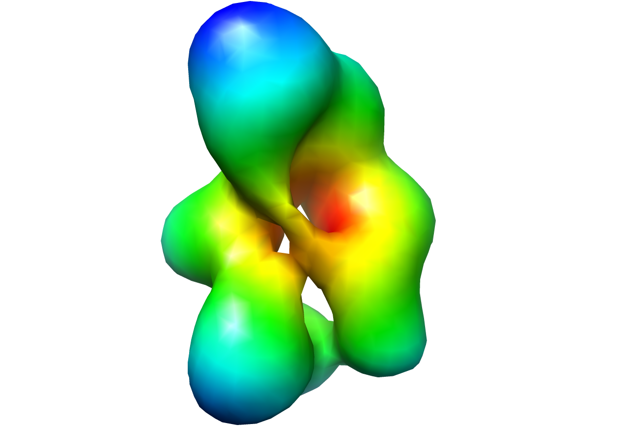 1638.png 1638.png | 218.6 KB | ||
| Archive directory |  http://ftp.pdbj.org/pub/emdb/structures/EMD-1638ftp://ftp.pdbj.org/pub/emdb/structures/EMD-1638 http://ftp.pdbj.org/pub/emdb/structures/EMD-1638ftp://ftp.pdbj.org/pub/emdb/structures/EMD-1638 | HTTPS FTP |
-Related structure data


-Links
| EMDB pages | EMDB (EBI/PDBe) / EMDataResource |
|---|
-Map
| File | Download / File: emd_1638.map.gz / Format: CCP4 / Size: 1001 KB / Type: IMAGE STORED AS FLOATING POINT NUMBER (4 BYTES) | ||||||||||||||||||||||||||||||||||||||||||||||||||||||||||||||||||||
|---|---|---|---|---|---|---|---|---|---|---|---|---|---|---|---|---|---|---|---|---|---|---|---|---|---|---|---|---|---|---|---|---|---|---|---|---|---|---|---|---|---|---|---|---|---|---|---|---|---|---|---|---|---|---|---|---|---|---|---|---|---|---|---|---|---|---|---|---|---|
| Annotation | Gyrase of mycoplasma pneumoniae | ||||||||||||||||||||||||||||||||||||||||||||||||||||||||||||||||||||
| Projections & slices | Image control
Images are generated by Spider. | ||||||||||||||||||||||||||||||||||||||||||||||||||||||||||||||||||||
| Voxel size | X=Y=Z: 5.2 Å | ||||||||||||||||||||||||||||||||||||||||||||||||||||||||||||||||||||
| Density |
| ||||||||||||||||||||||||||||||||||||||||||||||||||||||||||||||||||||
| Symmetry | Space group: 1 | ||||||||||||||||||||||||||||||||||||||||||||||||||||||||||||||||||||
| Details | EMDB XML:
CCP4 map header:
| ||||||||||||||||||||||||||||||||||||||||||||||||||||||||||||||||||||
 Z (Sec.)
 Y (Row.)
 X (Col.)


-Supplemental data
- Sample components
-Entire : Gyrase of Mycoplasma pneumoniae
| Entire | Name: Gyrase of Mycoplasma pneumoniae |
|---|---|
| Components |
|
-Supramolecule #1000: Gyrase of Mycoplasma pneumoniae
| Supramolecule | Name: Gyrase of Mycoplasma pneumoniae / type: sample / ID: 1000 / Details: Sample was fixed following the GRAFIX protocol / Number unique components: 1 |
|---|
-Macromolecule #1: Gyrase
| Macromolecule | Name: Gyrase / type: protein_or_peptide / ID: 1 / Name.synonym: Gyrase / Recombinant expression: Yes |
|---|---|
| Source (natural) | Organism: Mycoplasma pneumoniae (Filterable agent of primary atypical pneumonia) |
-Experimental details
-Structure determination
| Method | negative staining |
|---|---|
 Processing Processing | single particle reconstruction |
| Aggregation state | particle |
-Sample preparation
| Buffer | pH: 7.5 Details: 50mM Hepes, 20% glycerol, 0.075% glutaraldehyde, 100mM NaCl, 1.5 mM MgCl2 |
|---|---|
| Staining | Type: NEGATIVE Details: Grids were prepared by sandwich negative stain the sample was adsorbed to carbon on mica and floated on 1 % uranyl acetate the carbon was picked up with an uncoated grid then a second piece ...Details: Grids were prepared by sandwich negative stain the sample was adsorbed to carbon on mica and floated on 1 % uranyl acetate the carbon was picked up with an uncoated grid then a second piece of carbon, which was also floated on 1% uranyl acetate, was picked up with the same grid sandwiching the sample between the two layers of carbon |
| Grid | Details: 400 mesh copper grid |
| Vitrification | Cryogen name: NONE / Instrument: OTHER |
- Electron microscopy
| Microscope | FEI/PHILIPS CM200FEG |
|---|---|
| Electron beam | Acceleration voltage: 200 kV / Electron source: FIELD EMISSION GUN |
| Electron optics | Illumination mode: FLOOD BEAM / Imaging mode: BRIGHT FIELDBright-field microscopy / Cs: 2 mm / Nominal magnification: 27500 |
| Sample stage | Specimen holder: Eucentric / Specimen holder model: SIDE ENTRY, EUCENTRIC / Tilt angle min: -55 |
| Alignment procedure | Legacy - Astigmatism: Corrected at 200000 times magnification on graininess of carbon |
| Details | Collection of tilted pairs for random conical tilt reconstruction |
| Image recording | Category: CCD / Film or detector model: GENERIC CCD / Digitization - Sampling interval: 14.22 µm / Number real images: 68 Details: Images were recorded on CCD, no scanning sampling step size was adjusted to calibrated image size Bits/pixel: 12 |
| Tilt angle max | 0 |
-Image processing
| Final reconstruction | Applied symmetry - Point group: C1 (asymmetric) / Algorithm: OTHER / Resolution.type: BY AUTHOR / Resolution: 25.0 Å / Resolution method: FSC 0.5 CUT-OFF / Software - Name: IMAGIC, SPIDER, EMAN Details: Spider option BP 32F Back Projection - 3D, Sampled, Interpolated in Fourier space Number images used: 4767 |
|---|
