 Movie
Movie Controller


+ Open data

- Basic information
| Entry | 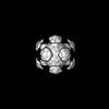 | |||||||||||||||||||||
|---|---|---|---|---|---|---|---|---|---|---|---|---|---|---|---|---|---|---|---|---|---|---|
| Title | In situ subtomogram average of GEM2 particles in human cells | |||||||||||||||||||||
 Map data Map data | FSC-weighted non-sharpened map | |||||||||||||||||||||
 Sample Sample |
| |||||||||||||||||||||
 Keywords Keywords |  Encapsulin / Protein Engineering / CYTOSOLIC PROTEIN Encapsulin / Protein Engineering / CYTOSOLIC PROTEIN | |||||||||||||||||||||
| Function / homology | Type 2A encapsulin shell protein SrpI-like / : / Type 2A encapsulin shell protein SrpI-like / encapsulin nanocompartment / Type 2A encapsulin shell protein SrpI Function and homology information Function and homology information | |||||||||||||||||||||
| Biological species |  Synechococcus elongatus PCC 7942 = FACHB-805 (bacteria) / Synechococcus elongatus PCC 7942 = FACHB-805 (bacteria) /  Homo sapiens (human) Homo sapiens (human) | |||||||||||||||||||||
| Method | subtomogram averaging / cryo EM / Resolution: 31.3 Å | |||||||||||||||||||||
 Authors Authors | Fung HKH / Hayashi Y / Salo VT / Babenko A / Zagoriy I / Brunner A / Ellenberg J / Mueller CW / Cuylen-Haering S / Mahamid J | |||||||||||||||||||||
| Funding support |  Germany, European Union, Germany, European Union,  France, 6 items France, 6 items
| |||||||||||||||||||||
 Citation Citation | Journal: Nat Methods / Year: 2023 Title: Genetically encoded multimeric tags for subcellular protein localization in cryo-EM. Authors: Herman K H Fung / Yuki Hayashi / Veijo T Salo / Anastasiia Babenko / Ievgeniia Zagoriy / Andreas Brunner / Jan Ellenberg / Christoph W Müller / Sara Cuylen-Haering / Julia Mahamid / Abstract: Cryo-electron tomography (cryo-ET) allows for label-free high-resolution imaging of macromolecular assemblies in their native cellular context. However, the localization of macromolecules of interest ...Cryo-electron tomography (cryo-ET) allows for label-free high-resolution imaging of macromolecular assemblies in their native cellular context. However, the localization of macromolecules of interest in tomographic volumes can be challenging. Here we present a ligand-inducible labeling strategy for intracellular proteins based on fluorescent, 25-nm-sized, genetically encoded multimeric particles (GEMs). The particles exhibit recognizable structural signatures, enabling their automated detection in cryo-ET data by convolutional neural networks. The coupling of GEMs to green fluorescent protein-tagged macromolecules of interest is triggered by addition of a small-molecule ligand, allowing for time-controlled labeling to minimize disturbance to native protein function. We demonstrate the applicability of GEMs for subcellular-level localization of endogenous and overexpressed proteins across different organelles in human cells using cryo-correlative fluorescence and cryo-ET imaging. We describe means for quantifying labeling specificity and efficiency, and for systematic optimization for rare and abundant protein targets, with emphasis on assessing the potential effects of labeling on protein function. | |||||||||||||||||||||
| History |
|
- Structure visualization
| Supplemental images |
|---|

- Downloads & links
-EMDB archive
| Map data | emd_16303.map.gz | 7.3 MB | EMDB map data format | |
|---|---|---|---|---|
| Header (meta data) | emd-16303-v30.xmlemd-16303.xml | 16.7 KB 16.7 KB | Display Display | EMDB header |
| FSC (resolution estimation) | emd_16303_fsc.xml | 4.7 KB | Display | FSC data file |
| Images | 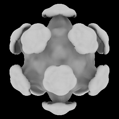 emd_16303.png emd_16303.png | 51 KB | ||
| Filedesc metadata | emd-16303.cif.gz | 5.6 KB | ||
| Others | emd_16303_half_map_1.map.gzemd_16303_half_map_2.map.gz | 6 MB 6 MB | ||
| Archive directory |  http://ftp.pdbj.org/pub/emdb/structures/EMD-16303ftp://ftp.pdbj.org/pub/emdb/structures/EMD-16303 http://ftp.pdbj.org/pub/emdb/structures/EMD-16303ftp://ftp.pdbj.org/pub/emdb/structures/EMD-16303 | HTTPS FTP |
-Related structure data
| Related structure data | C: citing same article ( |
|---|---|
| Similar structure data |

-Links
| EMDB pages | EMDB (EBI/PDBe) / EMDataResource |
|---|
-Map
| File | Download / File: emd_16303.map.gz / Format: CCP4 / Size: 8 MB / Type: IMAGE STORED AS FLOATING POINT NUMBER (4 BYTES) | ||||||||||||||||||||||||||||||||||||
|---|---|---|---|---|---|---|---|---|---|---|---|---|---|---|---|---|---|---|---|---|---|---|---|---|---|---|---|---|---|---|---|---|---|---|---|---|---|
| Annotation | FSC-weighted non-sharpened map | ||||||||||||||||||||||||||||||||||||
| Projections & slices | Image control
Images are generated by Spider. | ||||||||||||||||||||||||||||||||||||
| Voxel size | X=Y=Z: 6.85 Å | ||||||||||||||||||||||||||||||||||||
| Density |
| ||||||||||||||||||||||||||||||||||||
| Symmetry | Space group: 1 | ||||||||||||||||||||||||||||||||||||
| Details | EMDB XML:
|
 Z (Sec.)
 Y (Row.)
 X (Col.)


-Supplemental data
-Half map: Half map
| File | emd_16303_half_map_1.map | ||||||||||||
|---|---|---|---|---|---|---|---|---|---|---|---|---|---|
| Annotation | Half map | ||||||||||||
| Projections & Slices |
| ||||||||||||
| Density Histograms |


-Half map: Half map
| File | emd_16303_half_map_2.map | ||||||||||||
|---|---|---|---|---|---|---|---|---|---|---|---|---|---|
| Annotation | Half map | ||||||||||||
| Projections & Slices |
| ||||||||||||
| Density Histograms |


- Sample components
-Entire : Cryo-focused ion beam lamellae of human cells (HeLa, U2OS, Sum159...
| Entire | Name: Cryo-focused ion beam lamellae of human cells (HeLa, U2OS, Sum159) expressing the engineered genetically encoded multimeric tag GEM2 |
|---|---|
| Components |
|
-Supramolecule #1: Cryo-focused ion beam lamellae of human cells (HeLa, U2OS, Sum159...
| Supramolecule | Name: Cryo-focused ion beam lamellae of human cells (HeLa, U2OS, Sum159) expressing the engineered genetically encoded multimeric tag GEM2 type: cell / ID: 1 / Parent: 0 / Macromolecule list: all |
|---|---|
| Source (natural) | Organism: Synechococcus elongatus PCC 7942 = FACHB-805 (bacteria) |
-Macromolecule #1: Cryo-focused ion beam lamellae of human cells (HeLa, U2OS, Sum159...
| Macromolecule | Name: Cryo-focused ion beam lamellae of human cells (HeLa, U2OS, Sum159) expressing the engineered genetically encoded multimeric tag GEM2 type: protein_or_peptide / ID: 1 Details: Fusion of Synechococcus elongatus Srp1 with Halo-tag and the FKBP-rapamycin-binding (FRB) domain of mTOR Enantiomer: LEVO |
|---|---|
| Source (natural) | Organism: Homo sapiens (human) |
| Sequence | String: MGSTDNAPQL ALRDVAARQL ANATKTVPQL RTITPRWLVR LLHWTPVEAG IYRVNQVKDA SQITVACSER DESELPETFV DYIDNPREYL LSAVNTVVDV HTRISDLYSN PHDQIREQLR LTIEIMKERQ ESELINSREY GLLNNVAPGQ LVHTRNGAPT PDDLDELLIR ...String: MGSTDNAPQL ALRDVAARQL ANATKTVPQL RTITPRWLVR LLHWTPVEAG IYRVNQVKDA SQITVACSER DESELPETFV DYIDNPREYL LSAVNTVVDV HTRISDLYSN PHDQIREQLR LTIEIMKERQ ESELINSREY GLLNNVAPGQ LVHTRNGAPT PDDLDELLIR VWKEPAFFLA HPQAIAAFGR ECTRRGVPPA TVSLFGSSFI TWRGVPLIPS DKVPLENGKT KILLLRVGES RQGVVGLYQP NLPGEQGMGL SVRFMGINRK ALASYLVSLY CSLAVLTDDA LAVLDNVDVT QYHTYRYNGT GGGGSGGGSG GGSAEIGTGF PFDPHYVEVL GERMHYVDVG PRDGTPVLFL HGNPTSSYVW RNIIPHVAPT HRCIAPDLIG MGKSDKPDLG YFFDDHVRFM DAFIEALGLE EVVLVIHDWG SALGFHWAKR NPERVKGIAF MEFIRPIPTW DEWPEFARET FQAFRTTDVG RKLIIDQNVF IEGTLPMGVV RPLTEVEMDH YREPFLNPVD REPLWRFPNE LPIAGEPANI VALVEEYMDW LHQSPVPKLL FWGTPGVLIP PAEAARLAKS LPNCKAVDIG PGLNLLQEDN PDLIGSEIAR WLSTLEISGK LGSAGSAAGS GEGILWHEMW HEGLEEASRL YFGERNVKGM FEVLEPLHAM MERGPQTLKE TSFNQAYGRD LMEAQEWCRK YMKSGNVKDL LQAWDLYYHV FRRISK UniProtKB: Type 2A encapsulin shell protein SrpI |
-Experimental details
-Structure determination
| Method | cryo EM |
|---|---|
 Processing Processing | subtomogram averaging |
| Aggregation state | cell |
-Sample preparation
| Buffer | pH: 7.4 |
|---|---|
| Vitrification | Cryogen name: ETHANE |
- Electron microscopy
| Microscope | FEI TITAN KRIOS |
|---|---|
| Electron beam | Acceleration voltage: 300 kV / Electron source: FIELD EMISSION GUN |
| Electron optics | Illumination mode: FLOOD BEAM / Imaging mode: BRIGHT FIELDBright-field microscopy / Nominal defocus max: 4.0 µm / Nominal defocus min: 1.5 µm |
| Image recording | #0 - Image recording ID: 1 / #0 - Film or detector model: GATAN K3 BIOQUANTUM (6k x 4k) / #0 - Average electron dose: 2.0 e/Å2 / #1 - Image recording ID: 2 / #1 - Film or detector model: GATAN K2 SUMMIT (4k x 4k) / #1 - Average electron dose: 2.0 e/Å2 |
| Experimental equipment |  Model: Titan Krios / Image courtesy: FEI Company |
-Image processing
| Extraction | Number tomograms: 71 / Number images used: 1284 / Software - Name: Warp (ver. 1.0.9) |
|---|---|
| Final angle assignment | Type: MAXIMUM LIKELIHOOD / Software - Name: RELION (ver. 4.0.0-beta-2) |
| Final reconstruction | Applied symmetry - Point group: I (icosahedral) / Resolution.type: BY AUTHOR / Resolution: 31.3 Å / Resolution method: FSC 0.143 CUT-OFF / Software - Name: RELION (ver. 4.0.0-beta-2) / Number subtomograms used: 1284 |
| Details | All data acquired (with/without Volta phase plate, K2/K3) were pooled for subtomogram averaging. |
| Image recording ID | 1 |
| FSC plot (resolution estimation) |  |
