Netherlands Organisation for Scientific Research (NWO)
724.016.001
Netherlands
Citation
Journal: Nature / Year: 2023 Title: Visualization of translation and protein biogenesis at the ER membrane. Authors: Max Gemmer / Marten L Chaillet / Joyce van Loenhout / Rodrigo Cuevas Arenas / Dimitrios Vismpas / Mariska Gröllers-Mulderij / Fujiet A Koh / Pascal Albanese / Richard A Scheltema / Stuart C ...Authors: Max Gemmer / Marten L Chaillet / Joyce van Loenhout / Rodrigo Cuevas Arenas / Dimitrios Vismpas / Mariska Gröllers-Mulderij / Fujiet A Koh / Pascal Albanese / Richard A Scheltema / Stuart C Howes / Abhay Kotecha / Juliette Fedry / Friedrich Förster / Abstract: The dynamic ribosome-translocon complex, which resides at the endoplasmic reticulum (ER) membrane, produces a major fraction of the human proteome. It governs the synthesis, translocation, membrane ...The dynamic ribosome-translocon complex, which resides at the endoplasmic reticulum (ER) membrane, produces a major fraction of the human proteome. It governs the synthesis, translocation, membrane insertion, N-glycosylation, folding and disulfide-bond formation of nascent proteins. Although individual components of this machinery have been studied at high resolution in isolation, insights into their interplay in the native membrane remain limited. Here we use cryo-electron tomography, extensive classification and molecular modelling to capture snapshots of mRNA translation and protein maturation at the ER membrane at molecular resolution. We identify a highly abundant classical pre-translocation intermediate with eukaryotic elongation factor 1a (eEF1a) in an extended conformation, suggesting that eEF1a may remain associated with the ribosome after GTP hydrolysis during proofreading. At the ER membrane, distinct polysomes bind to different ER translocons specialized in the synthesis of proteins with signal peptides or multipass transmembrane proteins with the translocon-associated protein complex (TRAP) present in both. The near-complete atomic model of the most abundant ER translocon variant comprising the protein-conducting channel SEC61, TRAP and the oligosaccharyltransferase complex A (OSTA) reveals specific interactions of TRAP with other translocon components. We observe stoichiometric and sub-stoichiometric cofactors associated with OSTA, which are likely to include protein isomerases. In sum, we visualize ER-bound polysomes with their coordinated downstream machinery.
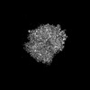
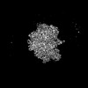
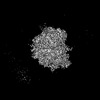
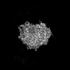
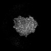
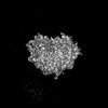
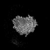
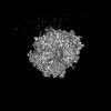
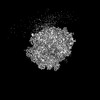
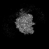
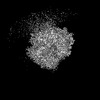
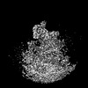
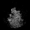
Organelle or cellular component: Ribosome in the Decoding-Sampling state
-
Supramolecule #1: Ribosome in the Decoding-Sampling state
Supramolecule
Name: Ribosome in the Decoding-Sampling state / type: organelle_or_cellular_component / ID: 1 / Parent: 0 / Macromolecule list: #1-#16 Details: Soluble and ER membrane-bound ribosomes in the Decoding-Sampling state
Film or detector model: GATAN K2 SUMMIT (4k x 4k) / Detector mode: COUNTING / Digitization - Frames/image: 7-7 / Number grids imaged: 8 / Average exposure time: 0.7 sec. / Average electron dose: 2.3 e/Å2
Experimental equipment
Model: Talos Arctica / Image courtesy: FEI Company
-
Image processing
Extraction
Number tomograms: 869 / Number images used: 134350 Reference model: Subtomogram average of ER membrane-associated ribosome Method: Template matching / Software - Name: PyTom (ver. 0.994)
Final 3D classification
Software - Name: RELION (ver. 3.1.1)
Final angle assignment
Type: OTHER
Final reconstruction
Resolution.type: BY AUTHOR / Resolution: 4.8 Å / Resolution method: FSC 0.143 CUT-OFF / Number subtomograms used: 26980
-
Atomic model buiding 1
Refinement
Space: REAL / Protocol: FLEXIBLE FIT
+
About Yorodumi
-
News
-
Feb 9, 2022. New format data for meta-information of EMDB entries
New format data for meta-information of EMDB entries
Version 3 of the EMDB header file is now the official format.
The previous official version 1.9 will be removed from the archive.
In the structure databanks used in Yorodumi, some data are registered as the other names, "COVID-19 virus" and "2019-nCoV". Here are the details of the virus and the list of structure data.
Jan 31, 2019. EMDB accession codes are about to change! (news from PDBe EMDB page)
EMDB accession codes are about to change! (news from PDBe EMDB page)
The allocation of 4 digits for EMDB accession codes will soon come to an end. Whilst these codes will remain in use, new EMDB accession codes will include an additional digit and will expand incrementally as the available range of codes is exhausted. The current 4-digit format prefixed with “EMD-” (i.e. EMD-XXXX) will advance to a 5-digit format (i.e. EMD-XXXXX), and so on. It is currently estimated that the 4-digit codes will be depleted around Spring 2019, at which point the 5-digit format will come into force.
The EM Navigator/Yorodumi systems omit the EMD- prefix.
Related info.:Q: What is EMD? / ID/Accession-code notation in Yorodumi/EM Navigator
Yorodumi is a browser for structure data from EMDB, PDB, SASBDB, etc.
This page is also the successor to EM Navigator detail page, and also detail information page/front-end page for Omokage search.
The word "yorodu" (or yorozu) is an old Japanese word meaning "ten thousand". "mi" (miru) is to see.
Related info.:EMDB / PDB / SASBDB / Comparison of 3 databanks / Yorodumi Search / Aug 31, 2016. New EM Navigator & Yorodumi / Yorodumi Papers / Jmol/JSmol / Function and homology information / Changes in new EM Navigator and Yorodumi
 Movie
Movie Controller

 Yorodumi
 Open data
 Basic information
 Map data
 Sample
 Homo sapiens (human)
 Authors
 Netherlands, 2 items
  Citation
 Structure visualization
 Downloads & links
 EMDB map data format
 emd_15871.png
 http://ftp.pdbj.org/pub/emdb/structures/EMD-15871
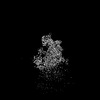


 Z (Sec.)
 Y (Row.)
 X (Col.)


 Sample components
 Processing
 Electron microscopy
