 Movie
Movie Controller

[English] 日本語
 Yorodumi
- EMDB-15808: In situ Dictyostelium discoideum 80S ribosome from dataset1 (2.17... -
+ Open data

- Basic information
| Entry |  | |||||||||
|---|---|---|---|---|---|---|---|---|---|---|
| Title | In situ Dictyostelium discoideum 80S ribosome from dataset1 (2.176A/px) | |||||||||
 Map data Map data | 80S ribosome from Dictyostelium discoideum cells from dataset1 (2.176A/px) Eukaryotic ribosome Eukaryotic ribosome | |||||||||
 Sample Sample |
| |||||||||
 Keywords Keywords | eukaryotic ribosome / translation / tRNA / elongation factor / expansion segments / RIBOSOME | |||||||||
| Biological species |  Dictyostelium discoideum AX2 (eukaryote) Dictyostelium discoideum AX2 (eukaryote) | |||||||||
| Method | subtomogram averaging / cryo EM / Resolution: 4.5 Å | |||||||||
 Authors Authors | Hoffmann PC / Kreysing JP / Khusainov I / Tuijtel MW / Welsch S / Beck M | |||||||||
| Funding support |  Germany, Germany,  United States, 2 items United States, 2 items
| |||||||||
 Citation Citation | Journal: Nat Commun / Year: 2022 Title: Structures of the eukaryotic ribosome and its translational states in situ. Authors: Patrick C Hoffmann / Jan Philipp Kreysing / Iskander Khusainov / Maarten W Tuijtel / Sonja Welsch / Martin Beck / Abstract: Ribosomes translate genetic information into primary structure. During translation, various cofactors transiently bind to the ribosome that undergoes prominent conformational and structural changes. ...Ribosomes translate genetic information into primary structure. During translation, various cofactors transiently bind to the ribosome that undergoes prominent conformational and structural changes. Different translational states of ribosomes have been well characterized in vitro. However, to which extent the known translational states are representative of the native situation inside cells has thus far only been addressed in prokaryotes. Here, we apply cryo-electron tomography to cryo-FIB milled Dictyostelium discoideum cells combined with subtomogram averaging and classification. We obtain an in situ structure that is locally resolved up to 3 Angstrom, the distribution of eukaryotic ribosome translational states, and unique arrangement of rRNA expansion segments. Our work demonstrates the use of in situ structural biology techniques for identifying distinct ribosome states within the cellular environment. | |||||||||
| History |
|
- Structure visualization
| Supplemental images |
|---|

- Downloads & links
-EMDB archive
| Map data | emd_15808.map.gz | 26.5 MB |  EMDB map data format EMDB map data format | |
|---|---|---|---|---|
| Header (meta data) | emd-15808-v30.xmlemd-15808.xml | 13.5 KB 13.5 KB | Display Display | EMDB header |
| FSC (resolution estimation) | emd_15808_fsc.xml | 13 KB | Display | FSC data file |
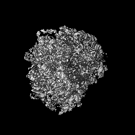
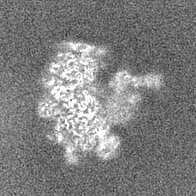
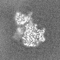
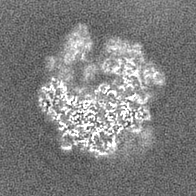
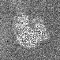
| Images | 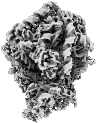 emd_15808.png emd_15808.png | 108.4 KB | ||
| Filedesc metadata | emd-15808.cif.gz | 4.1 KB | ||
| Others | emd_15808_half_map_1.map.gzemd_15808_half_map_2.map.gz | 26.7 MB 26.7 MB | ||
| Archive directory |  http://ftp.pdbj.org/pub/emdb/structures/EMD-15808ftp://ftp.pdbj.org/pub/emdb/structures/EMD-15808 http://ftp.pdbj.org/pub/emdb/structures/EMD-15808ftp://ftp.pdbj.org/pub/emdb/structures/EMD-15808 | HTTPS FTP |
-Related structure data
| Related structure data | C: citing same article ( |
|---|


-Links
| EMDB pages | EMDB (EBI/PDBe) / EMDataResource |
|---|
-Map
| File | Download / File: emd_15808.map.gz / Format: CCP4 / Size: 28.7 MB / Type: IMAGE STORED AS FLOATING POINT NUMBER (4 BYTES) | ||||||||||||||||||||||||||||||||||||
|---|---|---|---|---|---|---|---|---|---|---|---|---|---|---|---|---|---|---|---|---|---|---|---|---|---|---|---|---|---|---|---|---|---|---|---|---|---|
| Annotation | 80S ribosome from Dictyostelium discoideum cells from dataset1 (2.176A/px) | ||||||||||||||||||||||||||||||||||||
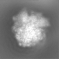
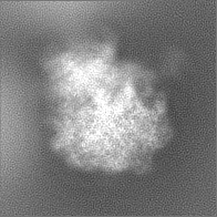
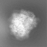
| Projections & slices | Image control
Images are generated by Spider. | ||||||||||||||||||||||||||||||||||||
| Voxel size | X=Y=Z: 2.176 Å | ||||||||||||||||||||||||||||||||||||
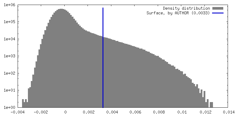
| Density |
| ||||||||||||||||||||||||||||||||||||
| Symmetry | Space group: 1 | ||||||||||||||||||||||||||||||||||||
| Details | EMDB XML:
|
 Z (Sec.)
 Y (Row.)
 X (Col.)


-Supplemental data
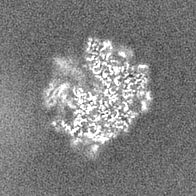
-Half map: 80S ribosome from Dictyostelium discoideum cells from dataset1...
| File | emd_15808_half_map_1.map | ||||||||||||
|---|---|---|---|---|---|---|---|---|---|---|---|---|---|
| Annotation | 80S ribosome from Dictyostelium discoideum cells from dataset1 (2.176A/px) | ||||||||||||
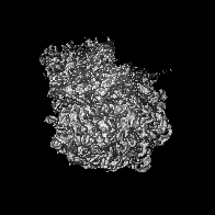
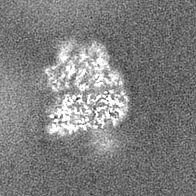
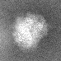
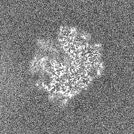
| Projections & Slices |
| ||||||||||||
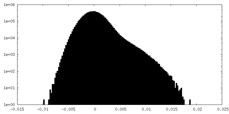
| Density Histograms |


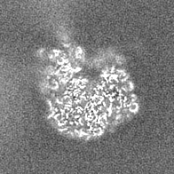
-Half map: 80S ribosome from Dictyostelium discoideum cells from dataset1...
| File | emd_15808_half_map_2.map | ||||||||||||
|---|---|---|---|---|---|---|---|---|---|---|---|---|---|
| Annotation | 80S ribosome from Dictyostelium discoideum cells from dataset1 (2.176A/px) | ||||||||||||
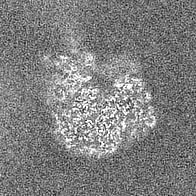
| Projections & Slices |
| ||||||||||||
| Density Histograms |


- Sample components
-Entire : 80S ribosome from inside Dictyostelium discoideum cells.
| Entire | Name: 80S ribosome from inside Dictyostelium discoideum cells.Eukaryotic ribosome |
|---|---|
| Components |
|
-Supramolecule #1: 80S ribosome from inside Dictyostelium discoideum cells.
| Supramolecule | Name: 80S ribosome from inside Dictyostelium discoideum cells. type: complex / ID: 1 / Parent: 0 Details: Cells from axenic Dictyostelium discoideum strain Ax2-214 with integrated GFP-Nup62 grown in HL5 medium were plunge frozen on Au 200mesh EM support grids. |
|---|---|
| Source (natural) | Organism: Dictyostelium discoideum AX2 (eukaryote) / Strain: Ax2-214 / Location in cell: cytoplasm of perinuclear region |
| Molecular weight | Theoretical: 4.2 MDa |
-Experimental details
-Structure determination
| Method | cryo EM |
|---|---|
 Processing Processing | subtomogram averaging |
| Aggregation state | cell |
-Sample preparation
| Buffer | pH: 6.5 |
|---|---|
| Grid | Model: Quantifoil / Material: GOLD / Mesh: 200 / Pretreatment - Type: GLOW DISCHARGE / Pretreatment - Time: 90 sec. |
| Vitrification | Cryogen name: ETHANE / Chamber temperature: 294 K / Instrument: LEICA EM GP |
- Electron microscopy
| Microscope | FEI TITAN KRIOS |
|---|---|
| Electron beam | Acceleration voltage: 300 kV / Electron source: FIELD EMISSION GUN |
| Electron optics | Illumination mode: FLOOD BEAM / Imaging mode: BRIGHT FIELDBright-field microscopy / Nominal defocus max: 5.0 µm / Nominal defocus min: 2.5 µm / Nominal magnification: 42000 |
| Image recording | Film or detector model: GATAN K3 BIOQUANTUM (6k x 4k) / Average electron dose: 2.3 e/Å2 |
| Experimental equipment |  Model: Titan Krios / Image courtesy: FEI Company |
-Image processing
| Extraction | Number tomograms: 127 / Number images used: 127000 |
|---|---|
| Final angle assignment | Type: OTHER |
| Final reconstruction | Applied symmetry - Point group: C1 (asymmetric) / Resolution.type: BY AUTHOR / Resolution: 4.5 Å / Resolution method: FSC 0.143 CUT-OFF / Number subtomograms used: 29858 |
| FSC plot (resolution estimation) |  |
