[English] 日本語
 Yorodumi
- EMDB-15520: Cryo-EM structure of human tankyrase 2 SAM-PARP filament (G1032W ... -
+ Open data
- Basic information
| Entry |  | |||||||||||||||
|---|---|---|---|---|---|---|---|---|---|---|---|---|---|---|---|---|
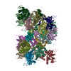
| Title | Cryo-EM structure of human tankyrase 2 SAM-PARP filament (G1032W mutant) | |||||||||||||||
 Map data Map data | ||||||||||||||||
 Sample Sample |
| |||||||||||||||
| Function / homology |  Function and homology information Function and homology informationXAV939 stabilizes AXIN /  NAD+ ADP-ribosyltransferase / protein localization to chromosome, telomeric region / negative regulation of telomere maintenance via telomere lengthening / protein auto-ADP-ribosylation / protein poly-ADP-ribosylation / NAD+ ADP-ribosyltransferase / protein localization to chromosome, telomeric region / negative regulation of telomere maintenance via telomere lengthening / protein auto-ADP-ribosylation / protein poly-ADP-ribosylation /  pericentriolar material / NAD+-protein ADP-ribosyltransferase activity / positive regulation of telomere capping / pericentriolar material / NAD+-protein ADP-ribosyltransferase activity / positive regulation of telomere capping /  NAD+ ADP-ribosyltransferase activity ...XAV939 stabilizes AXIN / NAD+ ADP-ribosyltransferase activity ...XAV939 stabilizes AXIN /  NAD+ ADP-ribosyltransferase / protein localization to chromosome, telomeric region / negative regulation of telomere maintenance via telomere lengthening / protein auto-ADP-ribosylation / protein poly-ADP-ribosylation / NAD+ ADP-ribosyltransferase / protein localization to chromosome, telomeric region / negative regulation of telomere maintenance via telomere lengthening / protein auto-ADP-ribosylation / protein poly-ADP-ribosylation /  pericentriolar material / NAD+-protein ADP-ribosyltransferase activity / positive regulation of telomere capping / pericentriolar material / NAD+-protein ADP-ribosyltransferase activity / positive regulation of telomere capping /  NAD+ ADP-ribosyltransferase activity / NAD+ ADP-ribosyltransferase activity /  Transferases; Glycosyltransferases; Pentosyltransferases / positive regulation of telomere maintenance via telomerase / Transferases; Glycosyltransferases; Pentosyltransferases / positive regulation of telomere maintenance via telomerase /  nucleotidyltransferase activity / TCF dependent signaling in response to WNT / Degradation of AXIN / nucleotidyltransferase activity / TCF dependent signaling in response to WNT / Degradation of AXIN /  Wnt signaling pathway / Regulation of PTEN stability and activity / protein polyubiquitination / positive regulation of canonical Wnt signaling pathway / Wnt signaling pathway / Regulation of PTEN stability and activity / protein polyubiquitination / positive regulation of canonical Wnt signaling pathway /  nuclear envelope / nuclear envelope /  chromosome, telomeric region / Ub-specific processing proteases / chromosome, telomeric region / Ub-specific processing proteases /  Golgi membrane / perinuclear region of cytoplasm / Golgi membrane / perinuclear region of cytoplasm /  enzyme binding / enzyme binding /  metal ion binding / metal ion binding /  nucleus / nucleus /  cytosol / cytosol /  cytoplasm cytoplasmSimilarity search - Function | |||||||||||||||
| Biological species |   Homo sapiens (human) Homo sapiens (human) | |||||||||||||||
| Method | helical reconstruction /  cryo EM / Resolution: 2.98 Å cryo EM / Resolution: 2.98 Å | |||||||||||||||
 Authors Authors | Mariotti L / Inian O / Desfosses A / Beuron F / Morris EP / Guettler S | |||||||||||||||
| Funding support |  United Kingdom, 4 items United Kingdom, 4 items
| |||||||||||||||
 Citation Citation |  Journal: Nature / Year: 2022 Journal: Nature / Year: 2022Title: Structural basis of tankyrase activation by polymerization. Authors: Nisha Pillay / Laura Mariotti / Mariola Zaleska / Oviya Inian / Matthew Jessop / Sam Hibbs / Ambroise Desfosses / Paul C R Hopkins / Catherine M Templeton / Fabienne Beuron / Edward P Morris ...Authors: Nisha Pillay / Laura Mariotti / Mariola Zaleska / Oviya Inian / Matthew Jessop / Sam Hibbs / Ambroise Desfosses / Paul C R Hopkins / Catherine M Templeton / Fabienne Beuron / Edward P Morris / Sebastian Guettler /   Abstract: The poly-ADP-ribosyltransferase tankyrase (TNKS, TNKS2) controls a wide range of disease-relevant cellular processes, including WNT-β-catenin signalling, telomere length maintenance, Hippo ...The poly-ADP-ribosyltransferase tankyrase (TNKS, TNKS2) controls a wide range of disease-relevant cellular processes, including WNT-β-catenin signalling, telomere length maintenance, Hippo signalling, DNA damage repair and glucose homeostasis. This has incentivized the development of tankyrase inhibitors. Notwithstanding, our knowledge of the mechanisms that control tankyrase activity has remained limited. Both catalytic and non-catalytic functions of tankyrase depend on its filamentous polymerization. Here we report the cryo-electron microscopy reconstruction of a filament formed by a minimal active unit of tankyrase, comprising the polymerizing sterile alpha motif (SAM) domain and its adjacent catalytic domain. The SAM domain forms a novel antiparallel double helix, positioning the protruding catalytic domains for recurring head-to-head and tail-to-tail interactions. The head interactions are highly conserved among tankyrases and induce an allosteric switch in the active site within the catalytic domain to promote catalysis. Although the tail interactions have a limited effect on catalysis, they are essential to tankyrase function in WNT-β-catenin signalling. This work reveals a novel SAM domain polymerization mode, illustrates how supramolecular assembly controls catalytic and non-catalytic functions, provides important structural insights into the regulation of a non-DNA-dependent poly-ADP-ribosyltransferase and will guide future efforts to modulate tankyrase and decipher its contribution to disease mechanisms. | |||||||||||||||
| History |
|
- Structure visualization
| Supplemental images |
|---|
- Downloads & links
-EMDB archive
| Map data |  emd_15520.map.gz emd_15520.map.gz | 105 MB |  EMDB map data format EMDB map data format | |
|---|---|---|---|---|
| Header (meta data) |  emd-15520-v30.xml emd-15520-v30.xml emd-15520.xml emd-15520.xml | 15.3 KB 15.3 KB | Display Display |  EMDB header EMDB header |
| FSC (resolution estimation) |  emd_15520_fsc.xml emd_15520_fsc.xml | 14.1 KB | Display |  FSC data file FSC data file |
| Images |  emd_15520.png emd_15520.png | 193.3 KB | ||
| Archive directory |  http://ftp.pdbj.org/pub/emdb/structures/EMD-15520 http://ftp.pdbj.org/pub/emdb/structures/EMD-15520 ftp://ftp.pdbj.org/pub/emdb/structures/EMD-15520 ftp://ftp.pdbj.org/pub/emdb/structures/EMD-15520 | HTTPS FTP |
-Related structure data
| Related structure data |  8alyMC M: atomic model generated by this map C: citing same article ( |
|---|---|
| Similar structure data | Similarity search - Function & homology  F&H Search F&H Search |
- Links
Links
| EMDB pages |  EMDB (EBI/PDBe) / EMDB (EBI/PDBe) /  EMDataResource EMDataResource |
|---|---|
| Related items in Molecule of the Month |
- Map
Map
| File |  Download / File: emd_15520.map.gz / Format: CCP4 / Size: 244.1 MB / Type: IMAGE STORED AS FLOATING POINT NUMBER (4 BYTES) Download / File: emd_15520.map.gz / Format: CCP4 / Size: 244.1 MB / Type: IMAGE STORED AS FLOATING POINT NUMBER (4 BYTES) | ||||||||||||||||||||||||||||||||||||
|---|---|---|---|---|---|---|---|---|---|---|---|---|---|---|---|---|---|---|---|---|---|---|---|---|---|---|---|---|---|---|---|---|---|---|---|---|---|
| Projections & slices | Image control
Images are generated by Spider. | ||||||||||||||||||||||||||||||||||||
| Voxel size | X=Y=Z: 1.06 Å | ||||||||||||||||||||||||||||||||||||
| Density |
| ||||||||||||||||||||||||||||||||||||
| Symmetry | Space group: 1 | ||||||||||||||||||||||||||||||||||||
| Details | EMDB XML:
|
-Supplemental data
- Sample components
-Entire : TNKS2 SAM-PARP (867-1162) filament
| Entire | Name: TNKS2 SAM-PARP (867-1162) filament |
|---|---|
| Components |
|
-Supramolecule #1: TNKS2 SAM-PARP (867-1162) filament
| Supramolecule | Name: TNKS2 SAM-PARP (867-1162) filament / type: complex / Chimera: Yes / ID: 1 / Parent: 0 / Macromolecule list: #1 Details: double-helical filament of human TNKS2 SAM-PARP G1032W, residues 867-1162, N-terminal vector-derived SNA tripeptide, 20 protomers in refined structure |
|---|---|
| Source (natural) | Organism:   Homo sapiens (human) Homo sapiens (human) |
| Molecular weight | Theoretical: 34 kDa/nm |
-Macromolecule #1: Poly [ADP-ribose] polymerase tankyrase-2
| Macromolecule | Name: Poly [ADP-ribose] polymerase tankyrase-2 / type: protein_or_peptide / ID: 1 / Number of copies: 20 / Enantiomer: LEVO / EC number:  NAD+ ADP-ribosyltransferase NAD+ ADP-ribosyltransferase |
|---|---|
| Source (natural) | Organism:   Homo sapiens (human) Homo sapiens (human) |
| Molecular weight | Theoretical: 34.077668 KDa |
| Recombinant expression | Organism:   Escherichia coli BL21(DE3) (bacteria) Escherichia coli BL21(DE3) (bacteria) |
| Sequence | String: SNAEKKEVPG VDFSITQFVR NLGLEHLMDI FEREQITLDV LVEMGHKELK EIGINAYGHR HKLIKGVERL ISGQQGLNPY LTLNTSGSG TILIDLSPDD KEFQSVEEEM QSTVREHRDG GHAGGIFNRY NILKIQKVCN KKLWERYTHR RKEVSEENHN H ANERMLFH ...String: SNAEKKEVPG VDFSITQFVR NLGLEHLMDI FEREQITLDV LVEMGHKELK EIGINAYGHR HKLIKGVERL ISGQQGLNPY LTLNTSGSG TILIDLSPDD KEFQSVEEEM QSTVREHRDG GHAGGIFNRY NILKIQKVCN KKLWERYTHR RKEVSEENHN H ANERMLFH WSPFVNAIIH KGFDERHAYI GGMFGAGIYF AENSSKSNQY VYGIGGGTGC PVHKDRSCYI CHRQLLFCRV TL GKSFLQF SAMKMAHSPP GHHSVTGRPS VNGLALAEYV IYRGEQAYPE YLITYQIMRP EG |
-Macromolecule #2: ZINC ION
| Macromolecule | Name: ZINC ION / type: ligand / ID: 2 / Number of copies: 20 / Formula: ZN |
|---|---|
| Molecular weight | Theoretical: 65.409 Da |
-Experimental details
-Structure determination
| Method |  cryo EM cryo EM |
|---|---|
 Processing Processing | helical reconstruction |
| Aggregation state | filament |
- Sample preparation
| Concentration | 0.86 mg/mL | ||||||||||||
|---|---|---|---|---|---|---|---|---|---|---|---|---|---|
| Buffer | pH: 7.5 Component:
Details: After brief incubation on the grid, the sample was washed 10 times with water to gradually lower the salt concentration and improve sample contrast. | ||||||||||||
| Grid | Model: Quantifoil R1.2/1.3 / Material: COPPER / Mesh: 400 / Support film - Material: CARBON / Support film - topology: CONTINUOUS / Pretreatment - Type: GLOW DISCHARGE | ||||||||||||
| Vitrification | Cryogen name: ETHANE / Instrument: FEI VITROBOT MARK IV | ||||||||||||
| Details | The PARP domain is inactivated by a G1032W mutation. |
- Electron microscopy
| Microscope | TFS KRIOS |
|---|---|
| Electron beam | Acceleration voltage: 300 kV / Electron source:  FIELD EMISSION GUN FIELD EMISSION GUN |
| Electron optics | Illumination mode: FLOOD BEAM / Imaging mode: BRIGHT FIELD Bright-field microscopy / Cs: 2.7 mm / Nominal defocus max: 3.5 µm / Nominal defocus min: 1.2 µm / Nominal magnification: 81000 Bright-field microscopy / Cs: 2.7 mm / Nominal defocus max: 3.5 µm / Nominal defocus min: 1.2 µm / Nominal magnification: 81000 |
| Sample stage | Specimen holder model: FEI TITAN KRIOS AUTOGRID HOLDER / Cooling holder cryogen: NITROGEN |
| Temperature | Min: 80.0 K / Max: 80.0 K |
| Image recording | #0 - Image recording ID: 1 / #0 - Film or detector model: GATAN K2 SUMMIT (4k x 4k) / #0 - Detector mode: INTEGRATING / #0 - Average electron dose: 40.0 e/Å2 / #1 - Image recording ID: 2 / #1 - Film or detector model: GATAN K3 (6k x 4k) / #1 - Average electron dose: 40.0 e/Å2 |
| Experimental equipment |  Model: Titan Krios / Image courtesy: FEI Company |
 Movie
Movie Controller


 X (Sec.)
 Y (Row.)
 Z (Col.)


