- EMDB-15417: human MutSalpha (MSH2/MSH6) binding to DNA with a GT mismatch -
+
Open data
ID or keywords:
Loading...
-
Basic information
Entry
Database: EMDB / ID: EMD-15417
Title
human MutSalpha (MSH2/MSH6) binding to DNA with a GT mismatch
Map data
Sample
Complex: MutSalpha on mismatched DNA
Complex: MSH2
Protein or peptide: DNA mismatch repair protein Msh2
Complex: MSH6
Protein or peptide: DNA mismatch repair protein Msh6
Complex: DNA containing a G/T mismatch
DNA: DNA (50-MER)
DNA: DNA (50-MER)
Ligand: MAGNESIUM ION
Ligand: ADENOSINE-5'-DIPHOSPHATE
Function / homology
Function and homology information
somatic recombination of immunoglobulin genes involved in immune response / MutSbeta complex / Defective Mismatch Repair Associated With MSH3 / MutSalpha complex / Defective Mismatch Repair Associated With MSH2 / Defective Mismatch Repair Associated With MSH6 / guanine/thymine mispair binding / somatic recombination of immunoglobulin gene segments / maintenance of DNA repeat elements / B cell mediated immunity ...somatic recombination of immunoglobulin genes involved in immune response / MutSbeta complex / Defective Mismatch Repair Associated With MSH3 / MutSalpha complex / Defective Mismatch Repair Associated With MSH2 / Defective Mismatch Repair Associated With MSH6 / guanine/thymine mispair binding / somatic recombination of immunoglobulin gene segments / maintenance of DNA repeat elements / B cell mediated immunity / positive regulation of helicase activity / positive regulation of isotype switching to IgA isotypes / meiotic mismatch repair / centromeric DNA binding / mitotic recombination / positive regulation of isotype switching to IgG isotypes / mismatched DNA binding / negative regulation of DNA recombination / Mismatch repair (MMR) directed by MSH2:MSH3 (MutSbeta) / Mismatch repair (MMR) directed by MSH2:MSH6 (MutSalpha) / isotype switching / postreplication repair / oxidative phosphorylation / response to UV-B / mitotic intra-S DNA damage checkpoint signaling / ATP-dependent DNA damage sensor activity / germ cell development / response to X-ray / mismatch repair / intrinsic apoptotic signaling pathway in response to DNA damage by p53 class mediator / somatic hypermutation of immunoglobulin genes / ATP-dependent activity, acting on DNA / response to UV / protein localization to chromatin / methylated histone binding / intrinsic apoptotic signaling pathway / B cell differentiation / determination of adult lifespan / TP53 Regulates Transcription of DNA Repair Genes / male gonad development / intrinsic apoptotic signaling pathway in response to DNA damage / double-strand break repair / spermatogenesis / negative regulation of neuron apoptotic process / in utero embryonic development / damaged DNA binding / chromosome, telomeric region / DNA repair / intracellular membrane-bounded organelle / chromatin binding / chromatin / Golgi apparatus / enzyme binding / ATP hydrolysis activity / protein homodimerization activity / DNA binding / nucleoplasm / ATP binding / membrane / nucleus / cytosol Similarity search - Function
DNA mismatch repair Msh2-type / DNA mismatch repair protein Msh2 / DNA mismatch repair protein MutS/MSH / DNA mismatch repair protein MutS-like, N-terminal / DNA mismatch repair protein MutS, connector domain / DNA mismatch repair protein MutS, clamp / DNA mismatch repair protein MutS, N-terminal / MutS, connector domain superfamily / MutS domain I / MutS domain II ...DNA mismatch repair Msh2-type / DNA mismatch repair protein Msh2 / DNA mismatch repair protein MutS/MSH / DNA mismatch repair protein MutS-like, N-terminal / DNA mismatch repair protein MutS, connector domain / DNA mismatch repair protein MutS, clamp / DNA mismatch repair protein MutS, N-terminal / MutS, connector domain superfamily / MutS domain I / MutS domain II / MutS family domain IV / MutS domain III / DNA mismatch repair MutS family / DNA mismatch repair protein MutS, C-terminal / DNA mismatch repair protein MutS, core / DNA mismatch repair protein MutS, core domain superfamily / MutS domain V / DNA mismatch repair proteins mutS family signature. / DNA-binding domain of DNA mismatch repair MUTS family / ATPase domain of DNA mismatch repair MUTS family / domain with conserved PWWP motif / PWWP domain / PWWP domain profile. / PWWP domain / P-loop containing nucleoside triphosphate hydrolase Similarity search - Domain/homology
Netherlands Organisation for Scientific Research (NWO)
NOW-TOP 714.016.002
Netherlands
European Commission
H2020-MSCA-ITN-2016 722433 DNAREPAIRMAN
European Union
Citation
Journal: Nucleic Acids Res / Year: 2023 Title: Unexpected moves: a conformational change in MutSα enables high-affinity DNA mismatch binding. Authors: Susanne R Bruekner / Wietske Pieters / Alexander Fish / A Manuel Liaci / Serge Scheffers / Emily Rayner / Daphne Kaldenbach / Lisa Drost / Marleen Dekker / Sandrine van Hees-Stuivenberg / ...Authors: Susanne R Bruekner / Wietske Pieters / Alexander Fish / A Manuel Liaci / Serge Scheffers / Emily Rayner / Daphne Kaldenbach / Lisa Drost / Marleen Dekker / Sandrine van Hees-Stuivenberg / Elly Delzenne-Goette / Charlotte de Konink / Hellen Houlleberghs / Hendrikus Jan Dubbink / Abeer AlSaegh / Niels de Wind / Friedrich Förster / Hein Te Riele / Titia K Sixma / Abstract: The DNA mismatch repair protein MutSα recognizes wrongly incorporated DNA bases and initiates their correction during DNA replication. Dysfunctions in mismatch repair lead to a predisposition to ...The DNA mismatch repair protein MutSα recognizes wrongly incorporated DNA bases and initiates their correction during DNA replication. Dysfunctions in mismatch repair lead to a predisposition to cancer. Here, we study the homozygous mutation V63E in MSH2 that was found in the germline of a patient with suspected constitutional mismatch repair deficiency syndrome who developed colorectal cancer before the age of 30. Characterization of the mutant in mouse models, as well as slippage and repair assays, shows a mildly pathogenic phenotype. Using cryogenic electron microscopy and surface plasmon resonance, we explored the mechanistic effect of this mutation on MutSα function. We discovered that V63E disrupts a previously unappreciated interface between the mismatch binding domains (MBDs) of MSH2 and MSH6 and leads to reduced DNA binding. Our research identifies this interface as a 'safety lock' that ensures high-affinity DNA binding to increase replication fidelity. Our mechanistic model explains the hypomorphic phenotype of the V63E patient mutation and other variants in the MBD interface.
Model: Quantifoil R1.2/1.3 / Material: COPPER / Mesh: 300 / Support film - Material: CARBON / Support film - topology: HOLEY / Pretreatment - Type: GLOW DISCHARGE / Pretreatment - Time: 45 sec. / Pretreatment - Atmosphere: AIR / Pretreatment - Pressure: 0.01 kPa
Vitrification
Cryogen name: ETHANE / Chamber humidity: 100 % / Chamber temperature: 277 K / Instrument: FEI VITROBOT MARK IV / Details: 3 s blotting with blot force 10.
Details
2 uM MutSa with 2-fold excess DNA oligo
-
Electron microscopy
Microscope
FEI TITAN KRIOS
Electron beam
Acceleration voltage: 300 kV / Electron source: FIELD EMISSION GUN
collected on Krios 1 at Netherlands Center for Electron Nanoscopy (NeCEN)
Image recording
Film or detector model: GATAN K3 (6k x 4k) / Number grids imaged: 1 / Number real images: 4821 / Average exposure time: 1.9 sec. / Average electron dose: 50.0 e/Å2
Experimental equipment
Model: Titan Krios / Image courtesy: FEI Company
-
Image processing
Particle selection
Number selected: 5412220
Initial angle assignment
Type: MAXIMUM LIKELIHOOD / Software - Name: cryoSPARC
Final 3D classification
Number classes: 5 / Software - Name: RELION (ver. 4.0)
Final angle assignment
Type: MAXIMUM LIKELIHOOD / Software - Name: RELION (ver. 4.0)
Final reconstruction
Number classes used: 1 / Applied symmetry - Point group: C1 (asymmetric) / Resolution.type: BY AUTHOR / Resolution: 2.8 Å / Resolution method: FSC 0.143 CUT-OFF / Software - Name: RELION (ver. 4.0) / Number images used: 289289
In the structure databanks used in Yorodumi, some data are registered as the other names, "COVID-19 virus" and "2019-nCoV". Here are the details of the virus and the list of structure data.
Jan 31, 2019. EMDB accession codes are about to change! (news from PDBe EMDB page)
EMDB accession codes are about to change! (news from PDBe EMDB page)
The allocation of 4 digits for EMDB accession codes will soon come to an end. Whilst these codes will remain in use, new EMDB accession codes will include an additional digit and will expand incrementally as the available range of codes is exhausted. The current 4-digit format prefixed with “EMD-” (i.e. EMD-XXXX) will advance to a 5-digit format (i.e. EMD-XXXXX), and so on. It is currently estimated that the 4-digit codes will be depleted around Spring 2019, at which point the 5-digit format will come into force.
The EM Navigator/Yorodumi systems omit the EMD- prefix.
Related info.:Q: What is EMD? / ID/Accession-code notation in Yorodumi/EM Navigator
Yorodumi is a browser for structure data from EMDB, PDB, SASBDB, etc.
This page is also the successor to EM Navigator detail page, and also detail information page/front-end page for Omokage search.
The word "yorodu" (or yorozu) is an old Japanese word meaning "ten thousand". "mi" (miru) is to see.
Related info.:EMDB / PDB / SASBDB / Comparison of 3 databanks / Yorodumi Search / Aug 31, 2016. New EM Navigator & Yorodumi / Yorodumi Papers / Jmol/JSmol / Function and homology information / Changes in new EM Navigator and Yorodumi
 Movie
Movie Controller


 Open data
 Basic information
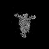
 Map data
 Sample
 Function and homology information
 mitotic recombination / positive regulation of isotype switching to IgG isotypes / mismatched DNA binding / negative regulation of DNA recombination / Mismatch repair (MMR) directed by MSH2:MSH3 (MutSbeta) / Mismatch repair (MMR) directed by MSH2:MSH6 (MutSalpha) /
 Authors
 Netherlands, European Union, 2 items
  Citation
 Structure visualization
 Downloads & links
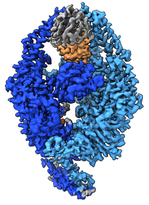 emd_15417.png
 http://ftp.pdbj.org/pub/emdb/structures/EMD-15417


 Z (Sec.)
 Y (Row.)
 X (Col.)


 Sample components


 Processing
 Electron microscopy


