Movie
Movie Controller

[English] 日本語
 Yorodumi
- EMDB-15358: Structure of dimeric yeast RNA polymerase II bound to a transcrip... -
+ Open data

- Basic information
| Entry |  | |||||||||
|---|---|---|---|---|---|---|---|---|---|---|
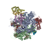
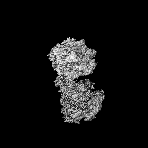
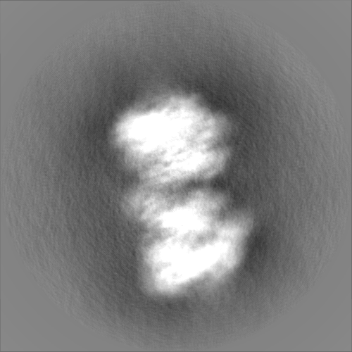
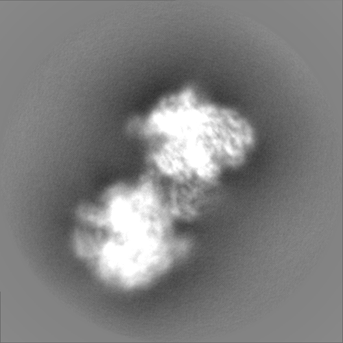
| Title | Structure of dimeric yeast RNA polymerase II bound to a transcription bubble (consensus map) | |||||||||
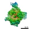
 Map data Map data | RNA Polymerase II dimer loaded with a DNA-RNA scaffold | |||||||||
 Sample Sample |
| |||||||||
 Keywords Keywords |  transcription regulation / RNA 3' end processing / transcription termination / GENE REGULATION transcription regulation / RNA 3' end processing / transcription termination / GENE REGULATION | |||||||||
| Biological species |  Saccharomyces cerevisiae (brewer's yeast) Saccharomyces cerevisiae (brewer's yeast) | |||||||||
| Method | single particle reconstruction / cryo EM / Resolution: 4.6 Å | |||||||||
 Authors Authors | Carminati M / Manav MC / Bellini D / Passmore LA | |||||||||
| Funding support | European Union, 2 items
| |||||||||
 Citation Citation | Journal: Mol Cell / Year: 2023 Title: A direct interaction between CPF and RNA Pol II links RNA 3' end processing to transcription. Authors: Manuel Carminati / Juan B Rodríguez-Molina / M Cemre Manav / Dom Bellini / Lori A Passmore /  Abstract: Transcription termination by RNA polymerase II (RNA Pol II) is linked to RNA 3' end processing by the cleavage and polyadenylation factor (CPF or CPSF). CPF contains endonuclease, poly(A) polymerase, ...Transcription termination by RNA polymerase II (RNA Pol II) is linked to RNA 3' end processing by the cleavage and polyadenylation factor (CPF or CPSF). CPF contains endonuclease, poly(A) polymerase, and protein phosphatase activities, which cleave and polyadenylate pre-mRNAs and dephosphorylate RNA Pol II to control transcription. Exactly how the RNA 3' end processing machinery is coupled to transcription remains unclear. Here, we combine in vitro reconstitution, structural studies, and genome-wide analyses to show that yeast CPF physically and functionally interacts with RNA Pol II. Surprisingly, CPF-mediated dephosphorylation promotes the formation of an RNA Pol II stalk-to-stalk homodimer in vitro. This dimer is compatible with transcription but not with the binding of transcription elongation factors. Disruption of the dimerization interface in cells causes transcription defects, including altered RNA Pol II abundance on protein-coding genes, tRNA genes, and intergenic regions. We hypothesize that RNA Pol II dimerization may provide a mechanistic basis for the allosteric model of transcription termination. | |||||||||
| History |
|
- Structure visualization
| Supplemental images |
|---|

- Downloads & links
-EMDB archive
| Map data | emd_15358.map.gz | 427.7 MB |  EMDB map data format EMDB map data format | |
|---|---|---|---|---|
| Header (meta data) | emd-15358-v30.xmlemd-15358.xml | 19.7 KB 19.7 KB | Display Display | EMDB header |
| FSC (resolution estimation) | emd_15358_fsc.xml | 17.8 KB | Display | FSC data file |
| Images | 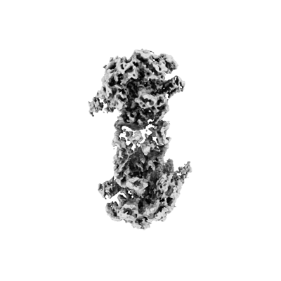 emd_15358.png emd_15358.png | 50.5 KB | ||
| Filedesc metadata | emd-15358.cif.gz | 5.1 KB | ||
| Others | emd_15358_half_map_1.map.gzemd_15358_half_map_2.map.gz | 380.4 MB 380.5 MB | ||
| Archive directory |  http://ftp.pdbj.org/pub/emdb/structures/EMD-15358ftp://ftp.pdbj.org/pub/emdb/structures/EMD-15358 http://ftp.pdbj.org/pub/emdb/structures/EMD-15358ftp://ftp.pdbj.org/pub/emdb/structures/EMD-15358 | HTTPS FTP |
-Related structure data

-Links
| EMDB pages | EMDB (EBI/PDBe) / EMDataResource |
|---|
-Map
| File | Download / File: emd_15358.map.gz / Format: CCP4 / Size: 476.8 MB / Type: IMAGE STORED AS FLOATING POINT NUMBER (4 BYTES) | ||||||||||||||||||||||||||||||||||||
|---|---|---|---|---|---|---|---|---|---|---|---|---|---|---|---|---|---|---|---|---|---|---|---|---|---|---|---|---|---|---|---|---|---|---|---|---|---|
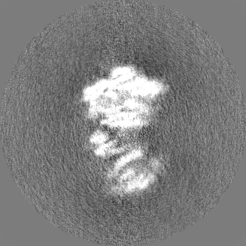
| Annotation | RNA Polymerase II dimer loaded with a DNA-RNA scaffold | ||||||||||||||||||||||||||||||||||||
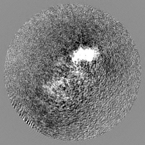
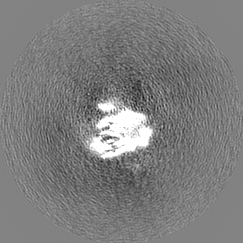
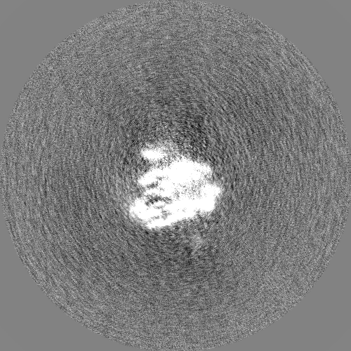
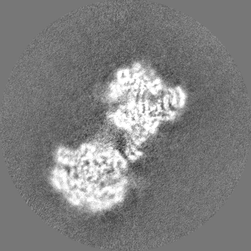
| Projections & slices | Image control
Images are generated by Spider. | ||||||||||||||||||||||||||||||||||||
| Voxel size | X=Y=Z: 0.83 Å | ||||||||||||||||||||||||||||||||||||
| Density |
| ||||||||||||||||||||||||||||||||||||
| Symmetry | Space group: 1 | ||||||||||||||||||||||||||||||||||||
| Details | EMDB XML:
|
 Z (Sec.)
 Y (Row.)
 X (Col.)


-Supplemental data
-Half map: #1
| File | emd_15358_half_map_1.map | ||||||||||||
|---|---|---|---|---|---|---|---|---|---|---|---|---|---|
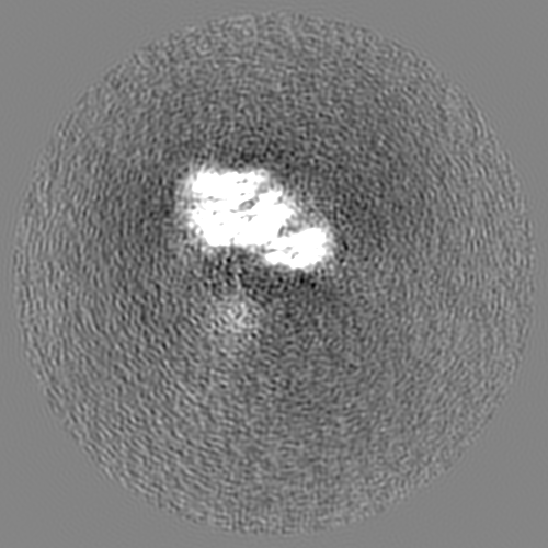
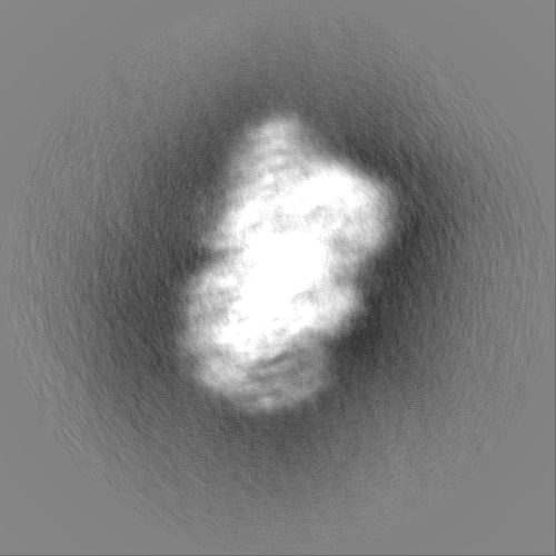
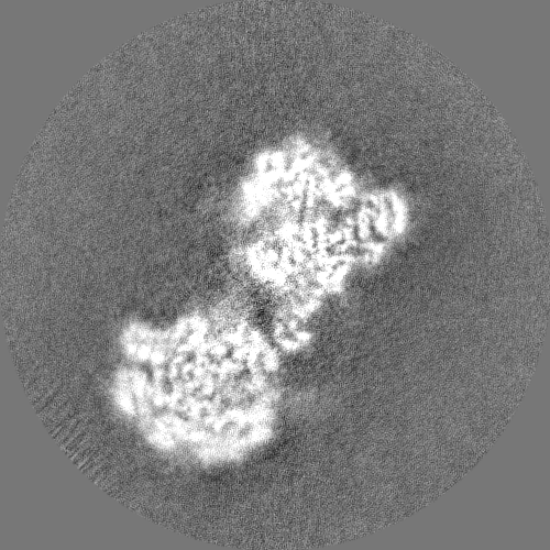
| Projections & Slices |
| ||||||||||||
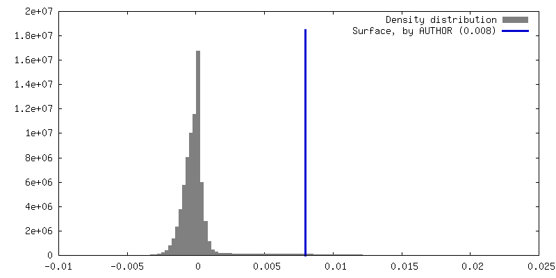
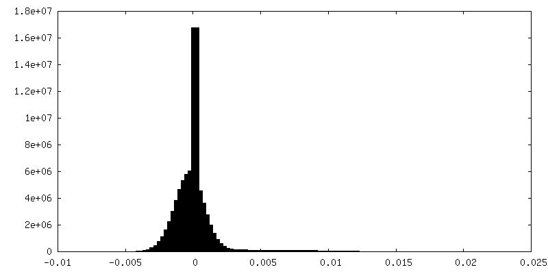
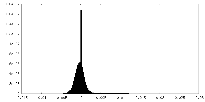
| Density Histograms |


-Half map: #2
| File | emd_15358_half_map_2.map | ||||||||||||
|---|---|---|---|---|---|---|---|---|---|---|---|---|---|
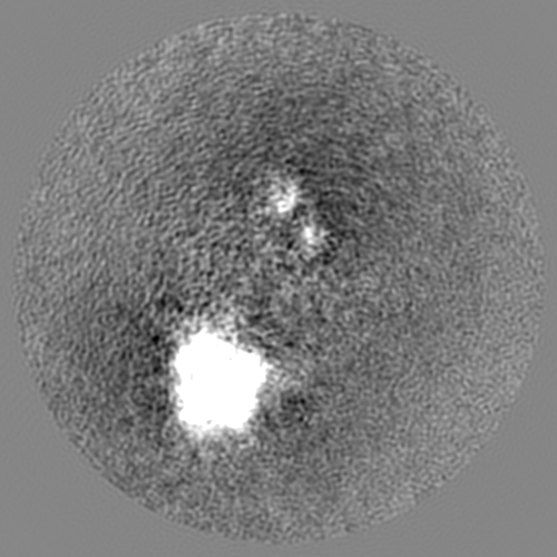
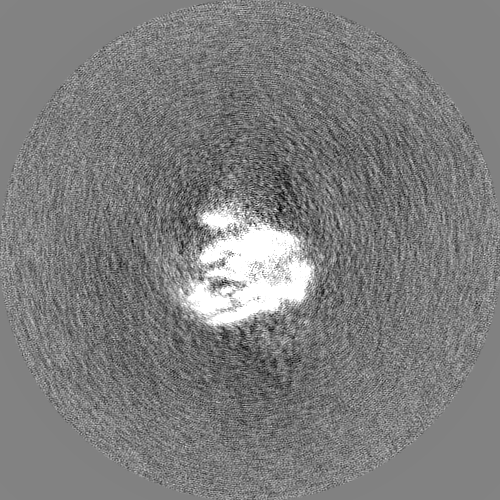
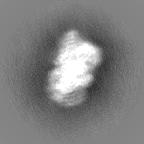
| Projections & Slices |
| ||||||||||||
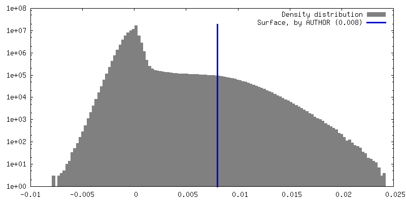
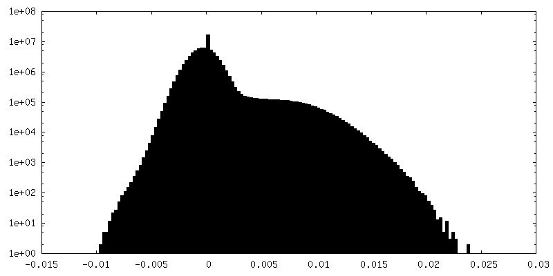
| Density Histograms |

- Sample components
-Entire : S. cerevisiae RNA polymerase II
| Entire | Name: S. cerevisiae RNA polymerase II |
|---|---|
| Components |
|
-Supramolecule #1: S. cerevisiae RNA polymerase II
| Supramolecule | Name: S. cerevisiae RNA polymerase II / type: complex / ID: 1 / Parent: 0 Details: RNA polymerase 'stalk-to-stalk' homodimer loaded with a DNA-RNA scaffold mimicking a transcription bubble |
|---|---|
| Source (natural) | Organism: Saccharomyces cerevisiae (brewer's yeast) |
| Molecular weight | Theoretical: 552 KDa |
-Experimental details
-Structure determination
| Method | cryo EM |
|---|---|
 Processing Processing | single particle reconstruction |
| Aggregation state | particle |
-Sample preparation
| Concentration | 1.1 mg/mL | ||||||||
|---|---|---|---|---|---|---|---|---|---|
| Buffer | pH: 8 Component:
Details: 0.005 % v/v Tween-20 was added to the sample just before vitrification to prevent preferred orientation problems. | ||||||||
| Grid | Model: UltrAuFoil R1.2/1.3 / Material: GOLD / Mesh: 300 / Pretreatment - Type: PLASMA CLEANING / Pretreatment - Time: 45 sec. | ||||||||
| Vitrification | Cryogen name: ETHANE / Chamber humidity: 100 % / Chamber temperature: 277.15 K / Instrument: FEI VITROBOT MARK IV / Details: blot for 4 seconds (force -12) before plunging. | ||||||||
| Details | The vitrified sample contained Pol II (with transcription bubble) bound to Ref2:Glc7:Swd2. We observed a dimeric Pol II population (~10 % of particles) which is reported in the present deposition. |
- Electron microscopy #1
| Microscope | FEI TITAN KRIOS |
|---|---|
| Electron beam | Acceleration voltage: 300 kV / Electron source: FIELD EMISSION GUN |
| Electron optics | C2 aperture diameter: 50.0 µm / Illumination mode: FLOOD BEAM / Imaging mode: BRIGHT FIELDBright-field microscopy / Cs: 2.7 mm / Nominal defocus max: 2.7 µm / Nominal defocus min: 1.5 µm / Nominal magnification: 105000 |
| Specialist optics | Energy filter - Slit width: 20 eV |
| Sample stage | Cooling holder cryogen: NITROGEN |
| Microscopy ID | 1 |
| Details | The movies were collected without tilt or with a tilt angle over a 33-45 degrees range. |
| Image recording | Image recording ID: 1 / Film or detector model: GATAN K3 BIOQUANTUM (6k x 4k) / Number grids imaged: 6 / Number real images: 25411 / Average electron dose: 40.0 e/Å2 |
| Experimental equipment |  Model: Titan Krios / Image courtesy: FEI Company |
-Electron microscopy #1~
| Microscope | FEI TITAN KRIOS |
|---|---|
| Electron beam | Acceleration voltage: 300 kV / Electron source: FIELD EMISSION GUN |
| Electron optics | C2 aperture diameter: 50.0 µm / Illumination mode: FLOOD BEAM / Imaging mode: BRIGHT FIELDBright-field microscopy / Cs: 2.7 mm / Nominal defocus max: 2.7 µm / Nominal defocus min: 1.5 µm / Nominal magnification: 75000 |
| Sample stage | Cooling holder cryogen: NITROGEN |
| Microscopy ID | 1 |
| Details | The movies were collected without tilt or with a tilt angle over a 30-40 degrees range. |
| Image recording | Image recording ID: 2 / Film or detector model: FEI FALCON III (4k x 4k) / Detector mode: COUNTING / Number grids imaged: 2 / Number real images: 2035 / Average electron dose: 40.0 e/Å2 |
| Experimental equipment | Model: Titan Krios / Image courtesy: FEI Company |
-Image processing
| Particle selection | Number selected: 5000000 Details: 800000 particles from the Falcon II datasets were used for the final reconstruction (please refer to the methods in the paper) |
|---|---|
| Startup model | Type of model: EMDB MAP EMDB ID: |
| Initial angle assignment | Type: MAXIMUM LIKELIHOOD / Software - Name: RELION (ver. 3.1) |
| Final 3D classification | Number classes: 6 / Software - Name: RELION (ver. 3.1) |
| Final angle assignment | Type: MAXIMUM LIKELIHOOD / Software - Name: RELION (ver. 3.1) |
| Final reconstruction | Number classes used: 1 / Applied symmetry - Point group: C1 (asymmetric) / Resolution.type: BY AUTHOR / Resolution: 4.6 Å / Resolution method: FSC 0.143 CUT-OFF / Software - Name: RELION (ver. 3.1) Details: Consensus refined map of homodimeric RNA Pol II before focused refinement on monomer 1 (EMD-15359) or monomer 2 (EMD-15360). Number images used: 151000 |
| Details | Consensus (refined) map of the intact Pol II dimer before focused refinement on monomer 1 (EMD-15359) and monomer 2 (EMD-15360). |
| Image recording ID | 1 |
| FSC plot (resolution estimation) | 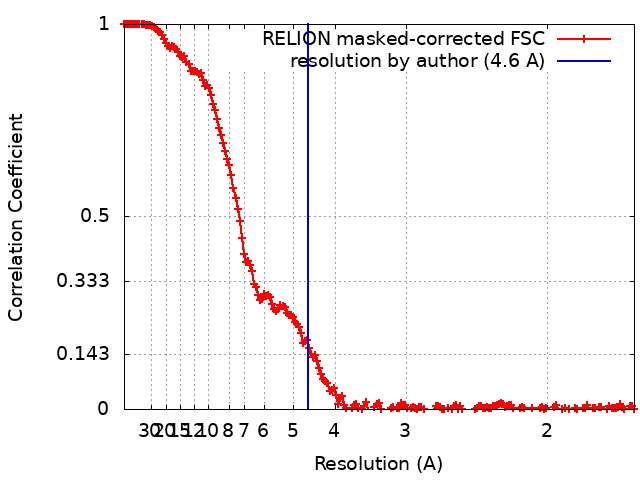 |
-Atomic model buiding 1
| Initial model | PDB ID: Chain - Source name: PDB / Chain - Initial model type: experimental model |
|---|---|
| Details | Two copies of the monomeric Pol II structure (PDB: 5C4X) were rigid fit into the dimeric Pol II EM density |
| Refinement | Protocol: RIGID BODY FIT |