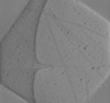
 Movie
Movie Controller


+ Open data

- Basic information
| Entry |  | |||||||||
|---|---|---|---|---|---|---|---|---|---|---|
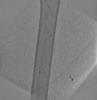
| Title | Microtubules in presence of CSPP-L and vinblastine Microtubule Microtubule | |||||||||
 Map data Map data | ||||||||||
 Sample Sample |
| |||||||||
| Biological species |  Sus scrofa (pig) Sus scrofa (pig) | |||||||||
| Method | electron tomography / cryo EM | |||||||||
 Authors Authors | Volkov VA | |||||||||
| Funding support | European Union, 1 items
| |||||||||
 Citation Citation | Journal: J Cell Biol / Year: 2023 Title: CSPP1 stabilizes growing microtubule ends and damaged lattices from the luminal side. Authors: Cyntha M van den Berg / Vladimir A Volkov / Sebastian Schnorrenberg / Ziqiang Huang / Kelly E Stecker / Ilya Grigoriev / Sania Gilani / Kari-Anne M Frikstad / Sebastian Patzke / Timo ...Authors: Cyntha M van den Berg / Vladimir A Volkov / Sebastian Schnorrenberg / Ziqiang Huang / Kelly E Stecker / Ilya Grigoriev / Sania Gilani / Kari-Anne M Frikstad / Sebastian Patzke / Timo Zimmermann / Marileen Dogterom / Anna Akhmanova /    Abstract: Microtubules are dynamic cytoskeletal polymers, and their organization and stability are tightly regulated by numerous cellular factors. While regulatory proteins controlling the formation of ...Microtubules are dynamic cytoskeletal polymers, and their organization and stability are tightly regulated by numerous cellular factors. While regulatory proteins controlling the formation of interphase microtubule arrays and mitotic spindles have been extensively studied, the biochemical mechanisms responsible for generating stable microtubule cores of centrioles and cilia are poorly understood. Here, we used in vitro reconstitution assays to investigate microtubule-stabilizing properties of CSPP1, a centrosome and cilia-associated protein mutated in the neurodevelopmental ciliopathy Joubert syndrome. We found that CSPP1 preferentially binds to polymerizing microtubule ends that grow slowly or undergo growth perturbations and, in this way, resembles microtubule-stabilizing compounds such as taxanes. Fluorescence microscopy and cryo-electron tomography showed that CSPP1 is deposited in the microtubule lumen and inhibits microtubule growth and shortening through two separate domains. CSPP1 also specifically recognizes and stabilizes damaged microtubule lattices. These data help to explain how CSPP1 regulates the elongation and stability of ciliary axonemes and other microtubule-based structures. #1: Journal: Biorxiv / Year: 2022Title: CSPP1 stabilizes growing microtubule ends and damaged lattices from the luminal side Authors: van den Berg CM / Volkov VA / Schnorrenberg S / Huang Z / Stecker KE / Grigoriev I / Patzke S / Zimmermann T / Dogterom M / Akhmanova A | |||||||||
| History |
|
- Structure visualization
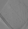
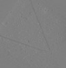
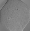
| Supplemental images |
|---|

- Downloads & links
-EMDB archive
| Map data | emd_15250.map.gz | 2.4 GB |  EMDB map data format EMDB map data format | |
|---|---|---|---|---|
| Header (meta data) | emd-15250-v30.xmlemd-15250.xml | 9.2 KB 9.2 KB | Display Display | EMDB header |
| Images | 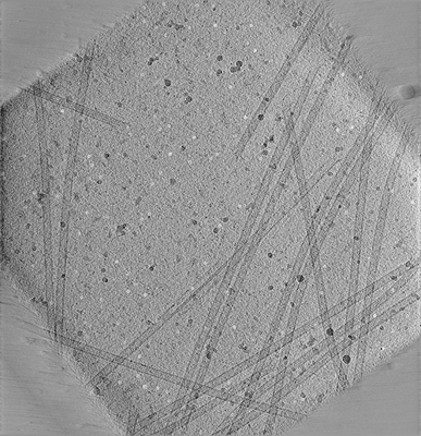 emd_15250.png emd_15250.png | 206.3 KB | ||
| Archive directory |  http://ftp.pdbj.org/pub/emdb/structures/EMD-15250ftp://ftp.pdbj.org/pub/emdb/structures/EMD-15250 http://ftp.pdbj.org/pub/emdb/structures/EMD-15250ftp://ftp.pdbj.org/pub/emdb/structures/EMD-15250 | HTTPS FTP |
-Related structure data
| Related structure data | C: citing same article ( |
|---|


-Links
| EMDB pages | EMDB (EBI/PDBe) / EMDataResource |
|---|---|
| Related items in Molecule of the Month |

-Map
| File | Download / File: emd_15250.map.gz / Format: CCP4 / Size: 2.6 GB / Type: IMAGE STORED AS FLOATING POINT NUMBER (4 BYTES) | ||||||||||||||||||||||||||||||||
|---|---|---|---|---|---|---|---|---|---|---|---|---|---|---|---|---|---|---|---|---|---|---|---|---|---|---|---|---|---|---|---|---|---|
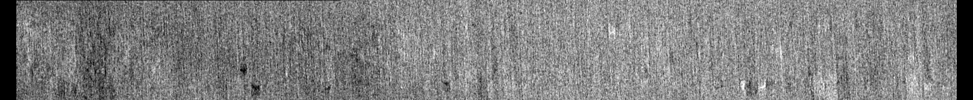
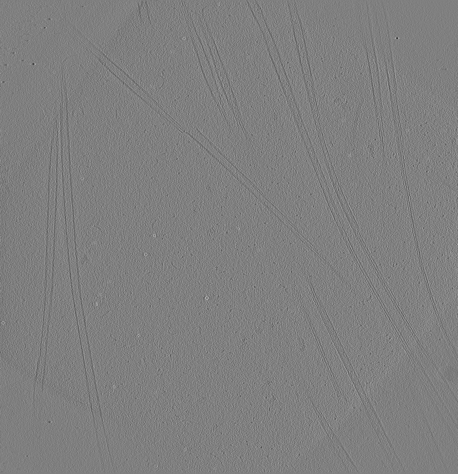
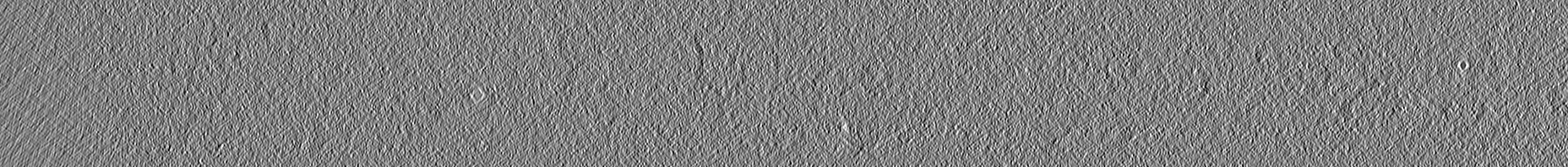
| Projections & slices | Image control
Images are generated by Spider. generated in cubic-lattice coordinate | ||||||||||||||||||||||||||||||||
| Voxel size | X=Y=Z: 0.734 Å | ||||||||||||||||||||||||||||||||
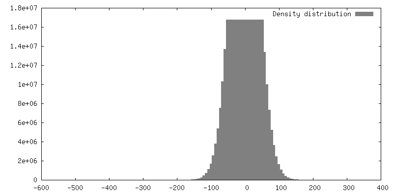
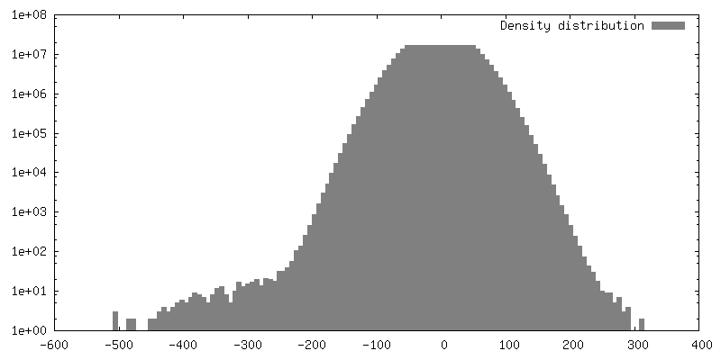
| Density |
| ||||||||||||||||||||||||||||||||
| Symmetry | Space group: 1 | ||||||||||||||||||||||||||||||||
| Details | EMDB XML:
|
 Z (Sec.)
 Y (Row.)
 X (Col.)


-Supplemental data
- Sample components
-Entire : Microtubules
| Entire | Name: MicrotubulesMicrotubule |
|---|---|
| Components |
|
-Supramolecule #1: Microtubules
| Supramolecule | Name: Microtubules / type: complex / ID: 1 / Chimera: Yes / Parent: 0 |
|---|---|
| Source (natural) | Organism: Sus scrofa (pig) |
-Experimental details
-Structure determination
| Method | cryo EM |
|---|---|
 Processing Processing | electron tomography |
| Aggregation state | filament |
-Sample preparation
| Buffer | pH: 6.9 |
|---|---|
| Vitrification | Cryogen name: ETHANE |
| Sectioning | Other: NO SECTIONING |
| Fiducial marker | Manufacturer: Sigma / Diameter: 5 nm |
- Electron microscopy
| Microscope | JEOL 3200FSC |
|---|---|
| Electron beam | Acceleration voltage: 300 kV / Electron source: FIELD EMISSION GUN |
| Electron optics | Illumination mode: FLOOD BEAM / Imaging mode: BRIGHT FIELDBright-field microscopy / Nominal defocus max: 4.0 µm / Nominal defocus min: 4.0 µm / Nominal magnification: 10000 |
| Specialist optics | Energy filter - Slit width: 30 eV |
| Image recording | Film or detector model: GATAN K2 SUMMIT (4k x 4k) / Detector mode: COUNTING / Average electron dose: 80.0 e/Å2 |
-Image processing
| Final reconstruction | Number images used: 61 |
|---|
