 Movie
Movie Controller


+ Open data

- Basic information
| Entry |  | |||||||||
|---|---|---|---|---|---|---|---|---|---|---|
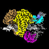
| Title | Complex of RecF and DNA from Thermus thermophilus. | |||||||||
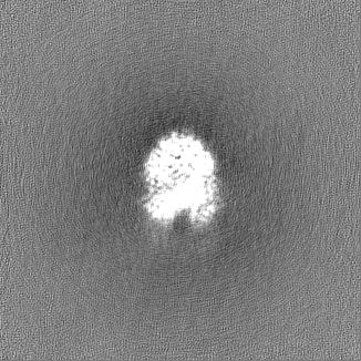
 Map data Map data | Sharpened map | |||||||||
 Sample Sample |
| |||||||||
| Function / homology |  Function and homology information Function and homology information SOS response / single-stranded DNA binding / DNA replication / DNA repair / ATP binding / cytoplasm SOS response / single-stranded DNA binding / DNA replication / DNA repair / ATP binding / cytoplasmSimilarity search - Function | |||||||||
| Biological species |   Thermus thermophilus HB8 (bacteria) / synthetic construct (others) Thermus thermophilus HB8 (bacteria) / synthetic construct (others) | |||||||||
| Method | single particle reconstruction / cryo EM / Resolution: 3.1 Å | |||||||||
 Authors Authors | Nirwal S / Czarnocki-Cieciura M / Chaudhary A / Zajko W / Skowronek K / Chamera S / Figiel M / Nowotny M | |||||||||
| Funding support |  Poland, 1 items Poland, 1 items
| |||||||||
 Citation Citation | Journal: Nat Struct Mol Biol / Year: 2023 Title: Mechanism of RecF-RecO-RecR cooperation in bacterial homologous recombination. Authors: Shivlee Nirwal / Mariusz Czarnocki-Cieciura / Anuradha Chaudhary / Weronika Zajko / Krzysztof Skowronek / Sebastian Chamera / Małgorzata Figiel / Marcin Nowotny / Abstract: In bacteria, one type of homologous-recombination-based DNA-repair pathway involves RecFOR proteins that bind at the junction between single-stranded (ss) and double-stranded (ds) DNA. They ...In bacteria, one type of homologous-recombination-based DNA-repair pathway involves RecFOR proteins that bind at the junction between single-stranded (ss) and double-stranded (ds) DNA. They facilitate the replacement of SSB protein, which initially covers ssDNA, with RecA, which mediates the search for homologous sequences. However, the molecular mechanism of RecFOR cooperation remains largely unknown. We used Thermus thermophilus proteins to study this system. Here, we present a cryo-electron microscopy structure of the RecF-dsDNA complex, and another reconstruction that shows how RecF interacts with two different regions of the tetrameric RecR ring. Lower-resolution reconstructions of the RecR-RecO subcomplex and the RecFOR-DNA assembly explain how RecO is positioned to interact with ssDNA and SSB, which is proposed to lock the complex on a ssDNA-dsDNA junction. Our results integrate the biochemical data available for the RecFOR system and provide a framework for its complete understanding. | |||||||||
| History |
|
- Structure visualization
| Supplemental images |
|---|

- Downloads & links
-EMDB archive
| Map data | emd_15231.map.gz | 124.6 MB | EMDB map data format | |
|---|---|---|---|---|
| Header (meta data) | emd-15231-v30.xmlemd-15231.xml | 22.6 KB 22.6 KB | Display Display | EMDB header |
| FSC (resolution estimation) | emd_15231_fsc.xml | 10.8 KB | Display | FSC data file |
| Images |  emd_15231.png emd_15231.png | 109.6 KB | ||
| Masks | emd_15231_msk_1.map | 132.2 MB | Mask map | |
| Others | emd_15231_additional_1.map.gzemd_15231_half_map_1.map.gzemd_15231_half_map_2.map.gz | 66 MB 122.5 MB 122.5 MB | ||
| Archive directory |  http://ftp.pdbj.org/pub/emdb/structures/EMD-15231ftp://ftp.pdbj.org/pub/emdb/structures/EMD-15231 http://ftp.pdbj.org/pub/emdb/structures/EMD-15231ftp://ftp.pdbj.org/pub/emdb/structures/EMD-15231 | HTTPS FTP |
-Related structure data
| Related structure data |  8a8jMC  8a93C  8ab0C  8bprC C: citing same article ( M: atomic model generated by this map |
|---|---|
| Similar structure data |

-Links
| EMDB pages | EMDB (EBI/PDBe) / EMDataResource |
|---|
-Map
| File | Download / File: emd_15231.map.gz / Format: CCP4 / Size: 132.2 MB / Type: IMAGE STORED AS FLOATING POINT NUMBER (4 BYTES) | ||||||||||||||||||||
|---|---|---|---|---|---|---|---|---|---|---|---|---|---|---|---|---|---|---|---|---|---|
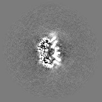
| Annotation | Sharpened map | ||||||||||||||||||||
| Voxel size | X=Y=Z: 0.86 Å | ||||||||||||||||||||
| Density |
| ||||||||||||||||||||
| Symmetry | Space group: 1 | ||||||||||||||||||||
| Details | EMDB XML:
|
-Supplemental data
-Mask #1
| File | emd_15231_msk_1.map | ||||||||||||
|---|---|---|---|---|---|---|---|---|---|---|---|---|---|
| Projections & Slices |
| ||||||||||||
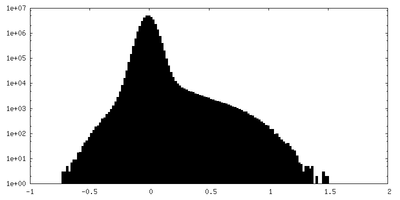
| Density Histograms |
 Z
Z Y
Y X
X


-Additional map: Raw map
| File | emd_15231_additional_1.map | ||||||||||||
|---|---|---|---|---|---|---|---|---|---|---|---|---|---|
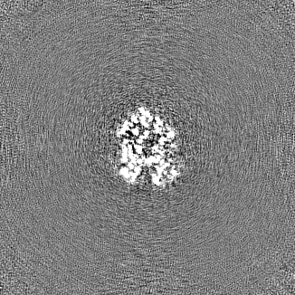
| Annotation | Raw map | ||||||||||||
| Projections & Slices |
| ||||||||||||
| Density Histograms |


-Half map: #2
| File | emd_15231_half_map_1.map | ||||||||||||
|---|---|---|---|---|---|---|---|---|---|---|---|---|---|
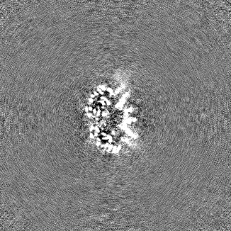
| Projections & Slices |
| ||||||||||||
| Density Histograms |


-Half map: #1
| File | emd_15231_half_map_2.map | ||||||||||||
|---|---|---|---|---|---|---|---|---|---|---|---|---|---|
| Projections & Slices |
| ||||||||||||
| Density Histograms |


- Sample components
-Entire : Complex of RecF and DNA from Thermus thermophilus.
| Entire | Name: Complex of RecF and DNA from Thermus thermophilus. |
|---|---|
| Components |
|
-Supramolecule #1: Complex of RecF and DNA from Thermus thermophilus.
| Supramolecule | Name: Complex of RecF and DNA from Thermus thermophilus. / type: complex / ID: 1 / Chimera: Yes / Parent: 0 / Macromolecule list: #1-#3 |
|---|---|
| Source (natural) | Organism: Thermus thermophilus HB8 (bacteria) |
| Molecular weight | Theoretical: 95.6 KDa |
-Macromolecule #1: DNA replication and repair protein RecF
| Macromolecule | Name: DNA replication and repair protein RecF / type: protein_or_peptide / ID: 1 / Number of copies: 2 / Enantiomer: LEVO |
|---|---|
| Source (natural) | Organism: Thermus thermophilus HB8 (bacteria) / Strain: ATCC 27634 / DSM 579 / HB8 |
| Molecular weight | Theoretical: 37.933715 KDa |
| Recombinant expression | Organism: Escherichia coli (E. coli) |
| Sequence | String: SMRLLLFRQR NFRNLALEAY RPPPGLSALV GANAQGKTSL LLGIHLALGG EVPLGLADLV RFGEEEAWLH AEVETELGAY RLEHRLGPG GREVLLNGKR VSLRTLWELP GSVLVSPLDL EAVLGPKEER RAYLDRLIAR FSRRYAALLS AYEKALRQRN A LLKAGGEG ...String: SMRLLLFRQR NFRNLALEAY RPPPGLSALV GANAQGKTSL LLGIHLALGG EVPLGLADLV RFGEEEAWLH AEVETELGAY RLEHRLGPG GREVLLNGKR VSLRTLWELP GSVLVSPLDL EAVLGPKEER RAYLDRLIAR FSRRYAALLS AYEKALRQRN A LLKAGGEG LSAWDRELAR YGDEIVALRR RFLRRFAPIL REVHAALAAK EAGLRLEETA GEGVLRALEA SRAEERERGQ TL VGPHRDD LVFLLEGRPA HRFASRGEAK TLALALRLAE HRLLGEHHGE PPLLLVDEWG EELDEARRRA VLAYAQALPQ AIL AGLEAP PGVPVCSVVR GVVLCPGA |
-Macromolecule #2: Oligo1
| Macromolecule | Name: Oligo1 / type: dna / ID: 2 / Number of copies: 1 / Classification: DNA |
|---|---|
| Source (natural) | Organism: synthetic construct (others) |
| Molecular weight | Theoretical: 7.661912 KDa |
| Sequence | String: (DG)(DG)(DC)(DC)(DA)(DG)(DA)(DT)(DC)(DT) (DG)(DC)(DC)(DG)(DC)(DG)(DG)(DA)(DT)(DC) (DC)(DG)(DC)(DG)(DC) |
-Macromolecule #3: Oligo2
| Macromolecule | Name: Oligo2 / type: dna / ID: 3 / Number of copies: 1 / Classification: DNA |
|---|---|
| Source (natural) | Organism: synthetic construct (others) |
| Molecular weight | Theoretical: 12.395925 KDa |
| Sequence | String: (DG)(DC)(DG)(DC)(DG)(DG)(DA)(DT)(DC)(DC) (DG)(DC)(DG)(DG)(DC)(DA)(DG)(DA)(DT)(DC) (DT)(DG)(DG)(DC)(DC)(DT)(DG)(DA)(DT) (DT)(DG)(DC)(DG)(DG)(DT)(DA)(DC)(DA)(DG) (DA) |
-Macromolecule #4: PHOSPHOAMINOPHOSPHONIC ACID-ADENYLATE ESTER
| Macromolecule | Name: PHOSPHOAMINOPHOSPHONIC ACID-ADENYLATE ESTER / type: ligand / ID: 4 / Number of copies: 2 / Formula: ANP |
|---|---|
| Molecular weight | Theoretical: 506.196 Da |
| Chemical component information |  ChemComp-ANP: |
-Macromolecule #5: MAGNESIUM ION
| Macromolecule | Name: MAGNESIUM ION / type: ligand / ID: 5 / Number of copies: 2 / Formula: MG |
|---|---|
| Molecular weight | Theoretical: 24.305 Da |
-Experimental details
-Structure determination
| Method | cryo EM |
|---|---|
 Processing Processing | single particle reconstruction |
| Aggregation state | particle |
-Sample preparation
| Buffer | pH: 7.5 Component:
| ||||||||||||
|---|---|---|---|---|---|---|---|---|---|---|---|---|---|
| Grid | Model: C-flat-2/1 / Material: COPPER / Mesh: 200 / Support film - Material: CARBON / Support film - topology: HOLEY / Pretreatment - Type: GLOW DISCHARGE | ||||||||||||
| Vitrification | Cryogen name: ETHANE / Chamber humidity: 95 % / Chamber temperature: 277 K / Instrument: FEI VITROBOT MARK IV | ||||||||||||
| Details | Sample fixed with 0.05% glutaraldehyde and concentrated prior to vitrification; exact concentration cannot be estimated accurately. |
- Electron microscopy
| Microscope | FEI TITAN KRIOS |
|---|---|
| Electron beam | Acceleration voltage: 300 kV / Electron source: FIELD EMISSION GUN |
| Electron optics | C2 aperture diameter: 50.0 µm / Illumination mode: FLOOD BEAM / Imaging mode: BRIGHT FIELDBright-field microscopy / Cs: 2.7 mm / Nominal defocus max: 2.7 µm / Nominal defocus min: 0.9 µm / Nominal magnification: 105000 |
| Specialist optics | Energy filter - Name: GIF Bioquantum / Energy filter - Slit width: 20 eV |
| Sample stage | Specimen holder model: FEI TITAN KRIOS AUTOGRID HOLDER / Cooling holder cryogen: NITROGEN |
| Image recording | Film or detector model: GATAN K3 BIOQUANTUM (6k x 4k) / Number grids imaged: 1 / Number real images: 7217 / Average electron dose: 41.71 e/Å2 |
| Experimental equipment |  Model: Titan Krios / Image courtesy: FEI Company |
-Image processing
| Particle selection | Number selected: 2381100 |
|---|---|
| Startup model | Type of model: OTHER / Details: Ab-initio reconstruction from cryoSPARC. |
| Initial angle assignment | Type: MAXIMUM LIKELIHOOD |
| Final angle assignment | Type: MAXIMUM LIKELIHOOD |
| Final reconstruction | Applied symmetry - Point group: C1 (asymmetric) / Resolution.type: BY AUTHOR / Resolution: 3.1 Å / Resolution method: FSC 0.143 CUT-OFF / Software - Name: cryoSPARC (ver. 3.3.2) / Number images used: 122950 |
| FSC plot (resolution estimation) |  |
-Atomic model buiding 1
| Refinement | Protocol: AB INITIO MODEL |
|---|---|
| Output model | PDB-8a8j: |
