Movie
Movie Controller


+ Open data

- Basic information
| Entry | 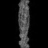 | |||||||||
|---|---|---|---|---|---|---|---|---|---|---|
| Title | Non-muscle F-actin decorated with non-muscle tropomyosin 1.6 | |||||||||
 Map data Map data | Description: Non-muscle F-actin decorated with non-muscle tropomyosin 1.6 | |||||||||
 Sample Sample |
| |||||||||
| Function / homology |  Function and homology information Function and homology information | |||||||||
| Biological species |   Homo sapiens (human) / human (human) Homo sapiens (human) / human (human) | |||||||||
| Method | helical reconstruction / cryo EM / Resolution: 3.9 Å | |||||||||
 Authors Authors | Selvaraj M / Kokate S / Kogan K / Kotila T / Kremneva E / Lappalainen P / Huiskonen JT | |||||||||
| Funding support |  Finland, 2 items Finland, 2 items
| |||||||||
 Citation Citation | Journal: Cell Rep / Year: 2023 Title: Structural basis underlying specific biochemical activities of non-muscle tropomyosin isoforms. Authors: Muniyandi Selvaraj / Shrikant B Kokate / Gabriella Reggiano / Konstantin Kogan / Tommi Kotila / Elena Kremneva / Frank DiMaio / Pekka Lappalainen / Juha T Huiskonen /  Abstract: The actin cytoskeleton is critical for cell migration, morphogenesis, endocytosis, organelle dynamics, and cytokinesis. To support diverse cellular processes, actin filaments form a variety of ...The actin cytoskeleton is critical for cell migration, morphogenesis, endocytosis, organelle dynamics, and cytokinesis. To support diverse cellular processes, actin filaments form a variety of structures with specific architectures and dynamic properties. Key proteins specifying actin filaments are tropomyosins. Non-muscle cells express several functionally non-redundant tropomyosin isoforms, which differentially control the interactions of other proteins, including myosins and ADF/cofilin, with actin filaments. However, the underlying molecular mechanisms have remained elusive. By determining the cryogenic electron microscopy structures of actin filaments decorated by two functionally distinct non-muscle tropomyosin isoforms, Tpm1.6 and Tpm3.2, we reveal that actin filament conformation remains unaffected upon binding. However, Tpm1.6 and Tpm3.2 follow different paths along the actin filament major groove, providing an explanation for their incapability to co-polymerize on actin filaments. We also elucidate the molecular basis underlying specific roles of Tpm1.6 and Tpm3.2 in myosin II activation and protecting actin filaments from ADF/cofilin-catalyzed severing. | |||||||||
| History |
|
- Structure visualization
| Supplemental images |
|---|

- Downloads & links
-EMDB archive
| Map data | emd_14957.map.gz | 229 MB | EMDB map data format | |
|---|---|---|---|---|
| Header (meta data) | emd-14957-v30.xmlemd-14957.xml | 17.9 KB 17.9 KB | Display Display | EMDB header |
| FSC (resolution estimation) | emd_14957_fsc.xml | 14.2 KB | Display | FSC data file |
| Images | 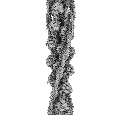 emd_14957.png emd_14957.png | 78.7 KB | ||
| Masks | emd_14957_msk_1.map | 244.1 MB | Mask map | |
| Others | emd_14957_half_map_1.map.gzemd_14957_half_map_2.map.gz | 194.2 MB 194.3 MB | ||
| Archive directory |  http://ftp.pdbj.org/pub/emdb/structures/EMD-14957ftp://ftp.pdbj.org/pub/emdb/structures/EMD-14957 http://ftp.pdbj.org/pub/emdb/structures/EMD-14957ftp://ftp.pdbj.org/pub/emdb/structures/EMD-14957 | HTTPS FTP |
-Related structure data
| Related structure data |  7ztcMC  7ztdC M: atomic model generated by this map C: citing same article ( |
|---|---|
| Similar structure data |
-Links
| EMDB pages | EMDB (EBI/PDBe) / EMDataResource |
|---|---|
| Related items in Molecule of the Month |


-Map
| File | Download / File: emd_14957.map.gz / Format: CCP4 / Size: 244.1 MB / Type: IMAGE STORED AS FLOATING POINT NUMBER (4 BYTES) | ||||||||||||||||||||||||||||||||||||
|---|---|---|---|---|---|---|---|---|---|---|---|---|---|---|---|---|---|---|---|---|---|---|---|---|---|---|---|---|---|---|---|---|---|---|---|---|---|
| Annotation | Description: Non-muscle F-actin decorated with non-muscle tropomyosin 1.6 | ||||||||||||||||||||||||||||||||||||
| Projections & slices | Image control
Images are generated by Spider. | ||||||||||||||||||||||||||||||||||||
| Voxel size | X=Y=Z: 0.97 Å | ||||||||||||||||||||||||||||||||||||
| Density |
| ||||||||||||||||||||||||||||||||||||
| Symmetry | Space group: 1 | ||||||||||||||||||||||||||||||||||||
| Details | EMDB XML:
|
 Z (Sec.)
 Y (Row.)
 X (Col.)


-Supplemental data
-Mask #1
| File | emd_14957_msk_1.map | ||||||||||||
|---|---|---|---|---|---|---|---|---|---|---|---|---|---|
| Projections & Slices |
| ||||||||||||
| Density Histograms |


-Half map: Halfmap 1
| File | emd_14957_half_map_1.map | ||||||||||||
|---|---|---|---|---|---|---|---|---|---|---|---|---|---|
| Annotation | Halfmap 1 | ||||||||||||
| Projections & Slices |
| ||||||||||||
| Density Histograms |


-Half map: Halfmap 2
| File | emd_14957_half_map_2.map | ||||||||||||
|---|---|---|---|---|---|---|---|---|---|---|---|---|---|
| Annotation | Halfmap 2 | ||||||||||||
| Projections & Slices |
| ||||||||||||
| Density Histograms |


- Sample components
-Entire : Non-muscle F-actin decorated with non-muscle tropomyosin 1.6
| Entire | Name: Non-muscle F-actin decorated with non-muscle tropomyosin 1.6 |
|---|---|
| Components |
|
-Supramolecule #1: Non-muscle F-actin decorated with non-muscle tropomyosin 1.6
| Supramolecule | Name: Non-muscle F-actin decorated with non-muscle tropomyosin 1.6 type: complex / ID: 1 / Chimera: Yes / Parent: 0 / Macromolecule list: #1-#2 |
|---|---|
| Source (natural) | Organism: Homo sapiens (human) |
-Supramolecule #2: Beta actin (non-muscle)
| Supramolecule | Name: Beta actin (non-muscle) / type: organelle_or_cellular_component / ID: 2 / Parent: 1 / Macromolecule list: #1 |
|---|---|
| Source (natural) | Organism: Homo sapiens (human) |
-Supramolecule #3: Non-muscle tropomyosin 1.6
| Supramolecule | Name: Non-muscle tropomyosin 1.6 / type: organelle_or_cellular_component / ID: 3 / Parent: 1 / Macromolecule list: #2 |
|---|---|
| Source (natural) | Organism: Homo sapiens (human) |
-Macromolecule #1: actin, cytoplasmic 1
| Macromolecule | Name: actin, cytoplasmic 1 / type: protein_or_peptide / ID: 1 / Number of copies: 8 / Enantiomer: LEVO |
|---|---|
| Source (natural) | Organism: human (human) / Tissue: Platelets |
| Molecular weight | Theoretical: 41.78266 KDa |
| Sequence | String: MDDDIAALVV DNGSGMCKAG FAGDDAPRAV FPSIVGRPRH QGVMVGMGQK DSYVGDEAQS KRGILTLKYP IEHGIVTNWD DMEKIWHHT FYNELRVAPE EHPVLLTEAP LNPKANREKM TQIMFETFNT PAMYVAIQAV LSLYASGRTT GIVMDSGDGV T HTVPIYEG ...String: MDDDIAALVV DNGSGMCKAG FAGDDAPRAV FPSIVGRPRH QGVMVGMGQK DSYVGDEAQS KRGILTLKYP IEHGIVTNWD DMEKIWHHT FYNELRVAPE EHPVLLTEAP LNPKANREKM TQIMFETFNT PAMYVAIQAV LSLYASGRTT GIVMDSGDGV T HTVPIYEG YALPHAILRL DLAGRDLTDY LMKILTERGY SFTTTAEREI VRDIKEKLCY VALDFEQEMA TAASSSSLEK SY ELPDGQV ITIGNERFRC PEALFQPSFL GMESCGIHET TFNSIMKCDV DIRKDLYANT VLSGGTTMYP GIADRMQKEI TAL APSTMK IKIIAPPERK YSVWIGGSIL ASLSTFQQMW ISKQEYDESG PSIVHRKCF |
-Macromolecule #2: Non-muscle tropomyosin 1.6
| Macromolecule | Name: Non-muscle tropomyosin 1.6 / type: protein_or_peptide / ID: 2 / Number of copies: 4 / Enantiomer: LEVO |
|---|---|
| Source (natural) | Organism: Homo sapiens (human) |
| Molecular weight | Theoretical: 11.507176 KDa |
| Recombinant expression | Organism:  Escherichia coli BL21(DE3) (bacteria) Escherichia coli BL21(DE3) (bacteria) |
| Sequence | String: (UNK)(UNK)(UNK)(UNK)(UNK)(UNK)(UNK)(UNK)(UNK)(UNK) (UNK)(UNK)(UNK)(UNK)(UNK)(UNK) (UNK)(UNK)(UNK) (UNK)(UNK)(UNK)(UNK)(UNK)(UNK)(UNK)(UNK)(UNK)(UNK) (UNK)(UNK)(UNK) (UNK)(UNK)(UNK)(UNK)(UNK) ...String: (UNK)(UNK)(UNK)(UNK)(UNK)(UNK)(UNK)(UNK)(UNK)(UNK) (UNK)(UNK)(UNK)(UNK)(UNK)(UNK) (UNK)(UNK)(UNK) (UNK)(UNK)(UNK)(UNK)(UNK)(UNK)(UNK)(UNK)(UNK)(UNK) (UNK)(UNK)(UNK) (UNK)(UNK)(UNK)(UNK)(UNK)(UNK) (UNK)(UNK)(UNK)(UNK)(UNK)(UNK)(UNK)(UNK)(UNK)(UNK) (UNK)(UNK)(UNK)(UNK)(UNK)(UNK)(UNK)(UNK)(UNK) (UNK)(UNK)(UNK)(UNK)(UNK)(UNK)(UNK) (UNK)(UNK) (UNK)(UNK)(UNK)(UNK)(UNK)(UNK)(UNK)(UNK)(UNK)(UNK) (UNK)(UNK)(UNK)(UNK) (UNK)(UNK)(UNK)(UNK)(UNK) (UNK)(UNK)(UNK)(UNK)(UNK)(UNK)(UNK)(UNK)(UNK)(UNK) (UNK) (UNK)(UNK)(UNK)(UNK)(UNK)(UNK)(UNK)(UNK) (UNK)(UNK)(UNK)(UNK)(UNK)(UNK)(UNK)(UNK) (UNK) (UNK)(UNK)(UNK)(UNK)(UNK)(UNK)(UNK)(UNK)(UNK)(UNK) (UNK)(UNK)(UNK)(UNK)(UNK) (UNK)(UNK)(UNK)(UNK) (UNK)(UNK)(UNK) |
-Macromolecule #3: ADENOSINE-5'-DIPHOSPHATE
| Macromolecule | Name: ADENOSINE-5'-DIPHOSPHATE / type: ligand / ID: 3 / Number of copies: 8 / Formula: ADP |
|---|---|
| Molecular weight | Theoretical: 427.201 Da |
| Chemical component information |  ChemComp-ADP: |
-Experimental details
-Structure determination
| Method | cryo EM |
|---|---|
 Processing Processing | helical reconstruction |
| Aggregation state | filament |
-Sample preparation
| Buffer | pH: 7.4 |
|---|---|
| Vitrification | Cryogen name: ETHANE |
- Electron microscopy
| Microscope | FEI TALOS ARCTICA |
|---|---|
| Electron beam | Acceleration voltage: 200 kV / Electron source: FIELD EMISSION GUN |
| Electron optics | Illumination mode: FLOOD BEAM / Imaging mode: BRIGHT FIELDBright-field microscopy / Nominal defocus max: 3.0 µm / Nominal defocus min: 1.0 µm |
| Image recording | Film or detector model: FEI FALCON III (4k x 4k) / Number real images: 1700 / Average exposure time: 45.0 sec. / Average electron dose: 45.0 e/Å2 |
| Experimental equipment |  Model: Talos Arctica / Image courtesy: FEI Company |
-Image processing
| Final angle assignment | Type: NOT APPLICABLE |
|---|---|
| Final reconstruction | Applied symmetry - Helical parameters - Δz: 27.8 Å Applied symmetry - Helical parameters - Δ&Phi: -166.5 ° Applied symmetry - Helical parameters - Axial symmetry: C1 (asymmetric) Resolution.type: BY AUTHOR / Resolution: 3.9 Å / Resolution method: FSC 0.143 CUT-OFF / Software - Name: RELION / Number images used: 79298 |
| FSC plot (resolution estimation) |  |