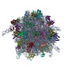
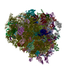
- EMDB-14751: Rabbit 80S ribosome as it decodes the Sec-UGA codon -
+
Open data
ID or keywords:
Loading...
-
Basic information
Entry
Database: EMDB / ID: EMD-14751
Title
Rabbit 80S ribosome as it decodes the Sec-UGA codon
Map data
Sample
Complex: 80S-Selenosome
Complex: eEFSec and SECIS Binding Protein 2
Protein or peptide: x 2 types
Complex: Ser-tRNA-Sec
RNA: x 1 types
Complex: CrPV IRESCripavirus internal ribosome entry site
RNA: x 1 types
Complex: GPX4 SECIS element
RNA: x 1 types
Complex: 80S RibosomeEukaryotic ribosome
RNA: x 4 types
Protein or peptide: x 77 types
Ligand: x 7 types
Keywords
Selenocysteine / recoding / 80S / RIBOSOME
Function / homology
Function and homology information
forebrain neuron development / Translation initiation complex formation / Formation of the ternary complex, and subsequently, the 43S complex / Ribosomal scanning and start codon recognition / negative regulation of nuclear-transcribed mRNA catabolic process, nonsense-mediated decay / selenocysteine incorporation / Major pathway of rRNA processing in the nucleolus and cytosol / GTP hydrolysis and joining of the 60S ribosomal subunit / selenocysteine insertion sequence binding / L13a-mediated translational silencing of Ceruloplasmin expression ...forebrain neuron development / Translation initiation complex formation / Formation of the ternary complex, and subsequently, the 43S complex / Ribosomal scanning and start codon recognition / negative regulation of nuclear-transcribed mRNA catabolic process, nonsense-mediated decay / selenocysteine incorporation / Major pathway of rRNA processing in the nucleolus and cytosol / GTP hydrolysis and joining of the 60S ribosomal subunit / selenocysteine insertion sequence binding / L13a-mediated translational silencing of Ceruloplasmin expression / SRP-dependent cotranslational protein targeting to membrane / Formation of a pool of free 40S subunits / Nonsense Mediated Decay (NMD) independent of the Exon Junction Complex (EJC) / Nonsense Mediated Decay (NMD) enhanced by the Exon Junction Complex (EJC) / striatum development / ribosomal subunit / rRNA modification in the nucleus and cytosol / Formation of the ternary complex, and subsequently, the 43S complex / Ribosomal scanning and start codon recognition / Translation initiation complex formation / SARS-CoV-1 modulates host translation machinery / Protein hydroxylation / TOR signaling / mTORC1-mediated signalling / Peptide chain elongation / Selenocysteine synthesis / Formation of a pool of free 40S subunits / Eukaryotic Translation Termination / organelle membrane / Response of EIF2AK4 (GCN2) to amino acid deficiency / translation regulator activity / SRP-dependent cotranslational protein targeting to membrane / Viral mRNA Translation / Nonsense Mediated Decay (NMD) independent of the Exon Junction Complex (EJC) / GTP hydrolysis and joining of the 60S ribosomal subunit / L13a-mediated translational silencing of Ceruloplasmin expression / Major pathway of rRNA processing in the nucleolus and cytosol / gastrulation / rough endoplasmic reticulum / Nonsense Mediated Decay (NMD) enhanced by the Exon Junction Complex (EJC) / ribonucleoprotein complex binding / translation elongation factor activity / Nuclear events stimulated by ALK signaling in cancer / maturation of SSU-rRNA from tricistronic rRNA transcript (SSU-rRNA, 5.8S rRNA, LSU-rRNA) / maturation of LSU-rRNA from tricistronic rRNA transcript (SSU-rRNA, 5.8S rRNA, LSU-rRNA) / maturation of LSU-rRNA / DNA-(apurinic or apyrimidinic site) lyase / small-subunit processome / cytosolic ribosome / mRNA 3'-UTR binding / Hydrolases; Acting on acid anhydrides; Acting on GTP to facilitate cellular and subcellular movement / ribosomal small subunit biogenesis / cytoplasmic ribonucleoprotein granule / rRNA processing / Regulation of expression of SLITs and ROBOs / cytosolic small ribosomal subunit / ribosome binding / large ribosomal subunit / glucose homeostasis / regulation of translation / heparin binding / cell body / cytoplasmic translation / small ribosomal subunit / 5S rRNA binding / cytosolic large ribosomal subunit / SARS-CoV-2 modulates host translation machinery / tRNA binding / postsynaptic density / rRNA binding / ribosome / structural constituent of ribosome / cadherin binding / ribonucleoprotein complex / translation / positive regulation of apoptotic process / focal adhesion / intracellular membrane-bounded organelle / GTPase activity / dendrite / synapse / positive regulation of cell population proliferation / GTP binding / nucleolus / protein kinase binding / endoplasmic reticulum / mitochondrion / DNA binding / RNA binding / nucleoplasm / membrane / metal ion binding / nucleus / cytosol / cytoplasm Similarity search - Function
Selenocysteine insertion sequence-binding protein 2 / : / : / eEFSec C-terminal RIFT domain / Selenocysteine-specific elongation factor, eukaryotes, 3rd domain / Ribosomal protein L28e / Ribosomal protein L2, archaeal-type / Ribosomal L28e/Mak16 / Ribosomal L28e protein family / : ...Selenocysteine insertion sequence-binding protein 2 / : / : / eEFSec C-terminal RIFT domain / Selenocysteine-specific elongation factor, eukaryotes, 3rd domain / Ribosomal protein L28e / Ribosomal protein L2, archaeal-type / Ribosomal L28e/Mak16 / Ribosomal L28e protein family / : / Small (40S) ribosomal subunit Asc1/RACK1 / Ribosomal protein S21e, conserved site / Ribosomal protein S26e / Ribosomal protein S26e superfamily / Ribosomal protein S19e, conserved site / S27a-like superfamily / Ribosomal protein S26e / Ribosomal protein S25 / Ribosomal protein S26e signature. / Ribosomal protein S27a / Ribosomal protein S27a / Ribosomal protein S21e / Ribosomal protein S21e superfamily / Ribosomal protein S21e / Ribosomal protein S8e subdomain, eukaryotes / S25 ribosomal protein / Ribosomal protein L27e, conserved site / Ribosomal protein S21e signature. / Ribosomal protein S12e signature. / Ribosomal protein S27a / Ribosomal protein S19e / Ribosomal_S19e / Ribosomal protein S5, eukaryotic/archaeal / Ribosomal protein S8e, conserved site / Ribosomal protein S6, eukaryotic / 40S ribosomal protein S4, C-terminal domain / Eukaryotic Ribosomal Protein L27, KOW domain / Ribosomal protein S4e, N-terminal, conserved site / Ribosomal protein S19e signature. / Ribosomal protein L44e / Ribosomal protein L38e / Ribosomal protein L38e superfamily / Ribosomal protein L27e / Ribosomal protein L27e superfamily / Ribosomal protein L22e / Ribosomal protein L22e superfamily / Ribosomal protein S19e / Ribosomal protein S27, zinc-binding domain superfamily / Ribosomal L38e protein family / Ribosomal L22e protein family / 40S Ribosomal protein S10 / Ribosomal protein S27 / Ribosomal protein S28e conserved site / Ribosomal protein S6/S6e/A/B/2, conserved site / Ribosomal protein S28e / 40S ribosomal protein S4 C-terminus / Ribosomal protein S4e, N-terminal / Plectin/S10, N-terminal / Ribosomal protein L44 / Plectin/S10 domain / Ribosomal protein S8e / Ribosomal L27e protein family / Ribosomal protein S4, KOW domain / Ribosomal protein S5/S7, eukaryotic/archaeal / Ribosomal protein S4e / Ribosomal protein S4e, central region / Ribosomal protein S4e, central domain superfamily / Ribosomal protein S6e / Ribosomal protein S13/S15, N-terminal / Ribosomal protein S15P / Ribosomal S13/S15 N-terminal domain / Ribosomal protein S6e / RS4NT (NUC023) domain / Ribosomal protein L1 signature. / Ribosomal protein L37ae / Ribosomal protein S27 / Ribosomal protein L19, eukaryotic / Ribosomal protein S17e signature. / Ribosomal protein L36e / Ribosomal protein L36e domain superfamily / Ribosomal L40e family / Ribosomal protein L36e / Ribosomal protein S28e / Ribosomal protein L44e signature. / Ribosomal_L40e / Ribosomal protein L40e / Ribosomal protein L40e superfamily / Ribosomal family S4e / Ribosomal protein L7A/L8 / Ribosomal S13/S15 N-terminal domain / Ribosomal protein L27e signature. / Ribosomal protein L10e signature. / Ribosomal protein S7e signature. / 60S ribosomal protein L18a/ L20, eukaryotes / Ribosomal protein L7, eukaryotic / Ribosomal protein L30, N-terminal / Ribosomal L30 N-terminal domain / Ribosomal protein L19/L19e conserved site / Ribosomal protein S6e / Ribosomal L37ae protein family Similarity search - Domain/homology
Small ribosomal subunit protein eS32 / Large ribosomal subunit protein uL13 / Large ribosomal subunit protein eL38 / 60S ribosomal protein L36 / Small ribosomal subunit protein eS21 / Large ribosomal subunit protein eL20 / Ribosomal protein L19 / 40S ribosomal protein S26 / Small ribosomal subunit protein RACK1 / 40S ribosomal protein S25 ...Small ribosomal subunit protein eS32 / Large ribosomal subunit protein uL13 / Large ribosomal subunit protein eL38 / 60S ribosomal protein L36 / Small ribosomal subunit protein eS21 / Large ribosomal subunit protein eL20 / Ribosomal protein L19 / 40S ribosomal protein S26 / Small ribosomal subunit protein RACK1 / 40S ribosomal protein S25 / Small ribosomal subunit protein eS19 / Small ribosomal subunit protein eS28 / Large ribosomal subunit protein uL14 / Large ribosomal subunit protein eL28 / Ribosomal protein S5 / Large ribosomal subunit protein uL24 / Small ribosomal subunit protein uS15 / 60S ribosomal protein L37a / 40S ribosomal protein S24 / Large ribosomal subunit protein eL42 / 40S ribosomal protein S27 / Large ribosomal subunit protein uL30 / Small ribosomal subunit protein uS11 / 60S ribosomal protein L27 / Large ribosomal subunit protein uL2 / Ubiquitin-ribosomal protein eS31 fusion protein / Ubiquitin-ribosomal protein eL40 fusion protein / Large ribosomal subunit protein uL22 / Large ribosomal subunit protein uL16 / Small ribosomal subunit protein uS4 / Large ribosomal subunit protein uL15/eL18 domain-containing protein / Large ribosomal subunit protein eL24 / Large ribosomal subunit protein uL23 / Large ribosomal subunit protein eL33 / Small ribosomal subunit protein eS12 / Large ribosomal subunit protein eL29 / Small ribosomal subunit protein uS9 / Large ribosomal subunit protein eL31 / Large ribosomal subunit protein eL21 / Large ribosomal subunit protein uL29 / Small ribosomal subunit protein uS10 / Large ribosomal subunit protein eL6 / Large ribosomal subunit protein uL1 / Large ribosomal subunit protein uL15 / Large ribosomal subunit protein uL10 / Small ribosomal subunit protein eS1 / Small ribosomal subunit protein eS7 / Large ribosomal subunit protein uL4 / Large ribosomal subunit protein uL6 / Large ribosomal subunit protein uL18 / Large ribosomal subunit protein eL14 / Small ribosomal subunit protein uS12 / Large ribosomal subunit protein eL15 / Ubiquitin-like FUBI-ribosomal protein eS30 fusion protein / Large ribosomal subunit protein eL30 / Small ribosomal subunit protein uS8 / Large ribosomal subunit protein eL13 / Large ribosomal subunit protein uL3 / Small ribosomal subunit protein uS2 / Small ribosomal subunit protein uS3 / Plectin/eS10 N-terminal domain-containing protein / Small ribosomal subunit protein uS17 / Large ribosomal subunit protein eL39 / Small ribosomal subunit protein eS17 / Large ribosomal subunit protein uL5 / Large ribosomal subunit protein eL34 / Small ribosomal subunit protein uS19 / Large ribosomal subunit protein eL32 / Small ribosomal subunit protein uS14 / Small ribosomal subunit protein uS13 / Small ribosomal subunit protein uS5 / Selenocysteine-specific elongation factor / Large ribosomal subunit protein eL8 / Small ribosomal subunit protein eS6 / Large ribosomal subunit protein eL22 / Small ribosomal subunit protein eS8 / Small ribosomal subunit protein eS4 / Selenocysteine insertion sequence-binding protein 2 / Large ribosomal subunit protein eL37 Similarity search - Component
National Institutes of Health/National Institute of General Medical Sciences (NIH/NIGMS)
GM097042, GM077073
United States
Citation
Journal: Science / Year: 2022 Title: Structure of the mammalian ribosome as it decodes the selenocysteine UGA codon. Authors: Tarek Hilal / Benjamin Y Killam / Milica Grozdanović / Malgorzata Dobosz-Bartoszek / Justus Loerke / Jörg Bürger / Thorsten Mielke / Paul R Copeland / Miljan Simonović / Christian M T Spahn / Abstract: The elongation of eukaryotic selenoproteins relies on a poorly understood process of interpreting in-frame UGA stop codons as selenocysteine (Sec). We used cryo-electron microscopy to visualize Sec ...The elongation of eukaryotic selenoproteins relies on a poorly understood process of interpreting in-frame UGA stop codons as selenocysteine (Sec). We used cryo-electron microscopy to visualize Sec UGA recoding in mammals. A complex between the noncoding Sec-insertion sequence (SECIS), SECIS-binding protein 2 (SBP2), and 40 ribosomal subunit enables Sec-specific elongation factor eEFSec to deliver Sec. eEFSec and SBP2 do not interact directly but rather deploy their carboxyl-terminal domains to engage with the opposite ends of the SECIS. By using its Lys-rich and carboxyl-terminal segments, the ribosomal protein eS31 simultaneously interacts with Sec-specific transfer RNA (tRNA) and SBP2, which further stabilizes the assembly. eEFSec is indiscriminate toward l-serine and facilitates its misincorporation at Sec UGA codons. Our results support a fundamentally distinct mechanism of Sec UGA recoding in eukaryotes from that in bacteria.
In the structure databanks used in Yorodumi, some data are registered as the other names, "COVID-19 virus" and "2019-nCoV". Here are the details of the virus and the list of structure data.
Jan 31, 2019. EMDB accession codes are about to change! (news from PDBe EMDB page)
EMDB accession codes are about to change! (news from PDBe EMDB page)
The allocation of 4 digits for EMDB accession codes will soon come to an end. Whilst these codes will remain in use, new EMDB accession codes will include an additional digit and will expand incrementally as the available range of codes is exhausted. The current 4-digit format prefixed with “EMD-” (i.e. EMD-XXXX) will advance to a 5-digit format (i.e. EMD-XXXXX), and so on. It is currently estimated that the 4-digit codes will be depleted around Spring 2019, at which point the 5-digit format will come into force.
The EM Navigator/Yorodumi systems omit the EMD- prefix.
Related info.:Q: What is EMD? / ID/Accession-code notation in Yorodumi/EM Navigator
Yorodumi is a browser for structure data from EMDB, PDB, SASBDB, etc.
This page is also the successor to EM Navigator detail page, and also detail information page/front-end page for Omokage search.
The word "yorodu" (or yorozu) is an old Japanese word meaning "ten thousand". "mi" (miru) is to see.
Related info.:EMDB / PDB / SASBDB / Comparison of 3 databanks / Yorodumi Search / Aug 31, 2016. New EM Navigator & Yorodumi / Yorodumi Papers / Jmol/JSmol / Function and homology information / Changes in new EM Navigator and Yorodumi
 Movie
Movie Controller


 Open data
 Basic information
 Map data
 Sample
 Keywords
 Selenocysteine /
  Function and homology information


 Authors
 Germany,
  United States, 2 items
  Citation
 Structure visualization
 Downloads & links
 emd_14751.png
 http://ftp.pdbj.org/pub/emdb/structures/EMD-14751
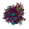


 Z (Sec.)
 Y (Row.)
 X (Col.)


 Sample components


 Processing
 Electron microscopy