 Movie
Movie Controller

[English] 日本語
 Yorodumi
- EMDB-14271: Structure of the SARS-CoV-2 spike glycoprotein in complex with th... -
+ Open data

- Basic information
| Entry |  | ||||||||||||
|---|---|---|---|---|---|---|---|---|---|---|---|---|---|
| Title | Structure of the SARS-CoV-2 spike glycoprotein in complex with the 87G7 antibody Fab fragment (local refinement) | ||||||||||||
 Map data Map data | DeepEMhancer sharpened map | ||||||||||||
 Sample Sample |
| ||||||||||||
 Keywords Keywords |  Coronavirus / Spike / Antibody / Complex / VIRAL PROTEIN Coronavirus / Spike / Antibody / Complex / VIRAL PROTEIN | ||||||||||||
| Biological species |   Severe acute respiratory syndrome coronavirus 2 / Severe acute respiratory syndrome coronavirus 2 /  Homo sapiens (human) Homo sapiens (human) | ||||||||||||
| Method | single particle reconstruction / cryo EM / Resolution: 4.9 Å | ||||||||||||
 Authors Authors | Hurdiss DL / Drulyte I | ||||||||||||
| Funding support | European Union,  Germany, 3 items Germany, 3 items
| ||||||||||||
 Citation Citation | Journal: Sci Immunol / Year: 2022 Title: An ACE2-blocking antibody confers broad neutralization and protection against Omicron and other SARS-CoV-2 variants of concern. Authors: Wenjuan Du / Daniel L Hurdiss / Dubravka Drabek / Anna Z Mykytyn / Franziska K Kaiser / Mariana González-Hernández / Diego Muñoz-Santos / Mart M Lamers / Rien van Haperen / Wentao Li / ...Authors: Wenjuan Du / Daniel L Hurdiss / Dubravka Drabek / Anna Z Mykytyn / Franziska K Kaiser / Mariana González-Hernández / Diego Muñoz-Santos / Mart M Lamers / Rien van Haperen / Wentao Li / Ieva Drulyte / Chunyan Wang / Isabel Sola / Federico Armando / Georg Beythien / Malgorzata Ciurkiewicz / Wolfgang Baumgärtner / Kate Guilfoyle / Tony Smits / Joline van der Lee / Frank J M van Kuppeveld / Geert van Amerongen / Bart L Haagmans / Luis Enjuanes / Albert D M E Osterhaus / Frank Grosveld / Berend-Jan Bosch /    Abstract: The ongoing evolution of SARS-CoV-2 has resulted in the emergence of Omicron, which displays notable immune escape potential through mutations at key antigenic sites on the spike protein. Many of ...The ongoing evolution of SARS-CoV-2 has resulted in the emergence of Omicron, which displays notable immune escape potential through mutations at key antigenic sites on the spike protein. Many of these mutations localize to the spike protein ACE2 receptor binding domain, annulling the neutralizing activity of therapeutic antibodies that were effective against other variants of concern (VOCs) earlier in the pandemic. Here, we identified a receptor-blocking human monoclonal antibody, 87G7, that retained potent in vitro neutralizing activity against SARS-CoV-2 variants including the Alpha, Beta, Gamma, Delta, and Omicron (BA.1/BA.2) VOCs. Using cryo-electron microscopy and site-directed mutagenesis experiments, we showed that 87G7 targets a patch of hydrophobic residues in the ACE2-binding site that are highly conserved in SARS-CoV-2 variants, explaining its broad neutralization capacity. 87G7 protected mice and hamsters prophylactically against challenge with all current SARS-CoV-2 VOCs and showed therapeutic activity against SARS-CoV-2 challenge in both animal models. Our findings demonstrate that 87G7 holds promise as a prophylactic or therapeutic agent for COVID-19 that is more resilient to SARS-CoV-2 antigenic diversity. | ||||||||||||
| History |
|
- Structure visualization
| Supplemental images |
|---|

- Downloads & links
-EMDB archive
| Map data | emd_14271.map.gz | 92.5 MB |  EMDB map data format EMDB map data format | |
|---|---|---|---|---|
| Header (meta data) | emd-14271-v30.xmlemd-14271.xml | 29.2 KB 29.2 KB | Display Display | EMDB header |
| FSC (resolution estimation) | emd_14271_fsc.xml | 10.7 KB | Display | FSC data file |
| Images | 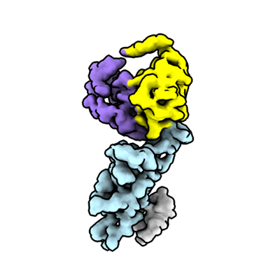 emd_14271.png emd_14271.png | 70.2 KB | ||
| Masks | emd_14271_msk_1.map | 103 MB | Mask map | |
| Filedesc metadata | emd-14271.cif.gz | 7.3 KB | ||
| Others | emd_14271_additional_1.map.gzemd_14271_additional_2.map.gzemd_14271_additional_3.map.gzemd_14271_half_map_1.map.gzemd_14271_half_map_2.map.gz | 60.9 MB 50.4 MB 97 MB 95.6 MB 95.6 MB | ||
| Archive directory |  http://ftp.pdbj.org/pub/emdb/structures/EMD-14271ftp://ftp.pdbj.org/pub/emdb/structures/EMD-14271 http://ftp.pdbj.org/pub/emdb/structures/EMD-14271ftp://ftp.pdbj.org/pub/emdb/structures/EMD-14271 | HTTPS FTP |
-Related structure data

-Links
| EMDB pages | EMDB (EBI/PDBe) / EMDataResource |
|---|
-Map
| File | Download / File: emd_14271.map.gz / Format: CCP4 / Size: 103 MB / Type: IMAGE STORED AS FLOATING POINT NUMBER (4 BYTES) | ||||||||||||||||||||||||||||||||||||
|---|---|---|---|---|---|---|---|---|---|---|---|---|---|---|---|---|---|---|---|---|---|---|---|---|---|---|---|---|---|---|---|---|---|---|---|---|---|
| Annotation | DeepEMhancer sharpened map | ||||||||||||||||||||||||||||||||||||
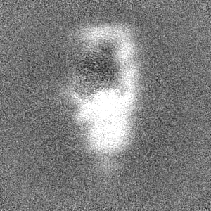
| Projections & slices | Image control
Images are generated by Spider. | ||||||||||||||||||||||||||||||||||||
| Voxel size | X=Y=Z: 1.24 Å | ||||||||||||||||||||||||||||||||||||
| Density |
| ||||||||||||||||||||||||||||||||||||
| Symmetry | Space group: 1 | ||||||||||||||||||||||||||||||||||||
| Details | EMDB XML:
|
 Z (Sec.)
 Y (Row.)
 X (Col.)


-Supplemental data
-Mask #1
| File | emd_14271_msk_1.map | ||||||||||||
|---|---|---|---|---|---|---|---|---|---|---|---|---|---|
| Projections & Slices |
| ||||||||||||
| Density Histograms |


-Additional map: Local resolution filtered map
| File | emd_14271_additional_1.map | ||||||||||||
|---|---|---|---|---|---|---|---|---|---|---|---|---|---|
| Annotation | Local resolution filtered map | ||||||||||||
| Projections & Slices |
| ||||||||||||
| Density Histograms |


-Additional map: Unsharpened map
| File | emd_14271_additional_2.map | ||||||||||||
|---|---|---|---|---|---|---|---|---|---|---|---|---|---|
| Annotation | Unsharpened map | ||||||||||||
| Projections & Slices |
| ||||||||||||
| Density Histograms |


-Additional map: Sharpened map
| File | emd_14271_additional_3.map | ||||||||||||
|---|---|---|---|---|---|---|---|---|---|---|---|---|---|
| Annotation | Sharpened map | ||||||||||||
| Projections & Slices |
| ||||||||||||
| Density Histograms |


-Half map: Half map 1
| File | emd_14271_half_map_1.map | ||||||||||||
|---|---|---|---|---|---|---|---|---|---|---|---|---|---|
| Annotation | Half map 1 | ||||||||||||
| Projections & Slices |
| ||||||||||||
| Density Histograms |


-Half map: Half map 2
| File | emd_14271_half_map_2.map | ||||||||||||
|---|---|---|---|---|---|---|---|---|---|---|---|---|---|
| Annotation | Half map 2 | ||||||||||||
| Projections & Slices |
| ||||||||||||
| Density Histograms |


- Sample components
-Entire : SARS-CoV-2 spike glycoprotein in complex with the 87G7 antibody F...
| Entire | Name: SARS-CoV-2 spike glycoprotein in complex with the 87G7 antibody Fab fragment |
|---|---|
| Components |
|
-Supramolecule #1: SARS-CoV-2 spike glycoprotein in complex with the 87G7 antibody F...
| Supramolecule | Name: SARS-CoV-2 spike glycoprotein in complex with the 87G7 antibody Fab fragment type: complex / ID: 1 / Parent: 0 / Macromolecule list: all |
|---|---|
| Molecular weight | Theoretical: 500 KDa |
-Supramolecule #2: SARS-CoV-2 spike protein
| Supramolecule | Name: SARS-CoV-2 spike protein / type: complex / ID: 2 / Parent: 1 / Macromolecule list: #1 |
|---|---|
| Source (natural) | Organism: Severe acute respiratory syndrome coronavirus 2 |
-Supramolecule #3: Fab
| Supramolecule | Name: Fab / type: complex / ID: 3 / Parent: 1 / Macromolecule list: #2-#3 |
|---|---|
| Source (natural) | Organism: Homo sapiens (human) |
-Macromolecule #1: SARS-CoV-2 spike protein
| Macromolecule | Name: SARS-CoV-2 spike protein / type: protein_or_peptide / ID: 1 / Enantiomer: LEVO |
|---|---|
| Source (natural) | Organism: Severe acute respiratory syndrome coronavirus 2 |
| Recombinant expression | Organism: Homo sapiens (human) |
| Sequence | String: MFVFLVLLPL VSSQCVNLTT RTQLPPAYTN SFTRGVYYPD KVFRSSVLHS TQDLFLPFFS NVTWFHAIHV SGTNGTKRFD NPVLPFNDGV YFASTEKSNI IRGWIFGTTL DSKTQSLLIV NNATNVVIKV CEFQFCNDPF LGVYYHKNNK SWMESEFRVY SSANNCTFEY ...String: MFVFLVLLPL VSSQCVNLTT RTQLPPAYTN SFTRGVYYPD KVFRSSVLHS TQDLFLPFFS NVTWFHAIHV SGTNGTKRFD NPVLPFNDGV YFASTEKSNI IRGWIFGTTL DSKTQSLLIV NNATNVVIKV CEFQFCNDPF LGVYYHKNNK SWMESEFRVY SSANNCTFEY VSQPFLMDLE GKQGNFKNLR EFVFKNIDGY FKIYSKHTPI NLVRDLPQGF SALEPLVDLP IGINITRFQT LLALHRSYLT PGDSSSGWTA GAAAYYVGYL QPRTFLLKYN ENGTITDAVD CALDPLSETK CTLKSFTVEK GIYQTSNFRV QPTESIVRFP NITNLCPFGE VFNATRFASV YAWNRKRISN CVADYSVLYN SASFSTFKCY GVSPTKLNDL CFTNVYADSF VIRGDEVRQI APGQTGKIAD YNYKLPDDFT GCVIAWNSNN LDSKVGGNYN YLYRLFRKSN LKPFERDIST EIYQAGSTPC NGVEGFNCYF PLQSYGFQPT NGVGYQPYRV VVLSFELLHA PATVCGPKKS TNLVKNKCVN FNFNGLTGTG VLTESNKKFL PFQQFGRDIA DTTDAVRDPQ TLEILDITPC SFGGVSVITP GTNTSNQVAV LYQDVNCTEV PVAIHADQLT PTWRVYSTGS NVFQTRAGCL IGAEHVNNSY ECDIPIGAGI CASYQTQTNS PAAARSVASQ SIIAYTMSLG AENSVAYSNN SIAIPTNFTI SVTTEILPVS MTKTSVDCTM YICGDSTECS NLLLQYGSFC TQLNRALTGI AVEQDKNTQE VFAQVKQIYK TPPIKDFGGF NFSQILPDPS KPSKRSPIED LLFNKVTLAD AGFIKQYGDC LGDIAARDLI CAQKFNGLTV LPPLLTDEMI AQYTSALLAG TITSGWTFGA GPALQIPFPM QMAYRFNGIG VTQNVLYENQ KLIANQFNSA IGKIQDSLSS TPSALGKLQD VVNQNAQALN TLVKQLSSNF GAISSVLNDI LSRLDPPEAE VQIDRLITGR LQSLQTYVTQ QLIRAAEIRA SANLAATKMS ECVLGQSKRV DFCGKGYHLM SFPQSAPHGV VFLHVTYVPA QEKNFTTAPA ICHDGKAHFP REGVFVSNGT HWFVTQRNFY EPQIITTDNT FVSGNCDVVI GIVNNTVYDP LQPELDSFKE ELDKYFKNHT SPDVDLGDIS GINASVVNIQ KEIDRLNEVA KNLNESLIDL QELGKYEQGS GYIPEAPRDG QAYVRKDGEW VLLSTFLGRS LEVLFQGPGH HHHHHHHSAW SHPQFEKGGG SGGGGSGGSA WSHPQFEK |
-Macromolecule #2: 87G7 heavy chain variable domain
| Macromolecule | Name: 87G7 heavy chain variable domain / type: protein_or_peptide / ID: 2 / Enantiomer: LEVO |
|---|---|
| Source (natural) | Organism: Homo sapiens (human) |
| Recombinant expression | Organism: Homo sapiens (human) |
| Sequence | String: EVQLLESGGG LVQPGGSLRL SCVASGFTFS SYVMSWVRQA PGKGLEWVAA ISGSGGNTYY ADSVKGRFTI SRDNSKNTLY LQMNSLRAED TAVYYCAKEG TYYYGSGSFW GQGTLVTVSS |
-Macromolecule #3: 87G7 light chain variable domain
| Macromolecule | Name: 87G7 light chain variable domain / type: protein_or_peptide / ID: 3 / Enantiomer: LEVO |
|---|---|
| Source (natural) | Organism: Homo sapiens (human) |
| Recombinant expression | Organism: Homo sapiens (human) |
| Sequence | String: EIVLTQSPAT LSLSPGERAT LSCRASQSFH NYLAWYQQKP GRAPRLLIFD ASNRATGIPA RFSGSGSGTD FTLTISSLEP EDFAVYYCQQ RFNWPLTFGG GTKVEIK |
-Experimental details
-Structure determination
| Method | cryo EM |
|---|---|
 Processing Processing | single particle reconstruction |
| Aggregation state | particle |
-Sample preparation
| Concentration | 4 mg/mL |
|---|---|
| Buffer | pH: 8 |
| Vitrification | Cryogen name: ETHANE / Instrument: FEI VITROBOT MARK IV |
- Electron microscopy
| Microscope | TFS KRIOS |
|---|---|
| Electron beam | Acceleration voltage: 300 kV / Electron source: FIELD EMISSION GUN |
| Electron optics | C2 aperture diameter: 70.0 µm / Illumination mode: FLOOD BEAM / Imaging mode: BRIGHT FIELDBright-field microscopy / Cs: 2.7 mm / Nominal defocus max: 1.5 µm / Nominal defocus min: 0.5 µm / Nominal magnification: 130000 |
| Specialist optics | Energy filter - Name: TFS Selectris X / Energy filter - Slit width: 10 eV |
| Sample stage | Specimen holder model: FEI TITAN KRIOS AUTOGRID HOLDER / Cooling holder cryogen: NITROGEN |
| Image recording | Film or detector model: FEI FALCON IV (4k x 4k) / Digitization - Dimensions - Width: 4096 pixel / Digitization - Dimensions - Height: 4096 pixel / Number grids imaged: 2 / Number real images: 3831 / Average electron dose: 51.5 e/Å2 Details: 1331 images collected from grid 1 (0.02% FOM) and 2500 images collected from grid 2 (0% FOM) |
| Experimental equipment |  Model: Titan Krios / Image courtesy: FEI Company |
-Image processing
| Particle selection | Number selected: 934811 Details: 313636 particles were picked from 1331 images from 0.02% FOM dataset and 621175 particles were picked from 2500 images without FOM. |
|---|---|
| Startup model | Type of model: OTHER / Details: Generated ab initio using cryoSPARC |
| Initial angle assignment | Type: RANDOM ASSIGNMENT |
| Final 3D classification | Number classes: 5 / Software - Name: RELION (ver. 3.1) Details: Particles were imported into Relion 3.1 and, using the relion particle symmetry expand tool, each particle from the C3 symmetry imposed reconstruction was assigned three orientations ...Details: Particles were imported into Relion 3.1 and, using the relion particle symmetry expand tool, each particle from the C3 symmetry imposed reconstruction was assigned three orientations corresponding to its symmetry related views. The soft mask was placed over a single RBD Fab region of the map, and the symmetry expanded particles were subjected to masked 3D classification without alignment using a regularization parameter (T number) of 20. Following a single round of focused classification, the best particle stack consisting of 72,118 particles was imported back to cryoSPARC for local refinement. |
| Final angle assignment | Type: MAXIMUM LIKELIHOOD / Software - Name: cryoSPARC / Software - details: Local refinement job |
| Final reconstruction | Number classes used: 1 / Applied symmetry - Point group: C1 (asymmetric) / Resolution.type: BY AUTHOR / Resolution: 4.9 Å / Resolution method: FSC 0.143 CUT-OFF / Software - Name: cryoSPARC / Number images used: 72118 |
| FSC plot (resolution estimation) |  |
