 Movie
Movie Controller


+ Open data

- Basic information
| Entry | Database: EMDB / ID: EMD-1396 | |||||||||
|---|---|---|---|---|---|---|---|---|---|---|
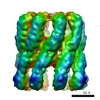
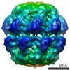
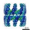
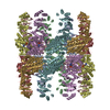
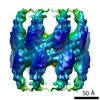
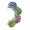
| Title | Multiple states of a nucleotide-bound group 2 chaperonin. | |||||||||
 Map data Map data | Reconstruction of the open conformation of the M. maripaludis group 2 chaperonin | |||||||||
 Sample Sample |
| |||||||||
| Function / homology | ATP binding Function and homology information Function and homology information | |||||||||
| Biological species |  Methanococcus maripaludis (archaea) Methanococcus maripaludis (archaea) | |||||||||
| Method | single particle reconstruction / cryo EM / Resolution: 10.0 Å | |||||||||
 Authors Authors | Clare DK / Stagg S / Quispe J / Farr GW / Horwich AL / Saibil HR | |||||||||
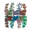
 Citation Citation | Journal: Structure / Year: 2008 Title: Multiple states of a nucleotide-bound group 2 chaperonin. Authors: Daniel K Clare / Scott Stagg / Joel Quispe / George W Farr / Arthur L Horwich / Helen R Saibil /  Abstract: Chaperonin action is controlled by cycles of nucleotide binding and hydrolysis. Here, we examine the effects of nucleotide binding on an archaeal group 2 chaperonin. In contrast to the ordered apo ...Chaperonin action is controlled by cycles of nucleotide binding and hydrolysis. Here, we examine the effects of nucleotide binding on an archaeal group 2 chaperonin. In contrast to the ordered apo state of the group 1 chaperonin GroEL, the unliganded form of the homo-16-mer Methanococcus maripaludis group 2 chaperonin is very open and flexible, with intersubunit contacts only in the central double belt of equatorial domains. The intermediate and apical domains are free of contacts and deviate significantly from the overall 8-fold symmetry. Nucleotide binding results in three distinct, ordered 8-fold symmetric conformations--open, partially closed, and fully closed. The partially closed ring encloses a 40% larger volume than does the GroEL-GroES folding chamber, enabling it to encapsulate proteins up to 80 kDa, in contrast to the fully closed form, whose cavities are 20% smaller than those of the GroEL-GroES chamber. | |||||||||
| History |
|
- Structure visualization
| Movie |
Movie viewer |
|---|---|
| Structure viewer | EM map: SurfViewMolmilJmol/JSmol |
| Supplemental images |

- Downloads & links
-EMDB archive
| Map data | emd_1396.map.gz | 175.5 KB | EMDB map data format | |
|---|---|---|---|---|
| Header (meta data) | emd-1396-v30.xmlemd-1396.xml | 10.6 KB 10.6 KB | Display Display | EMDB header |
| Images |  1396.gif 1396.gif | 140.7 KB | ||
| Archive directory |  http://ftp.pdbj.org/pub/emdb/structures/EMD-1396ftp://ftp.pdbj.org/pub/emdb/structures/EMD-1396 http://ftp.pdbj.org/pub/emdb/structures/EMD-1396ftp://ftp.pdbj.org/pub/emdb/structures/EMD-1396 | HTTPS FTP |
-Validation report
| Summary document | emd_1396_validation.pdf.gz | 213.5 KB | Display | EMDB validaton report |
|---|---|---|---|---|
| Full document | emd_1396_full_validation.pdf.gz | 212.6 KB | Display | |
| Data in XML | emd_1396_validation.xml.gz | 5.1 KB | Display | |
| Arichive directory | https://ftp.pdbj.org/pub/emdb/validation_reports/EMD-1396ftp://ftp.pdbj.org/pub/emdb/validation_reports/EMD-1396 | HTTPS FTP |
-Related structure data
-Links
| EMDB pages | EMDB (EBI/PDBe) / EMDataResource |
|---|
-Map
| File | Download / File: emd_1396.map.gz / Format: CCP4 / Size: 3.7 MB / Type: IMAGE STORED AS FLOATING POINT NUMBER (4 BYTES) | ||||||||||||||||||||||||||||||||||||||||||||||||||||||||||||||||||||
|---|---|---|---|---|---|---|---|---|---|---|---|---|---|---|---|---|---|---|---|---|---|---|---|---|---|---|---|---|---|---|---|---|---|---|---|---|---|---|---|---|---|---|---|---|---|---|---|---|---|---|---|---|---|---|---|---|---|---|---|---|---|---|---|---|---|---|---|---|---|
| Annotation | Reconstruction of the open conformation of the M. maripaludis group 2 chaperonin | ||||||||||||||||||||||||||||||||||||||||||||||||||||||||||||||||||||
| Projections & slices | Image control
Images are generated by Spider. | ||||||||||||||||||||||||||||||||||||||||||||||||||||||||||||||||||||
| Voxel size | X=Y=Z: 3.26 Å | ||||||||||||||||||||||||||||||||||||||||||||||||||||||||||||||||||||
| Density |
| ||||||||||||||||||||||||||||||||||||||||||||||||||||||||||||||||||||
| Symmetry | Space group: 1 | ||||||||||||||||||||||||||||||||||||||||||||||||||||||||||||||||||||
| Details | EMDB XML:
CCP4 map header:
| ||||||||||||||||||||||||||||||||||||||||||||||||||||||||||||||||||||
 Z (Sec.)
 X (Row.)
 Y (Col.)


-Supplemental data
- Sample components
-Entire : cpn60
| Entire | Name: cpn60 |
|---|---|
| Components |
|
-Supramolecule #1000: cpn60
| Supramolecule | Name: cpn60 / type: sample / ID: 1000 / Oligomeric state: hexadecamer / Number unique components: 2 |
|---|---|
| Molecular weight | Experimental: 900 KDa / Theoretical: 900 KDa |
-Macromolecule #1: cpn60
| Macromolecule | Name: cpn60 / type: protein_or_peptide / ID: 1 / Name.synonym: cpn60 / Details: chaperonin / Number of copies: 16 / Oligomeric state: hexadecamer / Recombinant expression: Yes |
|---|---|
| Source (natural) | Organism: Methanococcus maripaludis (archaea) / Strain: LL / synonym: M. maripaludis / Location in cell: cytoplasm |
| Molecular weight | Experimental: 900 KDa / Theoretical: 900 KDa |
| Recombinant expression | Organism: Escherichia coli BL21, DE3 / Recombinant plasmid: pET30b |
| Sequence | GO: ATP binding |
-Macromolecule #2: ADPAlF3
| Macromolecule | Name: ADPAlF3 / type: ligand / ID: 2 / Name.synonym: ADPAlF3 / Details: ATP analogue / Recombinant expression: No |
|---|---|
| Molecular weight | Experimental: 500 Da / Theoretical: 500 Da |
-Experimental details
-Structure determination
| Method | cryo EM |
|---|---|
 Processing Processing | single particle reconstruction |
| Aggregation state | particle |
-Sample preparation
| Concentration | 3 mg/mL |
|---|---|
| Buffer | pH: 7.4 Details: 50mM Tris-HCL, 150mM KCl, 10mM MgCl, 1mM DTT,1mM ADP, 5mMKF, 0.5mM Al |
| Grid | Details: 400 mesh c2/2 |
| Vitrification | Cryogen name: ETHANE / Chamber humidity: 100 % / Chamber temperature: 100 K / Instrument: OTHER Details: Vitrification instrument: vitrobot. vitobot chamber was maintained at 37C Method: 2 second blot |
- Electron microscopy
| Microscope | FEI TECNAI F20 |
|---|---|
| Temperature | Average: 100 K |
| Alignment procedure | Legacy - Astigmatism: objective astigmatism corrected by leginon |
| Date | May 12, 2006 |
| Image recording | Category: CCD / Film or detector model: GENERIC GATAN (4k x 4k) / Number real images: 531 / Average electron dose: 20 e/Å2 / Details: collected using the Leginon system at Scripps |
| Electron beam | Acceleration voltage: 120 kV / Electron source:  FIELD EMISSION GUN FIELD EMISSION GUN |
| Electron optics | Calibrated magnification: 92000 / Illumination mode: FLOOD BEAM / Imaging mode: BRIGHT FIELD / Cs: 2 mm / Nominal defocus max: 3.4 µm / Nominal defocus min: 0.9 µm / Nominal magnification: 50000 |
| Sample stage | Specimen holder: Side entry single tilt holder / Specimen holder model: GATAN LIQUID NITROGEN |
| Experimental equipment |  Model: Tecnai F20 / Image courtesy: FEI Company |
-Image processing
| Details | We used FindEM to pick the particle |
|---|---|
| CTF correction | Details: Each particle |
| Final reconstruction | Applied symmetry - Point group: C8 (8 fold cyclic) / Algorithm: OTHER / Resolution.type: BY AUTHOR / Resolution: 10.0 Å / Resolution method: FSC 0.5 CUT-OFF / Software - Name: Spider / Number images used: 2504 |
| Final angle assignment | Details: theta 80-100, phi 0-45 |
-Atomic model buiding 1
| Software | Name: URO |
|---|---|
| Details | Protocol: rigid body. the domains were separately fitted using pymol and then their postions were refined using URO |
| Refinement | Space: RECIPROCAL / Protocol: RIGID BODY FIT / Target criteria: correlation coefficient |
