 Movie
Movie Controller


+ Open data

- Basic information
| Entry |  | |||||||||
|---|---|---|---|---|---|---|---|---|---|---|
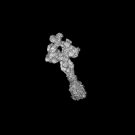
| Title | Structure of the MUCIN-2 Cterminal domains | |||||||||
 Map data Map data | ||||||||||
 Sample Sample |
| |||||||||
 Keywords Keywords | Mucus /  extracellular / net assemble / STRUCTURAL PROTEIN extracellular / net assemble / STRUCTURAL PROTEIN | |||||||||
| Function / homology |  Function and homology information Function and homology informationinner mucus layer / outer mucus layer / host-mediated regulation of intestinal microbiota composition / Defective GALNT3 causes HFTC / Defective C1GALT1C1 causes TNPS / Defective GALNT12 causes CRCS1 / Termination of O-glycan biosynthesis / O-linked glycosylation of mucins / maintenance of gastrointestinal epithelium / mucus secretion ...inner mucus layer / outer mucus layer / host-mediated regulation of intestinal microbiota composition / Defective GALNT3 causes HFTC / Defective C1GALT1C1 causes TNPS / Defective GALNT12 causes CRCS1 / Termination of O-glycan biosynthesis / O-linked glycosylation of mucins / maintenance of gastrointestinal epithelium / mucus secretion / detoxification of copper ion / cupric ion binding / Dectin-2 family / cuprous ion binding / extracellular matrix / Golgi lumen / collagen-containing extracellular matrix / extracellular space / plasma membraneSimilarity search - Function | |||||||||
| Biological species |  Homo sapiens (human) Homo sapiens (human) | |||||||||
| Method | single particle reconstruction / cryo EM / Resolution: 3.36 Å | |||||||||
 Authors Authors | Gallego P / Hansson GC | |||||||||
| Funding support |  Sweden, 1 items Sweden, 1 items
| |||||||||
 Citation Citation | Journal: Nat Commun / Year: 2023 Title: The intestinal MUC2 mucin C-terminus is stabilized by an extra disulfide bond in comparison to von Willebrand factor and other gel-forming mucins. Authors: Pablo Gallego / Maria-Jose Garcia-Bonete / Sergio Trillo-Muyo / Christian V Recktenwald / Malin E V Johansson / Gunnar C Hansson / Abstract: The MUC2 mucin polymer is the main building unit of the intestinal mucus layers separating intestinal microbiota from the host epithelium. The MUC2 mucin is a large glycoprotein with a C-terminal ...The MUC2 mucin polymer is the main building unit of the intestinal mucus layers separating intestinal microbiota from the host epithelium. The MUC2 mucin is a large glycoprotein with a C-terminal domain similar to the MUC5AC and MUC5B mucins and the von Willebrand factor (VWF). A structural model of the C-terminal part of MUC2, MUC2-C, was generated by combining Cryo-electron microscopy, AlphaFold prediction, information of its glycosylation, and small angle X-ray scattering information. The globular VWD4 assembly in the N-terminal of MUC2-C is followed by 3.5 linear VWC domains that form an extended flexible structure before the C-terminal cystine-knot. All gel-forming mucins and VWF form tail-tail disulfide-bonded dimers in their C-terminal cystine-knot domain, but interestingly the MUC2 mucin has an extra stabilizing disulfide bond on the N-terminal side of the VWD4 domain, likely essential for a stable intestinal mucus barrier. | |||||||||
| History |
|
- Structure visualization
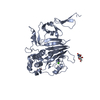
| Supplemental images |
|---|

- Downloads & links
-EMDB archive
| Map data | emd_13896.map.gz | 364 MB | EMDB map data format | |
|---|---|---|---|---|
| Header (meta data) | emd-13896-v30.xmlemd-13896.xml | 12.1 KB 12.1 KB | Display Display | EMDB header |
| Images | 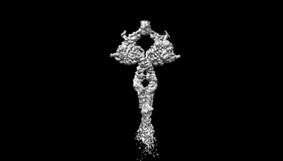 emd_13896.png emd_13896.png | 19.5 KB | ||
| Archive directory |  http://ftp.pdbj.org/pub/emdb/structures/EMD-13896ftp://ftp.pdbj.org/pub/emdb/structures/EMD-13896 http://ftp.pdbj.org/pub/emdb/structures/EMD-13896ftp://ftp.pdbj.org/pub/emdb/structures/EMD-13896 | HTTPS FTP |
-Related structure data
| Related structure data |  7qclMC  7qcnC  7qcuC M: atomic model generated by this map C: citing same article ( |
|---|---|
| Similar structure data |


-Links
| EMDB pages | EMDB (EBI/PDBe) / EMDataResource |
|---|---|
| Related items in Molecule of the Month |


-Map
| File | Download / File: emd_13896.map.gz / Format: CCP4 / Size: 729 MB / Type: IMAGE STORED AS FLOATING POINT NUMBER (4 BYTES) | ||||||||||||||||||||||||||||||||||||
|---|---|---|---|---|---|---|---|---|---|---|---|---|---|---|---|---|---|---|---|---|---|---|---|---|---|---|---|---|---|---|---|---|---|---|---|---|---|
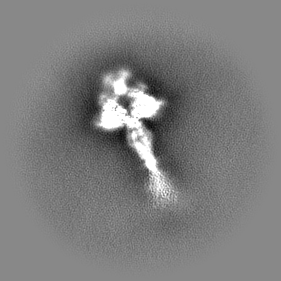
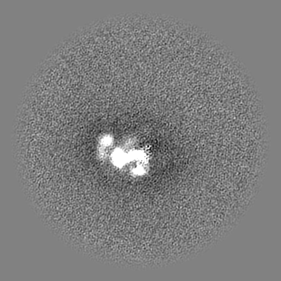
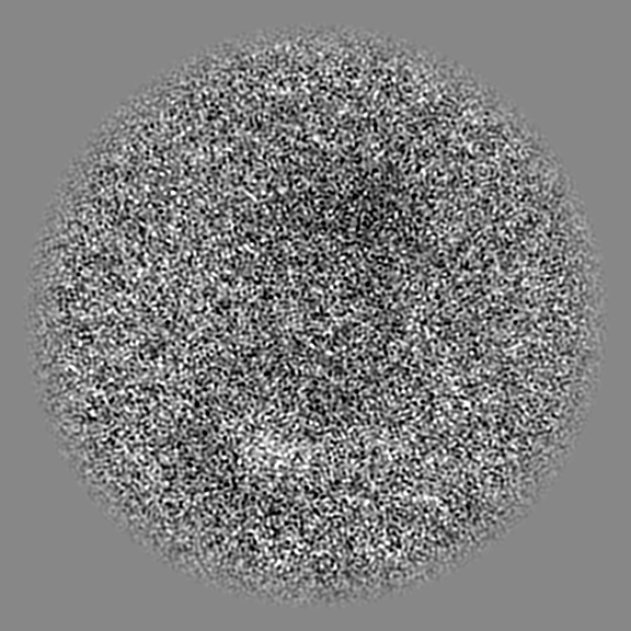
| Projections & slices | Image control
Images are generated by Spider. | ||||||||||||||||||||||||||||||||||||
| Voxel size | X=Y=Z: 0.86 Å | ||||||||||||||||||||||||||||||||||||
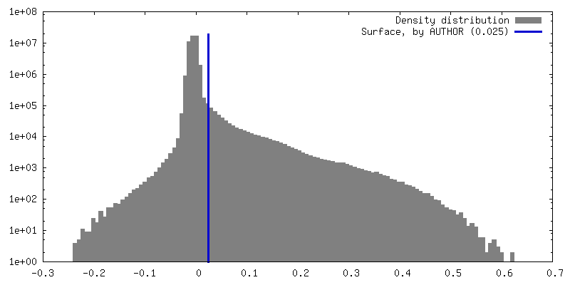
| Density |
| ||||||||||||||||||||||||||||||||||||
| Symmetry | Space group: 1 | ||||||||||||||||||||||||||||||||||||
| Details | EMDB XML:
|
 Z (Sec.)
 Y (Row.)
 X (Col.)


-Supplemental data
- Sample components
-Entire : Muc2 dimer, Mucus assemble
| Entire | Name: Muc2 dimer, Mucus assemble |
|---|---|
| Components |
|
-Supramolecule #1: Muc2 dimer, Mucus assemble
| Supramolecule | Name: Muc2 dimer, Mucus assemble / type: complex / ID: 1 / Parent: 0 / Macromolecule list: #1 |
|---|---|
| Source (natural) | Organism: Homo sapiens (human) |
-Macromolecule #1: Mucin-2
| Macromolecule | Name: Mucin-2 / type: protein_or_peptide / ID: 1 / Number of copies: 2 / Enantiomer: LEVO |
|---|---|
| Source (natural) | Organism: Homo sapiens (human) |
| Molecular weight | Theoretical: 64.296707 KDa |
| Recombinant expression | Organism:   Cricetulus griseus (Chinese hamster) Cricetulus griseus (Chinese hamster) |
| Sequence | String: KPPECPDFDP PRQENETWWL CDCFMATCKY NNTVEIVKVE CEPPPMPTCS NGLQPVRVED PDGCCWHWEC DCYCTGWGDP HYVTFDGLY YSYQGNCTYV LVEEISPSVD NFGVYIDNYH CDPNDKVSCP RTLIVRHETQ EVLIKTVHMM PMQVQVQVNR Q AVALPYKK ...String: KPPECPDFDP PRQENETWWL CDCFMATCKY NNTVEIVKVE CEPPPMPTCS NGLQPVRVED PDGCCWHWEC DCYCTGWGDP HYVTFDGLY YSYQGNCTYV LVEEISPSVD NFGVYIDNYH CDPNDKVSCP RTLIVRHETQ EVLIKTVHMM PMQVQVQVNR Q AVALPYKK YGLEVYQSGI NYVVDIPELG VLVSYNGLSF SVRLPYHRFG NNTKGQCGTC TNTTSDDCIL PSGEIVSNCE AA ADQWLVN DPSKPHCPHS SSTTKRPAVT VPGGGKTTPH KDCTPSPLCQ LIKDSLFAQC HALVPPQHYY DACVFDSCFM PGS SLECAS LQAYAALCAQ QNICLDWRNH THGACLVECP SHREYQACGP AEEPTCKSSS SQQNNTVLVE GCFCPEGTMN YAPG FDVCV KTCGCVGPDN VPREFGEHFE FDCKNCVCLE GGSGIICQPK RCSQKPVTHC VEDGTYLATE VNPADTCCNI TVCKC NTSL CKEKPSVCPL GFEVKSKMVP GRCCPFYWCE SKGVCVHGNA EYQPGSPVYS SKCQDCVCTD KVDNNTLLNV IACTHV PCN TSCSPGFELM EAPGECCKKC UniProtKB: Mucin-2 |
-Macromolecule #8: 2-acetamido-2-deoxy-beta-D-glucopyranose
| Macromolecule | Name: 2-acetamido-2-deoxy-beta-D-glucopyranose / type: ligand / ID: 8 / Number of copies: 4 / Formula: NAG |
|---|---|
| Molecular weight | Theoretical: 221.208 Da |
| Chemical component information |  ChemComp-NAG: |
-Macromolecule #9: CALCIUM ION
| Macromolecule | Name: CALCIUM ION / type: ligand / ID: 9 / Number of copies: 2 / Formula: CA |
|---|---|
| Molecular weight | Theoretical: 40.078 Da |
-Experimental details
-Structure determination
| Method | cryo EM |
|---|---|
 Processing Processing | single particle reconstruction |
| Aggregation state | particle |
-Sample preparation
| Concentration | 1 mg/mL | ||||||||||||
|---|---|---|---|---|---|---|---|---|---|---|---|---|---|
| Buffer | pH: 7.4 Component:
| ||||||||||||
| Grid | Model: Quantifoil R1.2/1.3 / Material: GOLD / Mesh: 300 / Pretreatment - Type: GLOW DISCHARGE / Pretreatment - Time: 40 sec. / Details: 15mA | ||||||||||||
| Vitrification | Cryogen name: ETHANE / Chamber humidity: 100 % / Chamber temperature: 277.15 K / Instrument: FEI VITROBOT MARK III |
- Electron microscopy
| Microscope | TFS KRIOS |
|---|---|
| Electron beam | Acceleration voltage: 300 kV / Electron source: FIELD EMISSION GUN |
| Electron optics | Illumination mode: FLOOD BEAM / Imaging mode: BRIGHT FIELDBright-field microscopy / Nominal defocus max: 5.0 µm / Nominal defocus min: 1.2 µm |
| Image recording | Film or detector model: GATAN K3 BIOQUANTUM (6k x 4k) / Average electron dose: 79.0 e/Å2 |
| Experimental equipment |  Model: Titan Krios / Image courtesy: FEI Company |
-Image processing
| Startup model | Type of model: PDB ENTRY PDB model - PDB ID: |
|---|---|
| Initial angle assignment | Type: MAXIMUM LIKELIHOOD |
| Final angle assignment | Type: MAXIMUM LIKELIHOOD / Software - Name: cryoSPARC (ver. 3) |
| Final reconstruction | Resolution.type: BY AUTHOR / Resolution: 3.36 Å / Resolution method: FSC 0.143 CUT-OFF / Number images used: 369150 |
-Atomic model buiding 1
| Refinement | Space: REAL / Protocol: RIGID BODY FIT |
|---|---|
| Output model | PDB-7qcl: |
