 Movie
Movie Controller


+ Open data

- Basic information
| Entry |  | |||||||||
|---|---|---|---|---|---|---|---|---|---|---|
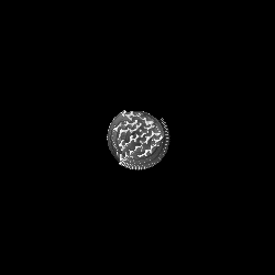
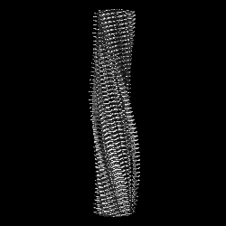
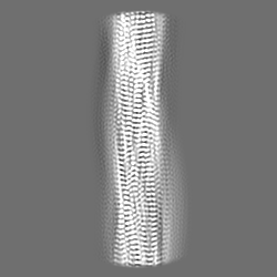
| Title | Cryo-em structure of the Nup98 fibril polymorph 3 | |||||||||
 Map data Map data | Cryo-em structure of the Nup98 fibril polymorph 3 | |||||||||
 Sample Sample |
| |||||||||
| Function / homology |  Function and homology information Function and homology informationtelomere tethering at nuclear periphery / nuclear pore organization / nuclear pore outer ring /  nuclear pore complex assembly / nuclear pore cytoplasmic filaments / post-transcriptional tethering of RNA polymerase II gene DNA at nuclear periphery / Nuclear Pore Complex (NPC) Disassembly / nuclear inclusion body / nuclear pore nuclear basket / Transport of Ribonucleoproteins into the Host Nucleus ...telomere tethering at nuclear periphery / nuclear pore organization / nuclear pore outer ring / nuclear pore complex assembly / nuclear pore cytoplasmic filaments / post-transcriptional tethering of RNA polymerase II gene DNA at nuclear periphery / Nuclear Pore Complex (NPC) Disassembly / nuclear inclusion body / nuclear pore nuclear basket / Transport of Ribonucleoproteins into the Host Nucleus / Regulation of Glucokinase by Glucokinase Regulatory Protein / Defective TPR may confer susceptibility towards thyroid papillary carcinoma (TPC) / Transport of the SLBP independent Mature mRNA / Transport of the SLBP Dependant Mature mRNA / NS1 Mediated Effects on Host Pathways / SUMOylation of SUMOylation proteins / Transport of Mature mRNA Derived from an Intronless Transcript / structural constituent of nuclear pore / Rev-mediated nuclear export of HIV RNA / SUMOylation of RNA binding proteins / Nuclear import of Rev protein / Transport of Mature mRNA derived from an Intron-Containing Transcript / NEP/NS2 Interacts with the Cellular Export Machinery / RNA export from nucleus / tRNA processing in the nucleus / positive regulation of mRNA splicing, via spliceosome / Postmitotic nuclear pore complex (NPC) reformation / nucleocytoplasmic transport / Viral Messenger RNA Synthesis / nuclear localization sequence binding / SUMOylation of ubiquitinylation proteins / Vpr-mediated nuclear import of PICs / SUMOylation of DNA replication proteins / Hydrolases; Acting on peptide bonds (peptidases); Serine endopeptidases / Regulation of HSF1-mediated heat shock response / mRNA transport / Amplification of signal from unattached kinetochores via a MAD2 inhibitory signal / SUMOylation of DNA damage response and repair proteins / nuclear pore / Mitotic Prometaphase / EML4 and NUDC in mitotic spindle formation / Resolution of Sister Chromatid Cohesion / serine-type peptidase activity / SUMOylation of chromatin organization proteins / nuclear periphery / HCMV Late Events / molecular condensate scaffold activity / RHO GTPases Activate Formins / promoter-specific chromatin binding / Transcriptional regulation by small RNAs / ISG15 antiviral mechanism / HCMV Early Events / Separation of Sister Chromatids / protein import into nucleus / nuclear envelope / snRNP Assembly / nuclear membrane / transcription coactivator activity / nuclear body / ribonucleoprotein complex / mRNA binding / SARS-CoV-2 activates/modulates innate and adaptive immune responses / proteolysis / RNA binding / nucleoplasm / cytosol nuclear pore complex assembly / nuclear pore cytoplasmic filaments / post-transcriptional tethering of RNA polymerase II gene DNA at nuclear periphery / Nuclear Pore Complex (NPC) Disassembly / nuclear inclusion body / nuclear pore nuclear basket / Transport of Ribonucleoproteins into the Host Nucleus ...telomere tethering at nuclear periphery / nuclear pore organization / nuclear pore outer ring / nuclear pore complex assembly / nuclear pore cytoplasmic filaments / post-transcriptional tethering of RNA polymerase II gene DNA at nuclear periphery / Nuclear Pore Complex (NPC) Disassembly / nuclear inclusion body / nuclear pore nuclear basket / Transport of Ribonucleoproteins into the Host Nucleus / Regulation of Glucokinase by Glucokinase Regulatory Protein / Defective TPR may confer susceptibility towards thyroid papillary carcinoma (TPC) / Transport of the SLBP independent Mature mRNA / Transport of the SLBP Dependant Mature mRNA / NS1 Mediated Effects on Host Pathways / SUMOylation of SUMOylation proteins / Transport of Mature mRNA Derived from an Intronless Transcript / structural constituent of nuclear pore / Rev-mediated nuclear export of HIV RNA / SUMOylation of RNA binding proteins / Nuclear import of Rev protein / Transport of Mature mRNA derived from an Intron-Containing Transcript / NEP/NS2 Interacts with the Cellular Export Machinery / RNA export from nucleus / tRNA processing in the nucleus / positive regulation of mRNA splicing, via spliceosome / Postmitotic nuclear pore complex (NPC) reformation / nucleocytoplasmic transport / Viral Messenger RNA Synthesis / nuclear localization sequence binding / SUMOylation of ubiquitinylation proteins / Vpr-mediated nuclear import of PICs / SUMOylation of DNA replication proteins / Hydrolases; Acting on peptide bonds (peptidases); Serine endopeptidases / Regulation of HSF1-mediated heat shock response / mRNA transport / Amplification of signal from unattached kinetochores via a MAD2 inhibitory signal / SUMOylation of DNA damage response and repair proteins / nuclear pore / Mitotic Prometaphase / EML4 and NUDC in mitotic spindle formation / Resolution of Sister Chromatid Cohesion / serine-type peptidase activity / SUMOylation of chromatin organization proteins / nuclear periphery / HCMV Late Events / molecular condensate scaffold activity / RHO GTPases Activate Formins / promoter-specific chromatin binding / Transcriptional regulation by small RNAs / ISG15 antiviral mechanism / HCMV Early Events / Separation of Sister Chromatids / protein import into nucleus / nuclear envelope / snRNP Assembly / nuclear membrane / transcription coactivator activity / nuclear body / ribonucleoprotein complex / mRNA binding / SARS-CoV-2 activates/modulates innate and adaptive immune responses / proteolysis / RNA binding / nucleoplasm / cytosolSimilarity search - Function | |||||||||
| Biological species |  Homo sapiens (human) Homo sapiens (human) | |||||||||
| Method | helical reconstruction / cryo EM / Resolution: 2.79 Å | |||||||||
 Authors Authors | Ibanez de Opakua A / Geraets JA / Frieg B / Dienemann C / Savastano A / Rankovic M / Cima-Omori M-S / Schroeder GF / Zweckstetter M | |||||||||
| Funding support |  Germany, 2 items Germany, 2 items
| |||||||||
 Citation Citation | Journal: Nat Chem / Year: 2022 Title: Molecular interactions of FG nucleoporin repeats at high resolution. Authors: Alain Ibáñez de Opakua / James A Geraets / Benedikt Frieg / Christian Dienemann / Adriana Savastano / Marija Rankovic / Maria-Sol Cima-Omori / Gunnar F Schröder / Markus Zweckstetter / Abstract: Proteins that contain repeat phenylalanine-glycine (FG) residues phase separate into oncogenic transcription factor condensates in malignant leukaemias, form the permeability barrier of the nuclear ...Proteins that contain repeat phenylalanine-glycine (FG) residues phase separate into oncogenic transcription factor condensates in malignant leukaemias, form the permeability barrier of the nuclear pore complex and mislocalize in neurodegenerative diseases. Insights into the molecular interactions of FG-repeat nucleoporins have, however, remained largely elusive. Using a combination of NMR spectroscopy and cryoelectron microscopy, we have identified uniformly spaced segments of transient β-structure and a stable preformed α-helix recognized by messenger RNA export factors in the FG-repeat domain of human nucleoporin 98 (Nup98). In addition, we have determined at high resolution the molecular organization of reversible FG-FG interactions in amyloid fibrils formed by a highly aggregation-prone segment in Nup98. We have further demonstrated that amyloid-like aggregates of the FG-repeat domain of Nup98 have low stability and are reversible. Our results provide critical insights into the molecular interactions underlying the self-association and phase separation of FG-repeat nucleoporins in physiological and pathological cell activities. | |||||||||
| History |
|
- Structure visualization
| Supplemental images |
|---|

- Downloads & links
-EMDB archive
| Map data | emd_13853.map.gz | 5.4 MB | EMDB map data format | |
|---|---|---|---|---|
| Header (meta data) | emd-13853-v30.xmlemd-13853.xml | 15.5 KB 15.5 KB | Display Display | EMDB header |
| FSC (resolution estimation) | emd_13853_fsc.xml | 8.9 KB | Display | FSC data file |
| Images | 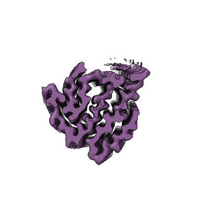 emd_13853.png emd_13853.png | 63.8 KB | ||
| Others | emd_13853_half_map_1.map.gzemd_13853_half_map_2.map.gz | 46.2 MB 46.2 MB | ||
| Archive directory |  http://ftp.pdbj.org/pub/emdb/structures/EMD-13853ftp://ftp.pdbj.org/pub/emdb/structures/EMD-13853 http://ftp.pdbj.org/pub/emdb/structures/EMD-13853ftp://ftp.pdbj.org/pub/emdb/structures/EMD-13853 | HTTPS FTP |
-Related structure data
| Related structure data |  7q66MC  7q64C  7q65C 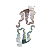 7q67C M: atomic model generated by this map C: citing same article ( |
|---|---|
| Similar structure data |


-Links
| EMDB pages | EMDB (EBI/PDBe) / EMDataResource |
|---|---|
| Related items in Molecule of the Month |


-Map
| File | Download / File: emd_13853.map.gz / Format: CCP4 / Size: 59.6 MB / Type: IMAGE STORED AS FLOATING POINT NUMBER (4 BYTES) | ||||||||||||||||||||||||||||||||||||
|---|---|---|---|---|---|---|---|---|---|---|---|---|---|---|---|---|---|---|---|---|---|---|---|---|---|---|---|---|---|---|---|---|---|---|---|---|---|
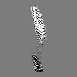
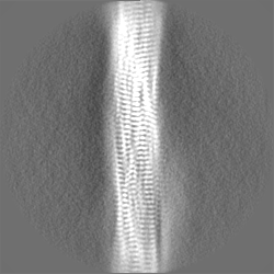
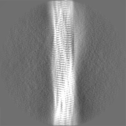
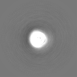
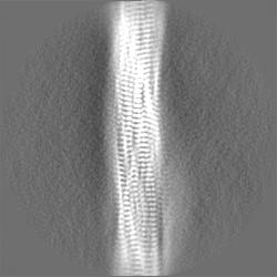
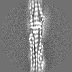
| Annotation | Cryo-em structure of the Nup98 fibril polymorph 3 | ||||||||||||||||||||||||||||||||||||
| Projections & slices | Image control
Images are generated by Spider. | ||||||||||||||||||||||||||||||||||||
| Voxel size | X=Y=Z: 1.05 Å | ||||||||||||||||||||||||||||||||||||
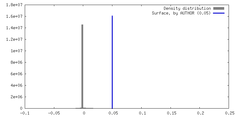
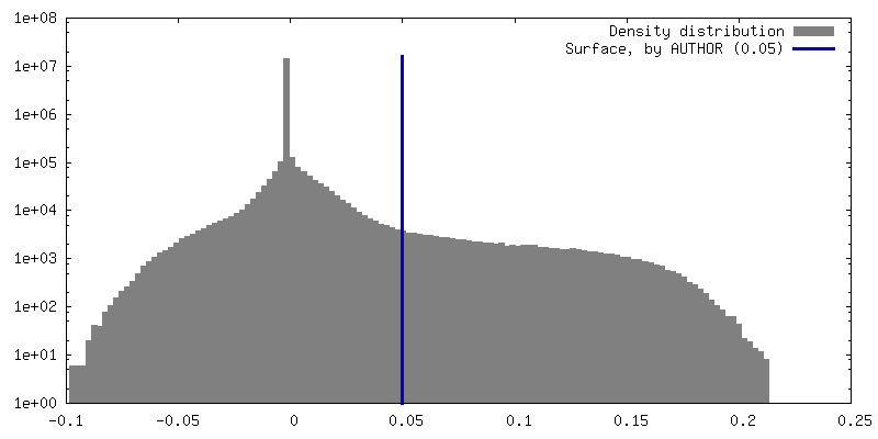
| Density |
| ||||||||||||||||||||||||||||||||||||
| Symmetry | Space group: 1 | ||||||||||||||||||||||||||||||||||||
| Details | EMDB XML:
|
 Z (Sec.)
 Y (Row.)
 X (Col.)


-Supplemental data
-Half map: Cryo-em structure of the Nup98 fibril polymorph 3, half map 1
| File | emd_13853_half_map_1.map | ||||||||||||
|---|---|---|---|---|---|---|---|---|---|---|---|---|---|
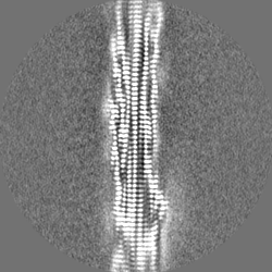
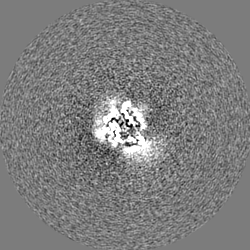
| Annotation | Cryo-em structure of the Nup98 fibril polymorph 3, half map 1 | ||||||||||||
| Projections & Slices |
| ||||||||||||
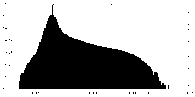
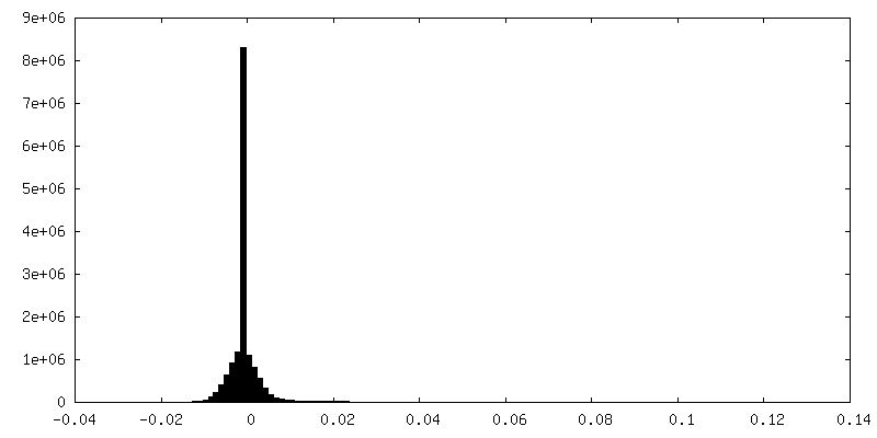
| Density Histograms |


-Half map: Cryo-em structure of the Nup98 fibril polymorph 3, half map 2
| File | emd_13853_half_map_2.map | ||||||||||||
|---|---|---|---|---|---|---|---|---|---|---|---|---|---|
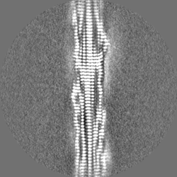
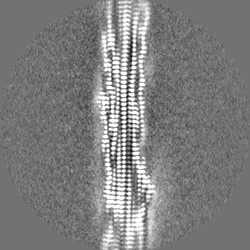
| Annotation | Cryo-em structure of the Nup98 fibril polymorph 3, half map 2 | ||||||||||||
| Projections & Slices |
| ||||||||||||
| Density Histograms |


- Sample components
-Entire : Cryo-em structure of the Nup98 fibril polymorph 3
| Entire | Name: Cryo-em structure of the Nup98 fibril polymorph 3 |
|---|---|
| Components |
|
-Supramolecule #1: Cryo-em structure of the Nup98 fibril polymorph 3
| Supramolecule | Name: Cryo-em structure of the Nup98 fibril polymorph 3 / type: complex / ID: 1 / Parent: 0 / Macromolecule list: all Details: The Nup98 polymorph 3 fibril is formed by two protofilaments, consisting of Nup98 monomers. |
|---|---|
| Source (natural) | Organism: Homo sapiens (human) |
| Recombinant expression | Organism:  Escherichia coli BL21(DE3) (bacteria) Escherichia coli BL21(DE3) (bacteria) |
-Macromolecule #1: Nuclear pore complex protein Nup98
| Macromolecule | Name: Nuclear pore complex protein Nup98 / type: protein_or_peptide / ID: 1 / Number of copies: 22 / Enantiomer: LEVO |
|---|---|
| Source (natural) | Organism: Homo sapiens (human) |
| Molecular weight | Theoretical: 4.005211 KDa |
| Recombinant expression | Organism: Escherichia coli BL21(DE3) (bacteria) |
| Sequence | String: TGTANTLFGT ASTGTSLFSS QNNAFAQNKP TGFGNFGTST |
-Experimental details
-Structure determination
| Method | cryo EM |
|---|---|
 Processing Processing | helical reconstruction |
| Aggregation state | filament |
-Sample preparation
| Buffer | pH: 7 / Component: | ||||
|---|---|---|---|---|---|
| Vitrification | Cryogen name: ETHANE / Chamber humidity: 100 % / Chamber temperature: 293 K / Instrument: FEI VITROBOT MARK IV |
- Electron microscopy
| Microscope | FEI TITAN KRIOS |
|---|---|
| Electron beam | Acceleration voltage: 300 kV / Electron source: FIELD EMISSION GUN |
| Electron optics | Illumination mode: FLOOD BEAM / Imaging mode: BRIGHT FIELDBright-field microscopy / Cs: 2.7 mm / Nominal defocus max: 2.0 µm / Nominal defocus min: 0.7000000000000001 µm / Nominal magnification: 81000 |
| Image recording | Film or detector model: GATAN K3 (6k x 4k) / Number real images: 4180 / Average exposure time: 2.557 sec. / Average electron dose: 41.0 e/Å2 |
| Experimental equipment |  Model: Titan Krios / Image courtesy: FEI Company |
-Image processing
| Segment selection | Number selected: 356482 |
|---|---|
| Startup model | Type of model: OTHER / Details: featureless cylinder |
| Final angle assignment | Type: NOT APPLICABLE |
| Final reconstruction | Applied symmetry - Helical parameters - Δz: 4.67 Å Applied symmetry - Helical parameters - Δ&Phi: -3.29 ° Applied symmetry - Helical parameters - Axial symmetry: C1 (asymmetric) Resolution.type: BY AUTHOR / Resolution: 2.79 Å / Resolution method: FSC 0.143 CUT-OFF / Software - Name: RELION (ver. 3.1) / Number images used: 28360 |
| FSC plot (resolution estimation) | 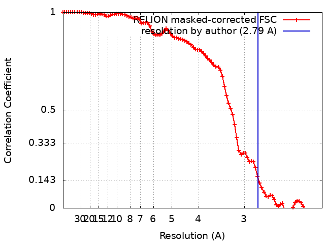 |
