 Movie
Movie Controller

[English] 日本語
 Yorodumi
- EMDB-13152: Negative stain EM 3D reconstruction of the Dam1 / DASH complex. -
+ Open data

- Basic information
| Entry | Database: EMDB / ID: EMD-13152 | |||||||||
|---|---|---|---|---|---|---|---|---|---|---|
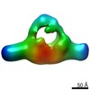
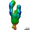
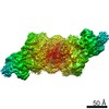
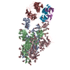
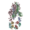
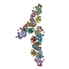
| Title | Negative stain EM 3D reconstruction of the Dam1 / DASH complex. | |||||||||
 Map data Map data | Dimer of the Dam1 / DASH complex. | |||||||||
 Sample Sample |
| |||||||||
| Function / homology |  Function and homology information Function and homology informationprotein localization to microtubule plus-end / mitotic spindle polar microtubule / mitotic spindle orientation checkpoint signaling / 2-micrometer plasmid partitioning / DASH complex / protein transport along microtubule to mitotic spindle pole body / mitotic sister chromatid biorientation / positive regulation of attachment of spindle microtubules to kinetochore / mitotic spindle pole body / mitotic spindle midzone ...protein localization to microtubule plus-end / mitotic spindle polar microtubule / mitotic spindle orientation checkpoint signaling / 2-micrometer plasmid partitioning / DASH complex / protein transport along microtubule to mitotic spindle pole body / mitotic sister chromatid biorientation / positive regulation of attachment of spindle microtubules to kinetochore / mitotic spindle pole body / mitotic spindle midzone / attachment of spindle microtubules to kinetochore / nuclear migration along microtubule / protein localization to microtubule / microtubule plus-end / negative regulation of microtubule depolymerization / microtubule plus-end binding /  microtubule organizing center / microtubule depolymerization / mitotic sister chromatid cohesion / spindle assembly / regulation of microtubule polymerization or depolymerization / spindle midzone / cytoplasmic microtubule / positive regulation of microtubule polymerization / spindle microtubule / mitotic spindle / spindle pole / microtubule / cell division / identical protein binding / cytoplasm microtubule organizing center / microtubule depolymerization / mitotic sister chromatid cohesion / spindle assembly / regulation of microtubule polymerization or depolymerization / spindle midzone / cytoplasmic microtubule / positive regulation of microtubule polymerization / spindle microtubule / mitotic spindle / spindle pole / microtubule / cell division / identical protein binding / cytoplasmSimilarity search - Function | |||||||||
| Biological species |  Saccharomyces cerevisiae (brewer's yeast) Saccharomyces cerevisiae (brewer's yeast) | |||||||||
| Method | single particle reconstruction / negative staining / Resolution: 35.0 Å | |||||||||
 Authors Authors | Engelhard L / Bourque C / Klink BU / Gatsogiannis C | |||||||||
| Funding support |  Germany, 1 items Germany, 1 items
| |||||||||
 Citation Citation | Journal: EMBO J / Year: 2021 Title: Phospho-regulated Bim1/EB1 interactions trigger Dam1c ring assembly at the budding yeast outer kinetochore. Authors: Alexander Dudziak / Lena Engelhard / Cole Bourque / Björn Udo Klink / Pascaline Rombaut / Nikolay Kornakov / Karolin Jänen / Franz Herzog / Christos Gatsogiannis / Stefan Westermann / Abstract: Kinetochores form the link between chromosomes and microtubules of the mitotic spindle. The heterodecameric Dam1 complex (Dam1c) is a major component of the Saccharomyces cerevisiae outer ...Kinetochores form the link between chromosomes and microtubules of the mitotic spindle. The heterodecameric Dam1 complex (Dam1c) is a major component of the Saccharomyces cerevisiae outer kinetochore, assembling into 3 MDa-sized microtubule-embracing rings, but how ring assembly is specifically initiated in vivo remains to be understood. Here, we describe a molecular pathway that provides local control of ring assembly during the establishment of sister kinetochore bi-orientation. We show that Dam1c and the general microtubule plus end-associated protein (+TIP) Bim1/EB1 form a stable complex depending on a conserved motif in the Duo1 subunit of Dam1c. EM analyses reveal that Bim1 crosslinks protrusion domains of adjacent Dam1c heterodecamers and promotes the formation of oligomers with defined curvature. Disruption of the Dam1c-Bim1 interaction impairs kinetochore localization of Dam1c in metaphase and delays mitosis. Phosphorylation promotes Dam1c-Bim1 binding by relieving an intramolecular inhibition of the Dam1 C-terminus. In addition, Bim1 recruits Bik1/CLIP-170 to Dam1c and induces formation of full rings even in the absence of microtubules. Our data help to explain how new kinetochore end-on attachments are formed during the process of attachment error correction. | |||||||||
| History |
|
- Structure visualization
| Movie |
Movie viewer |
|---|---|
| Structure viewer | EM map: SurfViewMolmilJmol/JSmol |
| Supplemental images |

- Downloads & links
-EMDB archive
| Map data | emd_13152.map.gz | 460 KB | EMDB map data format | |
|---|---|---|---|---|
| Header (meta data) | emd-13152-v30.xmlemd-13152.xml | 17.5 KB 17.5 KB | Display Display | EMDB header |
| Images | 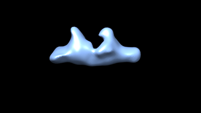 emd_13152.png emd_13152.png | 13.9 KB | ||
| Archive directory |  http://ftp.pdbj.org/pub/emdb/structures/EMD-13152ftp://ftp.pdbj.org/pub/emdb/structures/EMD-13152 http://ftp.pdbj.org/pub/emdb/structures/EMD-13152ftp://ftp.pdbj.org/pub/emdb/structures/EMD-13152 | HTTPS FTP |
-Related structure data
| Related structure data | C: citing same article ( |
|---|---|
| Similar structure data |
-Links
| EMDB pages | EMDB (EBI/PDBe) / EMDataResource |
|---|---|
| Related items in Molecule of the Month |

-Map
| File | Download / File: emd_13152.map.gz / Format: CCP4 / Size: 1.7 MB / Type: IMAGE STORED AS FLOATING POINT NUMBER (4 BYTES) | ||||||||||||||||||||||||||||||||||||||||||||||||||||||||||||
|---|---|---|---|---|---|---|---|---|---|---|---|---|---|---|---|---|---|---|---|---|---|---|---|---|---|---|---|---|---|---|---|---|---|---|---|---|---|---|---|---|---|---|---|---|---|---|---|---|---|---|---|---|---|---|---|---|---|---|---|---|---|
| Annotation | Dimer of the Dam1 / DASH complex. | ||||||||||||||||||||||||||||||||||||||||||||||||||||||||||||
| Voxel size | X=Y=Z: 4.92 Å | ||||||||||||||||||||||||||||||||||||||||||||||||||||||||||||
| Density |
| ||||||||||||||||||||||||||||||||||||||||||||||||||||||||||||
| Symmetry | Space group: 1 | ||||||||||||||||||||||||||||||||||||||||||||||||||||||||||||
| Details | EMDB XML:
CCP4 map header:
| ||||||||||||||||||||||||||||||||||||||||||||||||||||||||||||
-Supplemental data
- Sample components
+Entire : A dimer of the Dam1 / DASH complex.
+Supramolecule #1: A dimer of the Dam1 / DASH complex.
+Macromolecule #1: Ask1p of the Dam1c of S. cerevisiae
+Macromolecule #2: Dam1p of the Dam1c of S. cerevisiae
+Macromolecule #3: Spc34p of the Dam1c of S. cerevisiae
+Macromolecule #4: Duo1p of the Dam1c of S. cerevisiae
+Macromolecule #5: Spc19p of the Dam1c of S. cerevisiae
+Macromolecule #6: Dad2p of the Dam1c of S. cerevisiae
+Macromolecule #7: Dad1p of the Dam1c of S. cerevisiae
+Macromolecule #8: Dad3p of the Dam1c of S. cerevisiae
+Macromolecule #9: Dad4p of the Dam1c of S. cerevisiae
+Macromolecule #10: Hsk3p of the Dam1c of S. cerevisiae
-Experimental details
-Structure determination
| Method | negative staining |
|---|---|
 Processing Processing | single particle reconstruction |
| Aggregation state | particle |
-Sample preparation
| Concentration | 0.01 mg/mL |
|---|---|
| Buffer | pH: 7.4 / Component: (Formula: HEPES, NaClSodium chloride, TCEP) |
| Staining | Type: NEGATIVE / Material: Uranyl formate Details: 4 microliters of sample were applied onto freshly glow-discharged carbon-coated copper grids (Agar Scientific, 6400C). After an incubation of 2 minutes, the sample was blotted with Whatman ...Details: 4 microliters of sample were applied onto freshly glow-discharged carbon-coated copper grids (Agar Scientific, 6400C). After an incubation of 2 minutes, the sample was blotted with Whatman no. 4 filter paper, washed 2 times with dd H20 and stained with 75% uranyl formate, blotted immediately, and then stained again and incubated for 1 minute before final blotting. The sample was then air-dried for 4 minutes. |
| Grid | Model: Homemade / Support film - Material: CARBON / Pretreatment - Type: GLOW DISCHARGE |
| Details | 25 mM HEPES, pH 7.4, 200 mM NaCl, 1 mM MgCl2, and 0.5 mM TECP. |
- Electron microscopy
| Microscope | JEOL 1400 |
|---|---|
| Electron beam | Acceleration voltage: 120 kV / Electron source: LAB6 |
| Electron optics | Illumination mode: SPOT SCAN / Imaging mode: BRIGHT FIELDBright-field microscopy / Cs: 3.4 mm |
| Sample stage | Specimen holder model: JEOL |
| Image recording | Film or detector model: TVIPS TEMCAM-F416 (4k x 4k) / Average electron dose: 5.0 e/Å2 |
-Image processing
| Startup model | Type of model: INSILICO MODEL In silico model: Initial Model was computed using SPHIRE (VIPER model) |
|---|---|
| Initial angle assignment | Type: NOT APPLICABLE |
| Final angle assignment | Type: NOT APPLICABLE |
| Final reconstruction | Applied symmetry - Point group: C1 (asymmetric) / Resolution.type: BY AUTHOR / Resolution: 35.0 Å / Resolution method: OTHER / Number images used: 28140 |
