 Movie
Movie Controller

[English] 日本語
 Yorodumi
- EMDB-13056: Tomogram of a nuclease treated shVim HO H222P MEF nuclear lamina -
+ Open data

- Basic information
| Entry | Database: EMDB / ID: EMD-13056 | |||||||||
|---|---|---|---|---|---|---|---|---|---|---|
| Title | Tomogram of a nuclease treated shVim HO H222P MEF nuclear lamina | |||||||||
 Map data Map data | A tomogram of nuclease treated shvim HO H222P MEF nucleus. The tomogram is dose-weighted, CTF-corrected and reconstructed in IMOD. Fudicial markers are replaced by noise. | |||||||||
 Sample Sample |
| |||||||||
| Biological species |   Mus musculus (house mouse) Mus musculus (house mouse) | |||||||||
| Method | electron tomography / cryo EM | |||||||||
 Authors Authors | Tatli M / Medalia O | |||||||||
| Funding support |  Switzerland, 1 items Switzerland, 1 items
| |||||||||
 Citation Citation | Journal: J Cell Sci / Year: 2021 Title: A lamin A/C variant causing striated muscle disease provides insights into filament organization. Authors: Rafael Kronenberg-Tenga / Meltem Tatli / Matthias Eibauer / Wei Wu / Ji-Yeon Shin / Gisèle Bonne / Howard J Worman / Ohad Medalia /   Abstract: The gene encodes the A-type lamins, which polymerize into ∼3.5-nm-thick filaments and, together with B-type lamins and associated proteins, form the nuclear lamina. Mutations in cause a wide ...The gene encodes the A-type lamins, which polymerize into ∼3.5-nm-thick filaments and, together with B-type lamins and associated proteins, form the nuclear lamina. Mutations in cause a wide variety of pathologies. In this study, we analyzed the nuclear lamina of embryonic fibroblasts from mice, which develop cardiomyopathy and muscular dystrophy. Although the organization of the lamina appeared unaltered, there were changes in chromatin and B-type lamin expression. An increase in nuclear size and consequently a relative reduction in heterochromatin near the lamina allowed for a higher resolution structural analysis of lamin filaments using cryo-electron tomography. This was most apparent when visualizing lamin filaments and using a nuclear extraction protocol. Averaging of individual segments of filaments in mouse fibroblasts resolved two polymers that constitute the mature filaments. Our findings provide better views of the organization of lamin filaments and the effect of a striated muscle disease-causing mutation on nuclear structure. | |||||||||
| History |
|
- Structure visualization
| Movie |
 Movie viewer Movie viewer |
|---|---|
| Supplemental images |

- Downloads & links
-EMDB archive
| Map data | emd_13056.map.gz | 73.1 MB | EMDB map data format | |
|---|---|---|---|---|
| Header (meta data) | emd-13056-v30.xmlemd-13056.xml | 7.8 KB 7.8 KB | Display Display | EMDB header |
| Images | 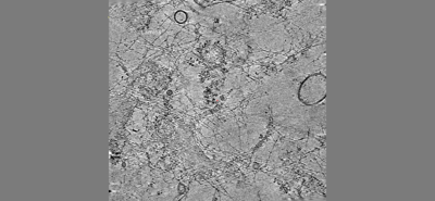 emd_13056.png emd_13056.png | 67.1 KB | ||
| Archive directory |  http://ftp.pdbj.org/pub/emdb/structures/EMD-13056ftp://ftp.pdbj.org/pub/emdb/structures/EMD-13056 http://ftp.pdbj.org/pub/emdb/structures/EMD-13056ftp://ftp.pdbj.org/pub/emdb/structures/EMD-13056 | HTTPS FTP |
-Related structure data
| Related structure data | C: citing same article ( |
|---|---|
| EM raw data | EMPIAR-10601 (Title: A lamin A/C variant causing striated muscle disease provides insights into filament organization Data size: 2.7 Data #1: Tilt-series of LmnaH222P/H222P revealing lamin meshwork in nuclease treated and FIB-milled nucleus [tilt series]) |
-Links
| EMDB pages | EMDB (EBI/PDBe) / EMDataResource |
|---|
-Map
| File | Download / File: emd_13056.map.gz / Format: CCP4 / Size: 165.7 MB / Type: IMAGE STORED AS SIGNED BYTE | ||||||||||||||||||||||||||||||||||||||||||||||||||||||||||||||||||||
|---|---|---|---|---|---|---|---|---|---|---|---|---|---|---|---|---|---|---|---|---|---|---|---|---|---|---|---|---|---|---|---|---|---|---|---|---|---|---|---|---|---|---|---|---|---|---|---|---|---|---|---|---|---|---|---|---|---|---|---|---|---|---|---|---|---|---|---|---|---|
| Annotation | A tomogram of nuclease treated shvim HO H222P MEF nucleus. The tomogram is dose-weighted, CTF-corrected and reconstructed in IMOD. Fudicial markers are replaced by noise. | ||||||||||||||||||||||||||||||||||||||||||||||||||||||||||||||||||||
| Voxel size | X=Y=Z: 8.82603 Å | ||||||||||||||||||||||||||||||||||||||||||||||||||||||||||||||||||||
| Density |
| ||||||||||||||||||||||||||||||||||||||||||||||||||||||||||||||||||||
| Symmetry | Space group: 1 | ||||||||||||||||||||||||||||||||||||||||||||||||||||||||||||||||||||
| Details | EMDB XML:
CCP4 map header:
| ||||||||||||||||||||||||||||||||||||||||||||||||||||||||||||||||||||
-Supplemental data
- Sample components
-Entire : Nuclear Lamina
| Entire | Name: Nuclear Lamina |
|---|---|
| Components |
|
-Supramolecule #1: Nuclear Lamina
| Supramolecule | Name: Nuclear Lamina / type: organelle_or_cellular_component / ID: 1 / Parent: 0 |
|---|---|
| Source (natural) | Organism: Mus musculus (house mouse) |
-Experimental details
-Structure determination
| Method | cryo EM |
|---|---|
 Processing Processing | electron tomography |
| Aggregation state | cell |
-Sample preparation
| Buffer | pH: 7 |
|---|---|
| Grid | Model: Quantifoil R2/1 / Support film - Material: CARBON / Support film - topology: HOLEY / Pretreatment - Type: GLOW DISCHARGE |
| Vitrification | Cryogen name: ETHANE |
| Sectioning | Other: NO SECTIONING |
| Fiducial marker | Manufacturer: Aurion / Diameter: 10 nm |
- Electron microscopy
| Microscope | FEI TITAN KRIOS |
|---|---|
| Electron beam | Acceleration voltage: 300 kV / Electron source: FIELD EMISSION GUN |
| Electron optics | Illumination mode: FLOOD BEAM / Imaging mode: BRIGHT FIELDBright-field microscopy |
| Image recording | Film or detector model: GATAN K2 SUMMIT (4k x 4k) / Average electron dose: 140.0 e/Å2 |
| Experimental equipment |  Model: Titan Krios / Image courtesy: FEI Company |
-Image processing
| Final reconstruction | Algorithm: BACK PROJECTION / Number images used: 61 |
|---|
