 Movie
Movie Controller


+ Open data

- Basic information
| Entry | Database: EMDB / ID: EMD-13035 | |||||||||
|---|---|---|---|---|---|---|---|---|---|---|
| Title | Cryo-EM structure of human TMEM45A | |||||||||
 Map data Map data | sharpened 3D reconstruction | |||||||||
 Sample Sample |
| |||||||||
| Function / homology | Protein of unknown function DUF716 /  Transmembrane protein 45 / Family of unknown function (DUF716) / membrane => GO:0016020 / Transmembrane protein 45A Transmembrane protein 45 / Family of unknown function (DUF716) / membrane => GO:0016020 / Transmembrane protein 45A Function and homology information Function and homology information | |||||||||
| Biological species |  Homo sapiens (human) Homo sapiens (human) | |||||||||
| Method | single particle reconstruction / cryo EM / Resolution: 3.27 Å | |||||||||
 Authors Authors | Grieben M / Pike ACW / Evans A / Shrestha L / Venkaya S / Mukhopadhyay SMM / Moreira T / Chalk R / MacLean EM / Marsden BD ...Grieben M / Pike ACW / Evans A / Shrestha L / Venkaya S / Mukhopadhyay SMM / Moreira T / Chalk R / MacLean EM / Marsden BD / Burgess-Brown NA / Bountra C / Carpenter EP | |||||||||
| Funding support |  United Kingdom, 2 items United Kingdom, 2 items
| |||||||||
 Citation Citation | Journal: To Be Published Title: CryoEM structure of human TMEM45A Authors: Grieben M / Pike ACW / Evans A / Shrestha L / Venkaya S / Mukhopadhyay SMM / Moreira T / Marsden BD / Burgess-Brown NA / Carpenter EP | |||||||||
| History |
|
- Structure visualization
| Movie |
Movie viewer |
|---|---|
| Structure viewer | EM map: SurfViewMolmilJmol/JSmol |
| Supplemental images |

- Downloads & links
-EMDB archive
| Map data | emd_13035.map.gz | 20.9 MB | EMDB map data format | |
|---|---|---|---|---|
| Header (meta data) | emd-13035-v30.xmlemd-13035.xml | 15.7 KB 15.7 KB | Display Display | EMDB header |
| FSC (resolution estimation) | emd_13035_fsc.xml | 7.7 KB | Display | FSC data file |
| Images | 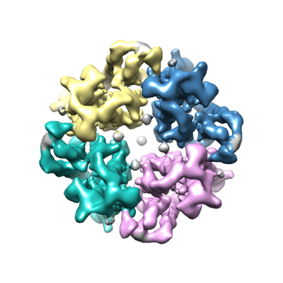 emd_13035.png emd_13035.png | 114.5 KB | ||
| Masks | emd_13035_msk_1.map | 40.6 MB | Mask map | |
| Others | emd_13035_half_map_1.map.gzemd_13035_half_map_2.map.gz | 37.3 MB 37.3 MB | ||
| Archive directory |  http://ftp.pdbj.org/pub/emdb/structures/EMD-13035ftp://ftp.pdbj.org/pub/emdb/structures/EMD-13035 http://ftp.pdbj.org/pub/emdb/structures/EMD-13035ftp://ftp.pdbj.org/pub/emdb/structures/EMD-13035 | HTTPS FTP |
-Related structure data
| Related structure data |  7oqzMC M: atomic model generated by this map C: citing same article ( |
|---|---|
| Similar structure data |


-Links
| EMDB pages | EMDB (EBI/PDBe) / EMDataResource |
|---|
-Map
| File | Download / File: emd_13035.map.gz / Format: CCP4 / Size: 40.6 MB / Type: IMAGE STORED AS FLOATING POINT NUMBER (4 BYTES) | ||||||||||||||||||||||||||||||||||||||||||||||||||||||||||||||||||||
|---|---|---|---|---|---|---|---|---|---|---|---|---|---|---|---|---|---|---|---|---|---|---|---|---|---|---|---|---|---|---|---|---|---|---|---|---|---|---|---|---|---|---|---|---|---|---|---|---|---|---|---|---|---|---|---|---|---|---|---|---|---|---|---|---|---|---|---|---|---|
| Annotation | sharpened 3D reconstruction | ||||||||||||||||||||||||||||||||||||||||||||||||||||||||||||||||||||
| Voxel size | X=Y=Z: 1.09 Å | ||||||||||||||||||||||||||||||||||||||||||||||||||||||||||||||||||||
| Density |
| ||||||||||||||||||||||||||||||||||||||||||||||||||||||||||||||||||||
| Symmetry | Space group: 1 | ||||||||||||||||||||||||||||||||||||||||||||||||||||||||||||||||||||
| Details | EMDB XML:
CCP4 map header:
| ||||||||||||||||||||||||||||||||||||||||||||||||||||||||||||||||||||
-Supplemental data
-Mask #1
| File | emd_13035_msk_1.map | ||||||||||||
|---|---|---|---|---|---|---|---|---|---|---|---|---|---|
| Projections & Slices |
| ||||||||||||
| Density Histograms |
 Z
Z Y
Y X
X


-Half map: half A
| File | emd_13035_half_map_1.map | ||||||||||||
|---|---|---|---|---|---|---|---|---|---|---|---|---|---|
| Annotation | half A | ||||||||||||
| Projections & Slices |
| ||||||||||||
| Density Histograms |


-Half map: half B
| File | emd_13035_half_map_2.map | ||||||||||||
|---|---|---|---|---|---|---|---|---|---|---|---|---|---|
| Annotation | half B | ||||||||||||
| Projections & Slices |
| ||||||||||||
| Density Histograms |


- Sample components
-Entire : Transmembrane Protein 45A
| Entire | Name: Transmembrane Protein 45ATransmembrane protein |
|---|---|
| Components |
|
-Supramolecule #1: Transmembrane Protein 45A
| Supramolecule | Name: Transmembrane Protein 45A / type: organelle_or_cellular_component / ID: 1 / Parent: 0 / Macromolecule list: #1 |
|---|---|
| Source (natural) | Organism: Homo sapiens (human) |
| Recombinant expression | Organism:   Spodoptera frugiperda (fall armyworm) Spodoptera frugiperda (fall armyworm) |
-Macromolecule #1: Transmembrane protein 45A
| Macromolecule | Name: Transmembrane protein 45A / type: protein_or_peptide / ID: 1 / Number of copies: 4 / Enantiomer: LEVO |
|---|---|
| Source (natural) | Organism: Homo sapiens (human) |
| Molecular weight | Theoretical: 32.51833 KDa |
| Recombinant expression | Organism: Spodoptera frugiperda (fall armyworm) |
| Sequence | String: MNFRGHALPG TFFFIIGLWW CTKSILKYIC KKQKRTCYLG SKTLFYRLEI LEGITIVGMA LTGMAGEQFI PGGPHLMLYD YKQGHWNQL LGWHHFTMYF FFGLLGVADI LCFTISSLPV SLTKLMLSNA LFVEAFIFYN HTHGREMLDI FVHQLLVLVV F LTGLVAFL ...String: MNFRGHALPG TFFFIIGLWW CTKSILKYIC KKQKRTCYLG SKTLFYRLEI LEGITIVGMA LTGMAGEQFI PGGPHLMLYD YKQGHWNQL LGWHHFTMYF FFGLLGVADI LCFTISSLPV SLTKLMLSNA LFVEAFIFYN HTHGREMLDI FVHQLLVLVV F LTGLVAFL EFLVRNNVLL ELLRSSLILL QGSWFFQIGF VLYPPSGGPA WDLMDHENIL FLTICFCWHY AVTIVIVGMN YA FITWLVK SRLKRLCSSE VGLLKNAERE QESEEEMAEN LYFQ |
-Macromolecule #2: 1,2-DIACYL-SN-GLYCERO-3-PHOSPHOCHOLINE
| Macromolecule | Name: 1,2-DIACYL-SN-GLYCERO-3-PHOSPHOCHOLINE / type: ligand / ID: 2 / Number of copies: 16 / Formula: PC1 |
|---|---|
| Molecular weight | Theoretical: 790.145 Da |
| Chemical component information |  ChemComp-PC1: |
-Experimental details
-Structure determination
| Method | cryo EM |
|---|---|
 Processing Processing | single particle reconstruction |
| Aggregation state | particle |
-Sample preparation
| Buffer | pH: 7.5 |
|---|---|
| Vitrification | Cryogen name: ETHANE / Instrument: FEI VITROBOT MARK IV |
- Electron microscopy
| Microscope | FEI TITAN KRIOS |
|---|---|
| Electron beam | Acceleration voltage: 300 kV / Electron source: FIELD EMISSION GUN |
| Electron optics | C2 aperture diameter: 50.0 µm / Illumination mode: FLOOD BEAM / Imaging mode: BRIGHT FIELDBright-field microscopy / Cs: 2.7 mm / Nominal defocus max: 2.6 µm / Nominal defocus min: 1.2 µm / Nominal magnification: 81000 |
| Sample stage | Specimen holder model: FEI TITAN KRIOS AUTOGRID HOLDER / Cooling holder cryogen: NITROGEN |
| Image recording | Film or detector model: GATAN K3 BIOQUANTUM (6k x 4k) / Number grids imaged: 1 / Average electron dose: 45.9 e/Å2 |
| Experimental equipment |  Model: Titan Krios / Image courtesy: FEI Company |
-Image processing
| CTF correction | Software - Name: cryoSPARC / Details: Patch CTF routine in cryoSPARC |
|---|---|
| Initial angle assignment | Type: MAXIMUM LIKELIHOOD |
| Final angle assignment | Type: MAXIMUM LIKELIHOOD |
| Final reconstruction | Number classes used: 1 / Applied symmetry - Point group: C4 (4 fold cyclic) / Algorithm: FOURIER SPACE / Resolution.type: BY AUTHOR / Resolution: 3.27 Å / Resolution method: FSC 0.143 CUT-OFF / Software - Name: cryoSPARC / Number images used: 94268 |
| FSC plot (resolution estimation) |  |
