 Movie
Movie Controller


+ Open data

- Basic information
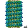
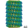
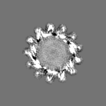
| Entry | Database: EMDB / ID: EMD-12733 | |||||||||
|---|---|---|---|---|---|---|---|---|---|---|
| Title | PspA helical structure in presence of lipids | |||||||||
 Map data Map data | symmetrized map from helical refinement in CryoSparc v3.1.0 | |||||||||
 Sample Sample |
| |||||||||
| Function / homology | PspA/IM30 / PspA/IM30 family / Chloroplast membrane-associated 30 kD protein Function and homology information Function and homology information | |||||||||
| Biological species |   Synechocystis sp. PCC 6803 (bacteria) / Synechocystis sp. PCC 6803 (bacteria) / | |||||||||
| Method | helical reconstruction / cryo EM / Resolution: 7.15 Å | |||||||||
 Authors Authors | Junglas B / Huber ST / Heidler T / Mann D / Henning R / Clarke M / Ortiz JO / Schneider D / Sachse C | |||||||||
 Citation Citation | Journal: Cell / Year: 2021 Title: PspA adopts an ESCRT-III-like fold and remodels bacterial membranes. Authors: Benedikt Junglas / Stefan T Huber / Thomas Heidler / Lukas Schlösser / Daniel Mann / Raoul Hennig / Mairi Clarke / Nadja Hellmann / Dirk Schneider / Carsten Sachse /  Abstract: PspA is the main effector of the phage shock protein (Psp) system and preserves the bacterial inner membrane integrity and function. Here, we present the 3.6 Å resolution cryoelectron microscopy ...PspA is the main effector of the phage shock protein (Psp) system and preserves the bacterial inner membrane integrity and function. Here, we present the 3.6 Å resolution cryoelectron microscopy (cryo-EM) structure of PspA assembled in helical rods. PspA monomers adopt a canonical ESCRT-III fold in an extended open conformation. PspA rods are capable of enclosing lipids and generating positive membrane curvature. Using cryo-EM, we visualized how PspA remodels membrane vesicles into μm-sized structures and how it mediates the formation of internalized vesicular structures. Hotspots of these activities are zones derived from PspA assemblies, serving as lipid transfer platforms and linking previously separated lipid structures. These membrane fusion and fission activities are in line with the described functional properties of bacterial PspA/IM30/LiaH proteins. Our structural and functional analyses reveal that bacterial PspA belongs to the evolutionary ancestry of ESCRT-III proteins involved in membrane remodeling. | |||||||||
| History |
|
- Structure visualization
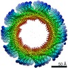
| Movie |
Movie viewer |
|---|---|
| Structure viewer | EM map: SurfViewMolmilJmol/JSmol |
| Supplemental images |

- Downloads & links
-EMDB archive
| Map data | emd_12733.map.gz | 42.3 MB | EMDB map data format | |
|---|---|---|---|---|
| Header (meta data) | emd-12733-v30.xmlemd-12733.xml | 19.6 KB 19.6 KB | Display Display | EMDB header |
| FSC (resolution estimation) | emd_12733_fsc.xml | 12.2 KB | Display | FSC data file |
| Images | 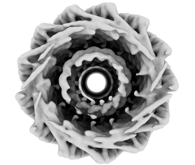 emd_12733.png emd_12733.png | 89.8 KB | ||
| Masks | emd_12733_msk_1.map | 163.6 MB | Mask map | |
| Others | emd_12733_additional_1.map.gzemd_12733_half_map_1.map.gzemd_12733_half_map_2.map.gz | 42.9 MB 150.7 MB 150.7 MB | ||
| Archive directory |  http://ftp.pdbj.org/pub/emdb/structures/EMD-12733ftp://ftp.pdbj.org/pub/emdb/structures/EMD-12733 http://ftp.pdbj.org/pub/emdb/structures/EMD-12733ftp://ftp.pdbj.org/pub/emdb/structures/EMD-12733 | HTTPS FTP |
-Related structure data


-Links
| EMDB pages | EMDB (EBI/PDBe) / EMDataResource |
|---|
-Map
| File | Download / File: emd_12733.map.gz / Format: CCP4 / Size: 163.6 MB / Type: IMAGE STORED AS FLOATING POINT NUMBER (4 BYTES) | ||||||||||||||||||||||||||||||||||||||||||||||||||||||||||||
|---|---|---|---|---|---|---|---|---|---|---|---|---|---|---|---|---|---|---|---|---|---|---|---|---|---|---|---|---|---|---|---|---|---|---|---|---|---|---|---|---|---|---|---|---|---|---|---|---|---|---|---|---|---|---|---|---|---|---|---|---|---|
| Annotation | symmetrized map from helical refinement in CryoSparc v3.1.0 | ||||||||||||||||||||||||||||||||||||||||||||||||||||||||||||
| Voxel size | X=Y=Z: 1.362 Å | ||||||||||||||||||||||||||||||||||||||||||||||||||||||||||||
| Density |
| ||||||||||||||||||||||||||||||||||||||||||||||||||||||||||||
| Symmetry | Space group: 1 | ||||||||||||||||||||||||||||||||||||||||||||||||||||||||||||
| Details | EMDB XML:
CCP4 map header:
| ||||||||||||||||||||||||||||||||||||||||||||||||||||||||||||
-Supplemental data
-Mask #1
| File | emd_12733_msk_1.map | ||||||||||||
|---|---|---|---|---|---|---|---|---|---|---|---|---|---|
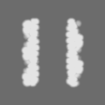
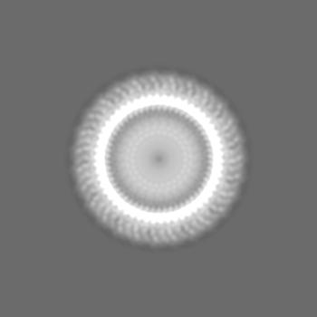
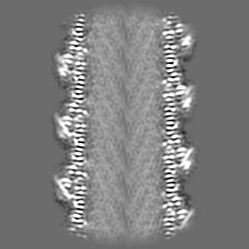
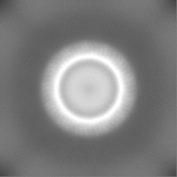
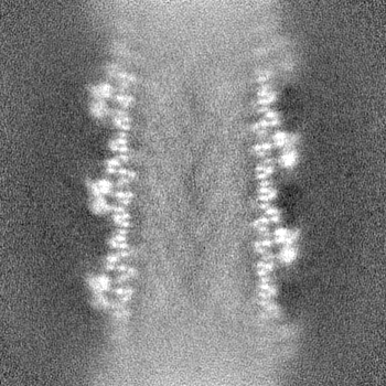
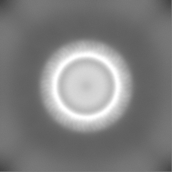
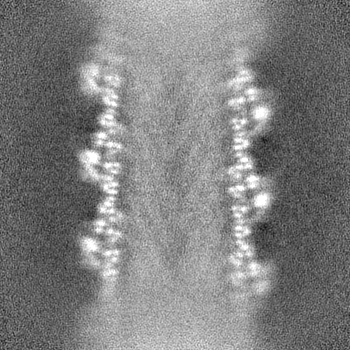
| Projections & Slices |
| ||||||||||||
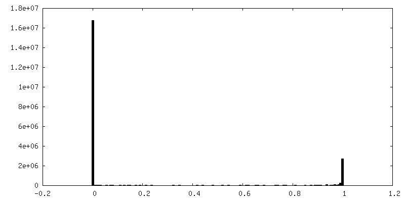
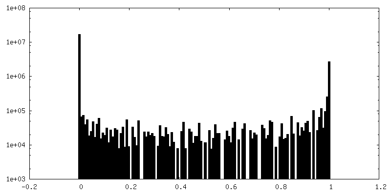
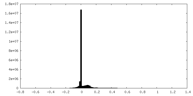
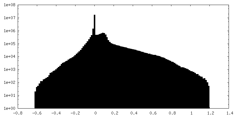
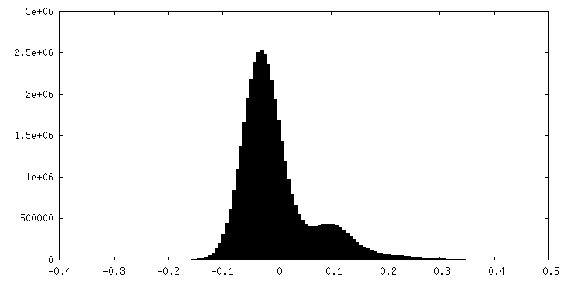
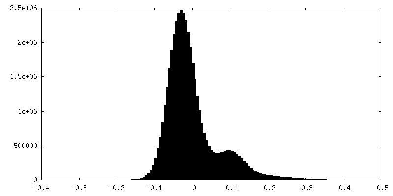
| Density Histograms |
 Z
Z Y
Y X
X


-Additional map: symmetrized and sharpened map from helical refinement in...
| File | emd_12733_additional_1.map | ||||||||||||
|---|---|---|---|---|---|---|---|---|---|---|---|---|---|
| Annotation | symmetrized and sharpened map from helical refinement in CryoSparc v3.1.0 | ||||||||||||
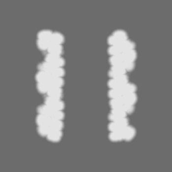
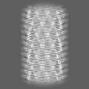
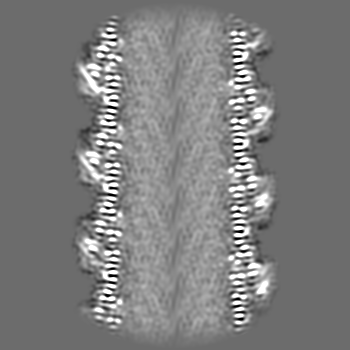
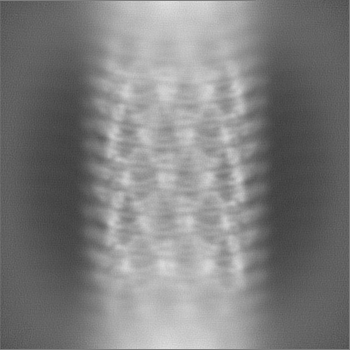
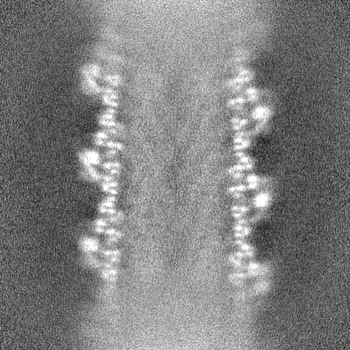
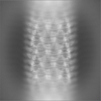
| Projections & Slices |
| ||||||||||||
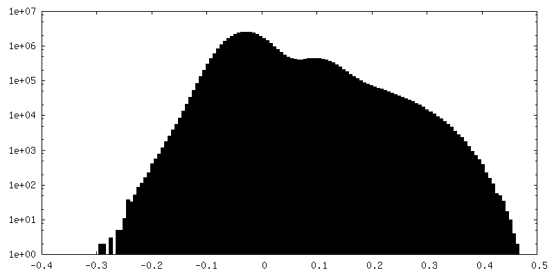
| Density Histograms |
-Half map: half map A
| File | emd_12733_half_map_1.map | ||||||||||||
|---|---|---|---|---|---|---|---|---|---|---|---|---|---|
| Annotation | half map A | ||||||||||||
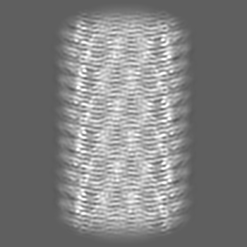
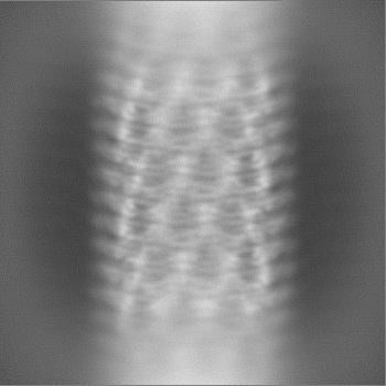
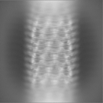
| Projections & Slices |
| ||||||||||||
| Density Histograms |

-Half map: half map B
| File | emd_12733_half_map_2.map | ||||||||||||
|---|---|---|---|---|---|---|---|---|---|---|---|---|---|
| Annotation | half map B | ||||||||||||
| Projections & Slices |
| ||||||||||||
| Density Histograms |


- Sample components
-Entire : PspA helical structure in presence of lipids
| Entire | Name: PspA helical structure in presence of lipids |
|---|---|
| Components |
|
-Supramolecule #1: PspA helical structure in presence of lipids
| Supramolecule | Name: PspA helical structure in presence of lipids / type: organelle_or_cellular_component / ID: 1 / Parent: 0 / Macromolecule list: all |
|---|---|
| Source (natural) | Organism: Synechocystis sp. PCC 6803 (bacteria) |
| Molecular weight | Experimental: 25 KDa |
| Recombinant expression | Organism: Escherichia coli BL21(DE3) (bacteria) / Recombinant plasmid: pRSET6 |
-Macromolecule #1: Chloroplast membrane-associated 30 kD protein
| Macromolecule | Name: Chloroplast membrane-associated 30 kD protein / type: protein_or_peptide / ID: 1 / Enantiomer: LEVO |
|---|---|
| Source (natural) | Organism: Strain: PCC 6803 / Kazusa |
| Recombinant expression | Organism: Escherichia coli BL21(DE3) (bacteria) |
| Sequence | String: MGHHHHHHHH HSSGHIDDDD KHMELFNRVG RVLKSQLTHW QQQQEAPEDL LERLLGEMEL ELIELRRALA QTIATFKSTE RQRDAQQLI AQRWYEKAQA ALDRGNEQLA REALGQRQSY QSHTEALGKS LGEQRALVEQ VRGQLQKLER KYLELKSQKN L YLARLKSA ...String: MGHHHHHHHH HSSGHIDDDD KHMELFNRVG RVLKSQLTHW QQQQEAPEDL LERLLGEMEL ELIELRRALA QTIATFKSTE RQRDAQQLI AQRWYEKAQA ALDRGNEQLA REALGQRQSY QSHTEALGKS LGEQRALVEQ VRGQLQKLER KYLELKSQKN L YLARLKSA IAAQKIEEIA GNLDNASASS LFERIETKIL ELEAERELLN PPPSPLDKKF EQWEEQQAVE ATLAAMKARR SL PPPSS |
-Experimental details
-Structure determination
| Method | cryo EM |
|---|---|
 Processing Processing | helical reconstruction |
| Aggregation state | filament |
-Sample preparation
| Concentration | 0.8 mg/mL | |||||||||
|---|---|---|---|---|---|---|---|---|---|---|
| Buffer | pH: 7.6 Component:
| |||||||||
| Grid | Model: PELCO Ultrathin Carbon with Lacey Carbon / Material: COPPER / Mesh: 200 / Pretreatment - Type: GLOW DISCHARGE / Details: Pelco easiGlow | |||||||||
| Vitrification | Cryogen name: ETHANE / Chamber humidity: 90 % / Chamber temperature: 283.15 K / Instrument: FEI VITROBOT MARK IV |
- Electron microscopy
| Microscope | TFS TALOS |
|---|---|
| Electron beam | Acceleration voltage: 200 kV / Electron source: FIELD EMISSION GUN |
| Electron optics | C2 aperture diameter: 50.0 µm / Illumination mode: FLOOD BEAM / Imaging mode: BRIGHT FIELDBright-field microscopy / Cs: 2.7 mm / Nominal defocus max: 3.0 µm / Nominal defocus min: 2.0 µm / Nominal magnification: 100000 |
| Specialist optics | Energy filter - Name: GIF Bioquantum / Energy filter - Slit width: 20 eV |
| Sample stage | Specimen holder model: FEI TITAN KRIOS AUTOGRID HOLDER / Cooling holder cryogen: NITROGEN |
| Image recording | Film or detector model: GATAN K3 BIOQUANTUM (6k x 4k) / Number real images: 1531 / Average exposure time: 3.0 sec. / Average electron dose: 31.0 e/Å2 |
-Image processing
| Segment selection | Number selected: 222901 / Software - Name: cryoSPARC (ver. 3.1.0) |
|---|---|
| CTF correction | Software - Name: cryoSPARC (ver. 3.1.0) |
| Final angle assignment | Type: NOT APPLICABLE / Software - Name: cryoSPARC (ver. 3.1.0) |
| Final reconstruction | Applied symmetry - Helical parameters - Δz: 4.425 Å Applied symmetry - Helical parameters - Δ&Phi: 26.629 ° Applied symmetry - Helical parameters - Axial symmetry: C1 (asymmetric) Resolution.type: BY AUTHOR / Resolution: 7.15 Å / Resolution method: FSC 0.143 CUT-OFF / Software - Name: cryoSPARC (ver. 3.1.0) / Number images used: 222901 |
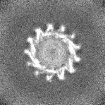
| FSC plot (resolution estimation) |  |
-Atomic model buiding 1
| Refinement | Protocol: AB INITIO MODEL |
|---|
