 Movie
Movie Controller

[English] 日本語
 Yorodumi
- EMDB-12704: Cooperation between the intrinsically disordered and ordered regi... -
+ Open data

- Basic information
| Entry |  | |||||||||||||||||||||
|---|---|---|---|---|---|---|---|---|---|---|---|---|---|---|---|---|---|---|---|---|---|---|
| Title | Cooperation between the intrinsically disordered and ordered regions of Spt6 regulates nucleosome and Pol II CTD binding, and nucleosome assembly | |||||||||||||||||||||
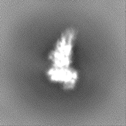
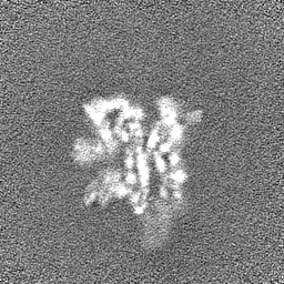
 Map data Map data | Cryosparc sharpened electron density map | |||||||||||||||||||||
 Sample Sample |
| |||||||||||||||||||||
| Function / homology |  Function and homology information Function and homology informationcarbon catabolite repression of transcription from RNA polymerase II promoter by glucose / regulation of transcriptional start site selection at RNA polymerase II promoter / transcription antitermination factor activity, DNA binding / regulation of mRNA 3'-end processing / nucleosome organization / transcription elongation-coupled chromatin remodeling / poly(A)+ mRNA export from nucleus /  nucleosome binding / transcription elongation factor complex / transcription antitermination ...carbon catabolite repression of transcription from RNA polymerase II promoter by glucose / regulation of transcriptional start site selection at RNA polymerase II promoter / transcription antitermination factor activity, DNA binding / regulation of mRNA 3'-end processing / nucleosome organization / transcription elongation-coupled chromatin remodeling / poly(A)+ mRNA export from nucleus / nucleosome binding / transcription elongation factor complex / transcription antitermination / transcription elongation by RNA polymerase II / positive regulation of transcription elongation by RNA polymerase II / euchromatin / nucleosome assembly / chromatin organization / histone binding / chromatin remodeling / regulation of transcription by RNA polymerase II / negative regulation of transcription by RNA polymerase II / positive regulation of transcription by RNA polymerase II / mitochondrion / DNA binding / nucleus nucleosome binding / transcription elongation factor complex / transcription antitermination ...carbon catabolite repression of transcription from RNA polymerase II promoter by glucose / regulation of transcriptional start site selection at RNA polymerase II promoter / transcription antitermination factor activity, DNA binding / regulation of mRNA 3'-end processing / nucleosome organization / transcription elongation-coupled chromatin remodeling / poly(A)+ mRNA export from nucleus / nucleosome binding / transcription elongation factor complex / transcription antitermination / transcription elongation by RNA polymerase II / positive regulation of transcription elongation by RNA polymerase II / euchromatin / nucleosome assembly / chromatin organization / histone binding / chromatin remodeling / regulation of transcription by RNA polymerase II / negative regulation of transcription by RNA polymerase II / positive regulation of transcription by RNA polymerase II / mitochondrion / DNA binding / nucleusSimilarity search - Function | |||||||||||||||||||||
| Biological species |  Saccharomyces cerevisiae (strain ATCC 204508 / S288c) (yeast) Saccharomyces cerevisiae (strain ATCC 204508 / S288c) (yeast) | |||||||||||||||||||||
| Method | single particle reconstruction / cryo EM / Resolution: 3.71 Å | |||||||||||||||||||||
 Authors Authors | Kasiliauskaite A / Kubicek K / Klumpler T / Zanova M / Zapletal D / Novacek J / Stefl R | |||||||||||||||||||||
| Funding support |  Czech Republic, 6 items Czech Republic, 6 items
| |||||||||||||||||||||
 Citation Citation | Journal: Nucleic Acids Res / Year: 2022 Title: Cooperation between intrinsically disordered and ordered regions of Spt6 regulates nucleosome and Pol II CTD binding, and nucleosome assembly. Authors: Aiste Kasiliauskaite / Karel Kubicek / Tomas Klumpler / Martina Zanova / David Zapletal / Eliska Koutna / Jiri Novacek / Richard Stefl / Abstract: Transcription elongation factor Spt6 associates with RNA polymerase II (Pol II) and acts as a histone chaperone, which promotes the reassembly of nucleosomes following the passage of Pol II. The ...Transcription elongation factor Spt6 associates with RNA polymerase II (Pol II) and acts as a histone chaperone, which promotes the reassembly of nucleosomes following the passage of Pol II. The precise mechanism of nucleosome reassembly mediated by Spt6 remains unclear. In this study, we used a hybrid approach combining cryo-electron microscopy and small-angle X-ray scattering to visualize the architecture of Spt6 from Saccharomyces cerevisiae. The reconstructed overall architecture of Spt6 reveals not only the core of Spt6, but also its flexible N- and C-termini, which are critical for Spt6's function. We found that the acidic N-terminal region of Spt6 prevents the binding of Spt6 not only to the Pol II CTD and Pol II CTD-linker, but also to pre-formed intact nucleosomes and nucleosomal DNA. The N-terminal region of Spt6 self-associates with the tSH2 domain and the core of Spt6 and thus controls binding to Pol II and nucleosomes. Furthermore, we found that Spt6 promotes the assembly of nucleosomes in vitro. These data indicate that the cooperation between the intrinsically disordered and structured regions of Spt6 regulates nucleosome and Pol II CTD binding, and also nucleosome assembly. | |||||||||||||||||||||
| History |
|
- Structure visualization
| Supplemental images |
|---|

- Downloads & links
-EMDB archive
| Map data | emd_12704.map.gz | 32.7 MB | EMDB map data format | |
|---|---|---|---|---|
| Header (meta data) | emd-12704-v30.xmlemd-12704.xml | 22.2 KB 22.2 KB | Display Display | EMDB header |
| Images | 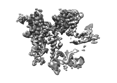 emd_12704.png emd_12704.png | 76.1 KB | ||
| Others | emd_12704_half_map_1.map.gzemd_12704_half_map_2.map.gz | 59.5 MB 59.5 MB | ||
| Archive directory |  http://ftp.pdbj.org/pub/emdb/structures/EMD-12704ftp://ftp.pdbj.org/pub/emdb/structures/EMD-12704 http://ftp.pdbj.org/pub/emdb/structures/EMD-12704ftp://ftp.pdbj.org/pub/emdb/structures/EMD-12704 | HTTPS FTP |
-Related structure data
| Related structure data | 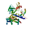 7o3dMC 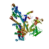 7o6bC M: atomic model generated by this map C: citing same article ( |
|---|---|
| Similar structure data |
-Links
| EMDB pages | EMDB (EBI/PDBe) / EMDataResource |
|---|---|
| Related items in Molecule of the Month |

-Map
| File | Download / File: emd_12704.map.gz / Format: CCP4 / Size: 64 MB / Type: IMAGE STORED AS FLOATING POINT NUMBER (4 BYTES) | ||||||||||||||||||||
|---|---|---|---|---|---|---|---|---|---|---|---|---|---|---|---|---|---|---|---|---|---|
| Annotation | Cryosparc sharpened electron density map | ||||||||||||||||||||
| Voxel size | X=Y=Z: 0.828 Å | ||||||||||||||||||||
| Density |
| ||||||||||||||||||||
| Symmetry | Space group: 1 | ||||||||||||||||||||
| Details | EMDB XML:
|
-Supplemental data
-Half map: #1
| File | emd_12704_half_map_1.map | ||||||||||||
|---|---|---|---|---|---|---|---|---|---|---|---|---|---|
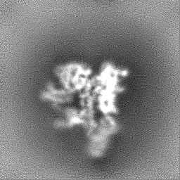
| Projections & Slices |
| ||||||||||||
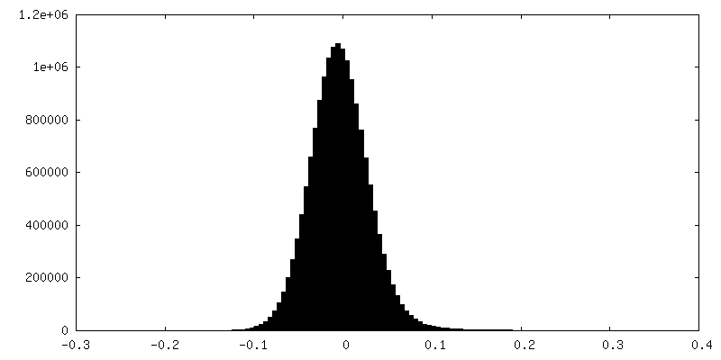
| Density Histograms |
 Z
Z Y
Y X
X


-Half map: #2
| File | emd_12704_half_map_2.map | ||||||||||||
|---|---|---|---|---|---|---|---|---|---|---|---|---|---|
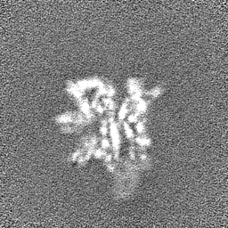
| Projections & Slices |
| ||||||||||||
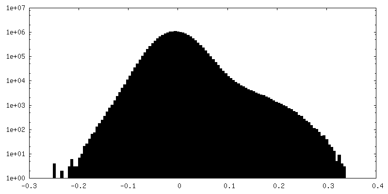
| Density Histograms |


- Sample components
-Entire : Structure of elongation factor Spt6
| Entire | Name: Structure of elongation factor Spt6 |
|---|---|
| Components |
|
-Supramolecule #1: Structure of elongation factor Spt6
| Supramolecule | Name: Structure of elongation factor Spt6 / type: organelle_or_cellular_component / ID: 1 / Parent: 0 / Macromolecule list: all |
|---|---|
| Source (natural) | Organism: Saccharomyces cerevisiae (strain ATCC 204508 / S288c) (yeast) |
| Recombinant expression | Organism:  Escherichia coli B (bacteria) Escherichia coli B (bacteria) |
-Macromolecule #1: Transcription elongation factor SPT6
| Macromolecule | Name: Transcription elongation factor SPT6 / type: protein_or_peptide / ID: 1 Details: the sequence stretches of the sample that do not make part of the coordinates are flexible regions that were not observed in the cryoEM map Number of copies: 1 / Enantiomer: LEVO |
|---|---|
| Source (natural) | Organism: Saccharomyces cerevisiae (strain ATCC 204508 / S288c) (yeast) Strain: ATCC 204508 / S288c |
| Molecular weight | Theoretical: 134.923031 KDa |
| Recombinant expression | Organism: Escherichia coli B (bacteria) |
| Sequence | String: GIDPFTDIYD LEDLKKNLMT EGDMKIRKTD IPERYQELRA GITDYGNMSS EDQELERNWI AEKISVDKNF DANYDLTEFK EAIGNAIKF ITKENLEVPF IYAYRRNYIS SREKDGFLLT EDDLWDIVSL DIEFHSLVNK KDYVQRFYAE LHIDDPIVTE Y FKNQNTAS ...String: GIDPFTDIYD LEDLKKNLMT EGDMKIRKTD IPERYQELRA GITDYGNMSS EDQELERNWI AEKISVDKNF DANYDLTEFK EAIGNAIKF ITKENLEVPF IYAYRRNYIS SREKDGFLLT EDDLWDIVSL DIEFHSLVNK KDYVQRFYAE LHIDDPIVTE Y FKNQNTAS IAELNSLQDI YDYLEFKYAN EINEMFINHT GKTGKKHLKN SSYEKFKASP LYQAVSDIGI SAEDVGENIS SQ HQIHPPV DHPSSKPVEV IESILNANSG DLQVFTSNTK LAIDTVQKYY SLELSKNTKI REKVRSDFSK YYLADVVLTA KGK KEIQKG SLYEDIKYAI NRTPMHFRRD PDVFLKMVEA ESLNLLSVKL HMSSQAQYIE HLFQIALETT NTSDIAIEWN NFRK LAFNQ AMDKIFQDIS QEVKDNLTKN CQKLVAKTVR HKFMTKLDQA PFIPNVRDPK IPKILSLTCG QGRFGADAII AVYVN RKGD FIRDYKIVDN PFDKTNPEKF EDTLDNIIQS CQPNAIGING PNPKTQKFYK RLQEVLHKKQ IVDSRGHTIP IIYVED EVA IRYQNSERAA QEFPNKPPLV KYCIALARYM HSPLLEYANL TSEEVRSLSI HPHQNLLSSE QLSWALETAF VDIVNLV SV EVNKATDNNY YASALKYISG FGKRKAIDFL QSLQRLNEPL LARQQLITHN ILHKTIFMNS AGFLYISWNE KRQKYEDL E HDQLDSTRIH PEDYHLATKV AADALEYDPD TIAEKEEQGT MSEFIELLRE DPDRRAKLES LNLESYAEEL EKNTGLRKL NNLNTIVLEL LDGFEELRND FHPLQGDEIF QSLTGESEKT FFKGSIIPVR VERFWHNDII CTTNSEVECV VNAQRHAGAQ LRRPANEIY EIGKTYPAKV IYIDYANITA EVSLLDHDVK QQYVPISYSK DPSIWDLKQE LEDAEEERKL MMAEARAKRT H RVINHPYY FPFNGRQAED YLRSKERGEF VIRQSSRGDD HLVITWKLDK DLFQHIDIQE LEKENPLALG KVLIVDNQKY ND LDQIIVE YLQNKVRLLN EMTSSEKFKS GTKKDVVKFI EDYSRVNPNK SVYYFSLNHD NPGWFYLMFK INANSKLYTW NVK LTNTGY FLVNYNYPSV IQLCNGFKTL LKSNSSKNRM NNYR |
-Experimental details
-Structure determination
| Method | cryo EM |
|---|---|
 Processing Processing | single particle reconstruction |
| Aggregation state | particle |
-Sample preparation
| Concentration | 0.15 mg/mL |
|---|---|
| Buffer | pH: 7.5 / Component - Concentration: 20.0 mM / Component - Formula: HEPES / Component - Name: HEPES |
| Grid | Model: Quantifoil R2/1 / Material: COPPER / Mesh: 300 / Pretreatment - Type: GLOW DISCHARGE / Pretreatment - Atmosphere: OTHER / Pretreatment - Pressure: 0.007 kPa |
| Vitrification | Cryogen name: ETHANE / Chamber humidity: 100 % / Chamber temperature: 277 K / Instrument: FEI VITROBOT MARK IV |
| Details | homogeneous monodisperse sample |
- Electron microscopy
| Microscope | TFS KRIOS |
|---|---|
| Electron beam | Acceleration voltage: 300 kV / Electron source: FIELD EMISSION GUN |
| Electron optics | C2 aperture diameter: 30.0 µm / Illumination mode: FLOOD BEAM / Imaging mode: BRIGHT FIELDBright-field microscopy / Cs: 2.7 mm |
| Sample stage | Specimen holder model: FEI TITAN KRIOS AUTOGRID HOLDER / Cooling holder cryogen: NITROGEN |
| Image recording | Film or detector model: GATAN K2 SUMMIT (4k x 4k) / Detector mode: COUNTING / Digitization - Dimensions - Width: 3840 pixel / Digitization - Dimensions - Height: 3710 pixel / Digitization - Frames/image: 1-40 / Number grids imaged: 1 / Number real images: 13807 / Average exposure time: 5.0 sec. / Average electron dose: 55.0 e/Å2 |
| Experimental equipment |  Model: Titan Krios / Image courtesy: FEI Company |
-Image processing
| Particle selection | Number selected: 2995892 |
|---|---|
| CTF correction | Software - Name: cryoSPARC (ver. 3.1) |
| Startup model | Type of model: PDB ENTRY PDB model - PDB ID: Details: Initial PDB coordinates of the Spt6 structure comprising residues 298-1248 were produced from PDBs of a crystal structure of Spt6, namely 3PSI and 3PSF. The PDB and the cryoSPARC sharpened ...Details: Initial PDB coordinates of the Spt6 structure comprising residues 298-1248 were produced from PDBs of a crystal structure of Spt6, namely 3PSI and 3PSF. The PDB and the cryoSPARC sharpened electron density map were imported into the program Coot and the tool 'Real Space Refine Zone' was used to achiev e optimal fit of the PDB coordinates within the map. Low-resolution regions of the map (regions corresponding to residues: 1226-1243 and the S1-domain) were also excluded from the PDB and were docked with a rigid body strategy using Phenix once the fit of the well-resolved regions exhibited no clashes and deviations. |
| Initial angle assignment | Type: MAXIMUM LIKELIHOOD / Software - Name: cryoSPARC (ver. 3.1) |
| Final 3D classification | Number classes: 313 / Avg.num./class: 2296 / Software - Name: cryoSPARC (ver. 3.1) |
| Final angle assignment | Type: MAXIMUM LIKELIHOOD / Software - Name: cryoSPARC (ver. 3.1) |
| Final reconstruction | Number classes used: 313 / Applied symmetry - Point group: C1 (asymmetric) / Resolution.type: BY AUTHOR / Resolution: 3.71 Å / Resolution method: FSC 0.143 CUT-OFF / Software - Name: cryoSPARC (ver. 3.1) / Number images used: 718639 |

-Atomic model buiding 1
| Initial model |
| ||||||
|---|---|---|---|---|---|---|---|
| Refinement | Space: REAL / Protocol: FLEXIBLE FIT / Target criteria: Correlation coefficients | ||||||
| Output model | PDB-7o3d: |

