 Movie
Movie Controller

[English] 日本語
 Yorodumi
- EMDB-12639: In situ subtomogram average of 13 protofilament microtubule from ... -
+ Open data

- Basic information
| Entry | Database: EMDB / ID: EMD-12639 | |||||||||
|---|---|---|---|---|---|---|---|---|---|---|
| Title | In situ subtomogram average of 13 protofilament microtubule from Mus musculus DRG axons | |||||||||
 Map data Map data | microtubule structure from mouse DRG axons | |||||||||
 Sample Sample |
| |||||||||
| Biological species |   Mus musculus (house mouse) Mus musculus (house mouse) | |||||||||
| Method | subtomogram averaging / cryo EM / Resolution: 12.0 Å | |||||||||
 Authors Authors | Foster HE / Ventura Santos C / Carter AP | |||||||||
| Funding support |  United Kingdom, 2 items United Kingdom, 2 items
| |||||||||
 Citation Citation | Journal: J Cell Biol / Year: 2022 Title: A cryo-ET survey of microtubules and intracellular compartments in mammalian axons. Authors: Helen E Foster / Camilla Ventura Santos / Andrew P Carter / Abstract: The neuronal axon is packed with cytoskeletal filaments, membranes, and organelles, many of which move between the cell body and axon tip. Here, we used cryo-electron tomography to survey the ...The neuronal axon is packed with cytoskeletal filaments, membranes, and organelles, many of which move between the cell body and axon tip. Here, we used cryo-electron tomography to survey the internal components of mammalian sensory axons. We determined the polarity of the axonal microtubules (MTs) by combining subtomogram classification and visual inspection, finding MT plus and minus ends are structurally similar. Subtomogram averaging of globular densities in the MT lumen suggests they have a defined structure, which is surprising given they likely contain the disordered protein MAP6. We found the endoplasmic reticulum in axons is tethered to MTs through multiple short linkers. We surveyed membrane-bound cargos and describe unexpected internal features such as granules and broken membranes. In addition, we detected proteinaceous compartments, including numerous virus-like capsid particles. Our observations outline novel features of axonal cargos and MTs, providing a platform for identification of their constituents. #1: Journal: Biorxiv / Year: 2021Title: A cryo-ET survey of intracellular compartments within mammalian axons Authors: Foster HE / Carter AP | |||||||||
| History |
|
- Structure visualization
| Movie |
 Movie viewer Movie viewer |
|---|---|
| Structure viewer | EM map: SurfViewMolmilJmol/JSmol |
| Supplemental images |

- Downloads & links
-EMDB archive
| Map data | emd_12639.map.gz | 15.3 MB | EMDB map data format | |
|---|---|---|---|---|
| Header (meta data) | emd-12639-v30.xmlemd-12639.xml | 18 KB 18 KB | Display Display | EMDB header |
| FSC (resolution estimation) | emd_12639_fsc.xml | 6.9 KB | Display | FSC data file |
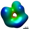
| Images | 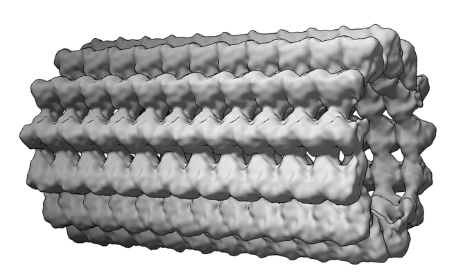 emd_12639.png emd_12639.png | 141.7 KB | ||
| Masks | emd_12639_msk_1.map | 27 MB | Mask map | |
| Others | emd_12639_half_map_1.map.gzemd_12639_half_map_2.map.gz | 25.2 MB 25.2 MB | ||
| Archive directory |  http://ftp.pdbj.org/pub/emdb/structures/EMD-12639ftp://ftp.pdbj.org/pub/emdb/structures/EMD-12639 http://ftp.pdbj.org/pub/emdb/structures/EMD-12639ftp://ftp.pdbj.org/pub/emdb/structures/EMD-12639 | HTTPS FTP |
-Related structure data
| Related structure data | C: citing same article ( |
|---|---|
| Similar structure data | |
| EM raw data | EMPIAR-10814 (Title: Cryo electron tomograms of mouse DRG axons (dataset 2) Data size: 206.2 Data #1: Raw image frames of mouse dorsal root ganglion axons (dataset 2) [micrographs - multiframe] Data #2: Corrected, aligned, dose-filtered and order-sorted tilt series for mouse dorsal root ganglion axons (dataset 2) [tilt series] Data #3: Tomograms of mouse dorsal root ganglion axons (dataset 2) binned by 4 and deconvolved [reconstructed volumes]) |

-Links
| EMDB pages | EMDB (EBI/PDBe) / EMDataResource |
|---|
-Map
| File | Download / File: emd_12639.map.gz / Format: CCP4 / Size: 27 MB / Type: IMAGE STORED AS FLOATING POINT NUMBER (4 BYTES) | ||||||||||||||||||||||||||||||||||||||||||||||||||||||||||||
|---|---|---|---|---|---|---|---|---|---|---|---|---|---|---|---|---|---|---|---|---|---|---|---|---|---|---|---|---|---|---|---|---|---|---|---|---|---|---|---|---|---|---|---|---|---|---|---|---|---|---|---|---|---|---|---|---|---|---|---|---|---|
| Annotation | microtubule structure from mouse DRG axons | ||||||||||||||||||||||||||||||||||||||||||||||||||||||||||||
| Voxel size | X=Y=Z: 2.75 Å | ||||||||||||||||||||||||||||||||||||||||||||||||||||||||||||
| Density |
| ||||||||||||||||||||||||||||||||||||||||||||||||||||||||||||
| Symmetry | Space group: 1 | ||||||||||||||||||||||||||||||||||||||||||||||||||||||||||||
| Details | EMDB XML:
CCP4 map header:
| ||||||||||||||||||||||||||||||||||||||||||||||||||||||||||||
-Supplemental data
-Mask #1
| File | emd_12639_msk_1.map | ||||||||||||
|---|---|---|---|---|---|---|---|---|---|---|---|---|---|
| Projections & Slices |
| ||||||||||||
| Density Histograms |
 Z
Z Y
Y X
X


-Half map: #2
| File | emd_12639_half_map_1.map | ||||||||||||
|---|---|---|---|---|---|---|---|---|---|---|---|---|---|
| Projections & Slices |
| ||||||||||||
| Density Histograms |


-Half map: #1
| File | emd_12639_half_map_2.map | ||||||||||||
|---|---|---|---|---|---|---|---|---|---|---|---|---|---|
| Projections & Slices |
| ||||||||||||
| Density Histograms |


- Sample components
-Entire : In situ subtomogram average of 13 protofilament microtubule from ...
| Entire | Name: In situ subtomogram average of 13 protofilament microtubule from Mus musculus DRG axons |
|---|---|
| Components |
|
-Supramolecule #1: In situ subtomogram average of 13 protofilament microtubule from ...
| Supramolecule | Name: In situ subtomogram average of 13 protofilament microtubule from Mus musculus DRG axons type: organelle_or_cellular_component / ID: 1 / Parent: 0 / Macromolecule list: #1 |
|---|---|
| Source (natural) | Organism: Mus musculus (house mouse) / Organ: Neurons / Tissue: Dorsal root ganglion / Organelle: microtubule cytoskeleton / Location in cell: axoplasm |
-Experimental details
-Structure determination
| Method | cryo EM |
|---|---|
 Processing Processing | subtomogram averaging |
| Aggregation state | cell |
-Sample preparation
| Buffer | pH: 7.4 |
|---|---|
| Grid | Model: Quantifoil R3.5/1 / Material: GOLD / Mesh: 200 / Support film - Material: CARBON / Support film - topology: CONTINUOUS / Pretreatment - Type: PLASMA CLEANING / Pretreatment - Atmosphere: OTHER Details: Grids were additionally coated in 0.1mg/mL poly-L-lysine then 0.01mg/mL laminin before cell plating |
| Vitrification | Cryogen name: ETHANE / Chamber humidity: 100 % / Chamber temperature: 310 K / Instrument: FEI VITROBOT MARK IV / Details: Manual blot for 3 s before plunging. |
| Details | Microtubules in axons of adult DRG neurons grown for 7 days in vitro |
- Electron microscopy
| Microscope | FEI TITAN KRIOS |
|---|---|
| Electron beam | Acceleration voltage: 300 kV / Electron source: FIELD EMISSION GUN |
| Electron optics | C2 aperture diameter: 50.0 µm / Illumination mode: FLOOD BEAM / Imaging mode: BRIGHT FIELDBright-field microscopy / Cs: 2.7 mm / Nominal defocus max: 5.0 µm / Nominal defocus min: 3.0 µm / Nominal magnification: 53000 |
| Specialist optics | Energy filter - Name: GIF Bioquantum / Energy filter - Slit width: 20 eV |
| Sample stage | Specimen holder model: FEI TITAN KRIOS AUTOGRID HOLDER / Cooling holder cryogen: NITROGEN |
| Image recording | Film or detector model: GATAN K2 SUMMIT (4k x 4k) / Detector mode: COUNTING / Digitization - Dimensions - Width: 3710 pixel / Digitization - Dimensions - Height: 3838 pixel / Digitization - Frames/image: 1-10 / Average exposure time: 1.7 sec. / Average electron dose: 1.85 e/Å2 Details: 61 images per tilt series with 112.85 e/A2 total dose. |
| Experimental equipment |  Model: Titan Krios / Image courtesy: FEI Company |
-Image processing
| Extraction | Number tomograms: 39 / Number images used: 476268 / Reference model: average of all particles (tube-shape) / Method: Dynamo filament extraction Software:
Details: Paths of microtubules were modelled in IMOD (version 4.10.32). After importing into Dynamo, initial particle positions were determined using the 'filamentWithTorsion' model workflow. Helical ...Details: Paths of microtubules were modelled in IMOD (version 4.10.32). After importing into Dynamo, initial particle positions were determined using the 'filamentWithTorsion' model workflow. Helical symmetry was applied at the particle level and subvolumes were extracted using subTOM. | ||||||
|---|---|---|---|---|---|---|---|
| CTF correction | Software: (Name: NOVACTF (ver. 1.0.0), IMOD (ver. 4.10.32)) Details: Defoci were estimated with CTFPLOTTER (IMOD version 4.10.32). CTF correction was done with novaCTF. | ||||||
| Final angle assignment | Type: OTHER Details: Alignment, classification and averaging was performed in subTOM (1.1.5) | ||||||
| Final reconstruction | Applied symmetry - Point group: C1 (asymmetric) / Algorithm: BACK PROJECTION / Resolution.type: BY AUTHOR / Resolution: 12.0 Å / Resolution method: FSC 0.143 CUT-OFF / Software - Name: RELION (ver. 3.0) / Software - details: postprocess Details: Particles were aligned together at bin4. Half maps were generated at bin4 and independently aligned at bin2 and bin1. Number subtomograms used: 64528 | ||||||
| FSC plot (resolution estimation) |  |
