 Movie
Movie Controller

[English] 日本語
 Yorodumi
- EMDB-12376: Mutant huntingtin containing organelle resembles MVB in 6-month-o... -
+ Open data

- Basic information
| Entry | Database: EMDB / ID: EMD-12376 | |||||||||||||||
|---|---|---|---|---|---|---|---|---|---|---|---|---|---|---|---|---|
| Title | Mutant huntingtin containing organelle resembles MVB in 6-month-old zQ175 mice | |||||||||||||||
 Map data Map data | Tomogram of mutant HTT containing organelle resembles MVB/amphisome in 6-month-old zQ175 mice | |||||||||||||||
 Sample Sample |
| |||||||||||||||
| Biological species |   Mus musculus (house mouse) Mus musculus (house mouse) | |||||||||||||||
| Method | electron tomography / negative staining | |||||||||||||||
 Authors Authors | Zhou Y / Saibil HR | |||||||||||||||
| Funding support |  United Kingdom, United Kingdom,  United States, 4 items United States, 4 items
| |||||||||||||||
 Citation Citation | Journal: Acta Neuropathol Commun / Year: 2021 Title: Correlative light and electron microscopy suggests that mutant huntingtin dysregulates the endolysosomal pathway in presymptomatic Huntington's disease. Authors: Ya Zhou / Thomas R Peskett / Christian Landles / John B Warner / Kirupa Sathasivam / Edward J Smith / Shu Chen / Ronald Wetzel / Hilal A Lashuel / Gillian P Bates / Helen R Saibil /  Abstract: Huntington's disease (HD) is a late onset, inherited neurodegenerative disorder for which early pathogenic events remain poorly understood. Here we show that mutant exon 1 HTT proteins are recruited ...Huntington's disease (HD) is a late onset, inherited neurodegenerative disorder for which early pathogenic events remain poorly understood. Here we show that mutant exon 1 HTT proteins are recruited to a subset of cytoplasmic aggregates in the cell bodies of neurons in brain sections from presymptomatic HD, but not wild-type, mice. This occurred in a disease stage and polyglutamine-length dependent manner. We successfully adapted a high-resolution correlative light and electron microscopy methodology, originally developed for mammalian and yeast cells, to allow us to correlate light microscopy and electron microscopy images on the same brain section within an accuracy of 100 nm. Using this approach, we identified these recruitment sites as single membrane bound, vesicle-rich endolysosomal organelles, specifically as (1) multivesicular bodies (MVBs), or amphisomes and (2) autolysosomes or residual bodies. The organelles were often found in close-proximity to phagophore-like structures. Immunogold labeling localized mutant HTT to non-fibrillar, electron lucent structures within the lumen of these organelles. In presymptomatic HD, the recruitment organelles were predominantly MVBs/amphisomes, whereas in late-stage HD, there were more autolysosomes or residual bodies. Electron tomograms indicated the fusion of small vesicles with the vacuole within the lumen, suggesting that MVBs develop into residual bodies. We found that markers of MVB-related exocytosis were depleted in presymptomatic mice and throughout the disease course. This suggests that endolysosomal homeostasis has moved away from exocytosis toward lysosome fusion and degradation, in response to the need to clear the chronically aggregating mutant HTT protein, and that this occurs at an early stage in HD pathogenesis. | |||||||||||||||
| History |
|
- Structure visualization
| Movie |
 Movie viewer Movie viewer |
|---|---|
| Supplemental images |

- Downloads & links
-EMDB archive
| Map data | emd_12376.map.gz | 1.4 GB | EMDB map data format | |
|---|---|---|---|---|
| Header (meta data) | emd-12376-v30.xmlemd-12376.xml | 10.5 KB 10.5 KB | Display Display | EMDB header |
| Images | 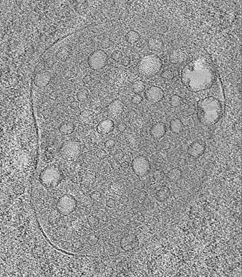 emd_12376.png emd_12376.png | 127 KB | ||
| Archive directory |  http://ftp.pdbj.org/pub/emdb/structures/EMD-12376ftp://ftp.pdbj.org/pub/emdb/structures/EMD-12376 http://ftp.pdbj.org/pub/emdb/structures/EMD-12376ftp://ftp.pdbj.org/pub/emdb/structures/EMD-12376 | HTTPS FTP |
-Related structure data
-Links
| EMDB pages | EMDB (EBI/PDBe) / EMDataResource |
|---|
-Map
| File | Download / File: emd_12376.map.gz / Format: CCP4 / Size: 2.4 GB / Type: IMAGE STORED AS SIGNED BYTE | ||||||||||||||||||||||||||||||||||||||||||||||||||||||||||||
|---|---|---|---|---|---|---|---|---|---|---|---|---|---|---|---|---|---|---|---|---|---|---|---|---|---|---|---|---|---|---|---|---|---|---|---|---|---|---|---|---|---|---|---|---|---|---|---|---|---|---|---|---|---|---|---|---|---|---|---|---|---|
| Annotation | Tomogram of mutant HTT containing organelle resembles MVB/amphisome in 6-month-old zQ175 mice | ||||||||||||||||||||||||||||||||||||||||||||||||||||||||||||
| Voxel size | X=Y=Z: 4.441 Å | ||||||||||||||||||||||||||||||||||||||||||||||||||||||||||||
| Density |
| ||||||||||||||||||||||||||||||||||||||||||||||||||||||||||||
| Symmetry | Space group: 1 | ||||||||||||||||||||||||||||||||||||||||||||||||||||||||||||
| Details | EMDB XML:
CCP4 map header:
| ||||||||||||||||||||||||||||||||||||||||||||||||||||||||||||
-Supplemental data
- Sample components
-Entire : Mutant huntingtin containing organelle in hippocampal CA1 neuron ...
| Entire | Name: Mutant huntingtin containing organelle in hippocampal CA1 neuron from zQ175 knock-in mice. |
|---|---|
| Components |
|
-Supramolecule #1: Mutant huntingtin containing organelle in hippocampal CA1 neuron ...
| Supramolecule | Name: Mutant huntingtin containing organelle in hippocampal CA1 neuron from zQ175 knock-in mice. type: tissue / ID: 1 / Parent: 0 Details: Cytoplasmic inclusion sites identified by correlative fluorescence/EM on freeze substituted brain sections. |
|---|---|
| Source (natural) | Organism: Mus musculus (house mouse) / Strain: C57BL/6J / Organ: Brain / Tissue: hippocampus CA1 |
-Experimental details
-Structure determination
| Method | negative staining |
|---|---|
 Processing Processing | electron tomography |
| Aggregation state | tissue |
-Sample preparation
| Buffer | pH: 7 / Details: MilliQ water was used. |
|---|---|
| Staining | Type: NEGATIVE / Material: Uranyl acetate |
| Sugar embedding | Material: Lowicryl HM20 Details: High pressure frozen samples were freeze substituted in a Leica AFS |
| Grid | Model: Quantifoil R3.5/1 / Material: GOLD / Mesh: 200 |
| Details | Brain slices were high pressure frozen and freeze substituted |
| High pressure freezing | Instrument: OTHER Details: High pressure freezing chamber was 100um thick, 3.0mm diameter.. The value given for _emd_high_pressure_freezing.instrument is Leica EM HP100. This is not in a list of allowed values {'EMS- ...Details: High pressure freezing chamber was 100um thick, 3.0mm diameter.. The value given for _emd_high_pressure_freezing.instrument is Leica EM HP100. This is not in a list of allowed values {'EMS-002 RAPID IMMERSION FREEZER', 'OTHER', 'LEICA EM PACT', 'LEICA EM HPM100', 'LEICA EM PACT2', 'BAL-TEC HPM 010'} so OTHER is written into the XML file. |
| Cryo protectant | BSA |
| Sectioning | Ultramicrotomy - Instrument: Leica EM UC7 / Ultramicrotomy - Temperature: 20 K / Ultramicrotomy - Final thickness: 300 nm |
| Fiducial marker | Manufacturer: EMS / Diameter: 10 nm |
- Electron microscopy
| Microscope | FEI POLARA 300 |
|---|---|
| Electron beam | Acceleration voltage: 300 kV / Electron source: FIELD EMISSION GUN |
| Electron optics | Illumination mode: FLOOD BEAM / Imaging mode: BRIGHT FIELDBright-field microscopy |
| Sample stage | Cooling holder cryogen: NITROGEN |
| Image recording | Film or detector model: GATAN K2 SUMMIT (4k x 4k) / Average electron dose: 19.38 e/Å2 |
| Experimental equipment |  Model: Tecnai Polara / Image courtesy: FEI Company |
-Image processing
| Final reconstruction | Algorithm: BACK PROJECTION / Software - Name: IMOD / Number images used: 30 |
|---|
