 Movie
Movie Controller

[English] 日本語
 Yorodumi
- EMDB-1205: Three-dimensional structure of a type III glutamine synthetase by... -
+ Open data

- Basic information
| Entry | Database: EMDB / ID: EMD-1205 | |||||||||
|---|---|---|---|---|---|---|---|---|---|---|
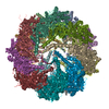
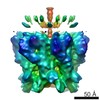
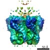
| Title | Three-dimensional structure of a type III glutamine synthetase by single-particle reconstruction. | |||||||||
 Map data Map data | GSIII from Bacteroides fragilis (negative stain reconstruction) | |||||||||
 Sample Sample |
| |||||||||
| Function / homology |  Glutamine synthetase, N-terminal domain Glutamine synthetase, N-terminal domain Function and homology information Function and homology information | |||||||||
| Biological species |  Bacteroides fragilis (bacteria) Bacteroides fragilis (bacteria) | |||||||||
| Method | single particle reconstruction / cryo EM / negative staining / Resolution: 21.0 Å | |||||||||
 Authors Authors | van Rooyen JM / Abratt VR / Sewell BT | |||||||||
 Citation Citation | Journal: J Mol Biol / Year: 2006 Title: Three-dimensional structure of a type III glutamine synthetase by single-particle reconstruction. Authors: Jason M van Rooyen / Valerie R Abratt / B Trevor Sewell /  Abstract: GlnN, the type III glutamine synthetase (GSIII) from the medically important, anaerobic, opportunistic pathogen Bacteroides fragilis, has 82.8 kDa subunits that share only 9% sequence identity with ...GlnN, the type III glutamine synthetase (GSIII) from the medically important, anaerobic, opportunistic pathogen Bacteroides fragilis, has 82.8 kDa subunits that share only 9% sequence identity with the type I glutamine synthetases (GSI), the only family for which a structure is known. Active GlnN was found predominantly in a single peak that eluted from a calibrated gel-filtration chromatography column at a position equaivalent to 0.86(+/-0.08) MDa. Negative-stain electron microscopy enabled the identification of double-ringed particles and single hexameric rings ("pinwheels") resulting from partial staining. A 2D average of these pinwheels showed marked similarity to the corresponding structures found in preparations of GSI, except that the arms of the subunits were 40% longer. Reconstructions from particles embedded in vitreous ice showed that GlnN has a double-ringed, dodecameric structure with a 6-fold dihedral space group (D6) symmetry and dimensions of 17.0 nm parallel with the 6-fold axis and 18.3 nm parallel with the 2-fold axes. The structures, combined with a sequence alignment based on structural principles, showed how many aspects of the structure of GSI, and most notably the alpha/beta barrel fold active site were preserved. There was evidence for the presence of this structure in the reconstructed volume, thus, identifying the indentations between the pinwheel spokes as putative active sites and suggesting conservation of the overall molecular geometry found in GSI despite their low level of global homology. Furthermore, docking of GSI into the reconstruction left sufficient plausibly located unoccupied density to account for the additional residues in GSIII, thus validating the structure. | |||||||||
| History |
|
- Structure visualization
| Movie |
Movie viewer |
|---|---|
| Structure viewer | EM map: SurfViewMolmilJmol/JSmol |
| Supplemental images |

- Downloads & links
-EMDB archive
| Map data | emd_1205.map.gz | 86.6 KB | EMDB map data format | |
|---|---|---|---|---|
| Header (meta data) | emd-1205-v30.xmlemd-1205.xml | 9.6 KB 9.6 KB | Display Display | EMDB header |
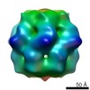
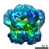
| Images | 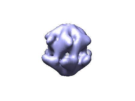 1205.gif 1205.gif | 15.2 KB | ||
| Archive directory |  http://ftp.pdbj.org/pub/emdb/structures/EMD-1205ftp://ftp.pdbj.org/pub/emdb/structures/EMD-1205 http://ftp.pdbj.org/pub/emdb/structures/EMD-1205ftp://ftp.pdbj.org/pub/emdb/structures/EMD-1205 | HTTPS FTP |
-Related structure data

-Links
| EMDB pages | EMDB (EBI/PDBe) / EMDataResource |
|---|
-Map
| File | Download / File: emd_1205.map.gz / Format: CCP4 / Size: 1.9 MB / Type: IMAGE STORED AS FLOATING POINT NUMBER (4 BYTES) | ||||||||||||||||||||||||||||||||||||||||||||||||||||||||||||||||||||
|---|---|---|---|---|---|---|---|---|---|---|---|---|---|---|---|---|---|---|---|---|---|---|---|---|---|---|---|---|---|---|---|---|---|---|---|---|---|---|---|---|---|---|---|---|---|---|---|---|---|---|---|---|---|---|---|---|---|---|---|---|---|---|---|---|---|---|---|---|---|
| Annotation | GSIII from Bacteroides fragilis (negative stain reconstruction) | ||||||||||||||||||||||||||||||||||||||||||||||||||||||||||||||||||||
| Voxel size | X=Y=Z: 4.25 Å | ||||||||||||||||||||||||||||||||||||||||||||||||||||||||||||||||||||
| Density |
| ||||||||||||||||||||||||||||||||||||||||||||||||||||||||||||||||||||
| Symmetry | Space group: 1 | ||||||||||||||||||||||||||||||||||||||||||||||||||||||||||||||||||||
| Details | EMDB XML:
CCP4 map header:
| ||||||||||||||||||||||||||||||||||||||||||||||||||||||||||||||||||||
-Supplemental data
- Sample components
-Entire : B.fragilis GlnN purified from E.coli
| Entire | Name: B.fragilis GlnN purified from E.coli |
|---|---|
| Components |
|
-Supramolecule #1000: B.fragilis GlnN purified from E.coli
| Supramolecule | Name: B.fragilis GlnN purified from E.coli / type: sample / ID: 1000 / Oligomeric state: One dodecamer of GlnN / Number unique components: 1 |
|---|---|
| Molecular weight | Experimental: 1.3 MDa / Theoretical: 990 KDa / Method: Calibrated gel-filtration |
-Macromolecule #1: GlnN
| Macromolecule | Name: GlnN / type: protein_or_peptide / ID: 1 / Name.synonym: GSIII Details: Recombinant GlnN purified from GlnA deficient E.coli mutant; SwissProt P15623 Number of copies: 12 / Oligomeric state: Dodecamer / Recombinant expression: Yes |
|---|---|
| Source (natural) | Organism: Bacteroides fragilis (bacteria) / Strain: B. fragilis BF-1 / Tissue: E.coli cytoplasm / Cell: E.coli |
| Molecular weight | Experimental: 990 KDa / Theoretical: 1.3 MDa |
| Recombinant expression | Organism: Escherichia coli (E. coli) / Recombinant plasmid: pJS139 |
| Sequence | InterPro: Glutamine synthetase, N-terminal domain |
-Experimental details
-Structure determination
| Method | negative staining, cryo EM |
|---|---|
 Processing Processing | single particle reconstruction |
| Aggregation state | particle |
-Sample preparation
| Concentration | 0.25 mg/mL |
|---|---|
| Buffer | pH: 7.15 / Details: 10 mM Imidazole-HCl, 10 mM MnCl2 |
| Staining | Type: NEGATIVE Details: Aliquots (10ul) were applied to 300 mesh copper grids, which had been coated with thin carbon support film and previously glow discharged in air for 20 seconds, before being stained with 2% ...Details: Aliquots (10ul) were applied to 300 mesh copper grids, which had been coated with thin carbon support film and previously glow discharged in air for 20 seconds, before being stained with 2% uranyl acetate solution using the droplet method. |
| Grid | Details: 300 mesh copper grid |
| Vitrification | Cryogen name: ETHANE |
- Electron microscopy
| Microscope | FEI TECNAI F20 |
|---|---|
| Electron beam | Acceleration voltage: 120 kV / Electron source: TUNGSTEN HAIRPIN |
| Electron optics | Calibrated magnification: 65882 / Illumination mode: FLOOD BEAM / Imaging mode: BRIGHT FIELDBright-field microscopy / Nominal magnification: 50000 |
| Specialist optics | Energy filter - Name: LEO OMEGA |
| Sample stage | Specimen holder: Eucentric / Specimen holder model: OTHER |
| Details | Leo 912 TEM |
| Date | May 13, 2004 |
| Image recording | Category: CCD / Film or detector model: PROSCAN TEM-PIV (2k x 2k) / Digitization - Sampling interval: 14 µm / Number real images: 160 Details: Images were digitized using an Ilford Leafscan and downsized by a factor of 2 Bits/pixel: 16 |
| Experimental equipment |  Model: Tecnai F20 / Image courtesy: FEI Company |
-Image processing
| Final two d classification | Number classes: 166 |
|---|---|
| Final reconstruction | Applied symmetry - Point group: C6 (6 fold cyclic) / Algorithm: OTHER / Resolution.type: BY AUTHOR / Resolution: 21.0 Å / Resolution method: FSC 0.5 CUT-OFF / Software - Name: SPIDER / Number images used: 12587 |
-Atomic model buiding 1
| Software | Name: Situs |
|---|---|
| Details | Protocol: rigid body |
| Refinement | Space: REAL / Protocol: RIGID BODY FIT / Target criteria: CC score |
