 Movie
Movie Controller


+ Open data

- Basic information
| Entry | Database: EMDB / ID: EMD-11986 | |||||||||||||||
|---|---|---|---|---|---|---|---|---|---|---|---|---|---|---|---|---|
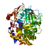
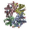
| Title | Cryo-EM structure of exoglucanase Cel48S | |||||||||||||||
 Map data Map data | ||||||||||||||||
 Sample Sample |
| |||||||||||||||
| Biological species |   Hungateiclostridium thermocellum DSM 1313 (bacteria) Hungateiclostridium thermocellum DSM 1313 (bacteria) | |||||||||||||||
| Method | single particle reconstruction / cryo EM / Resolution: 3.4 Å | |||||||||||||||
 Authors Authors | Tatli M / Morais S / Tovar-Herrera OE / Bomble YJ / Bayer EA / Medalia O / Mizrahi I | |||||||||||||||
| Funding support |  Switzerland, Switzerland,  Israel, Israel,  Germany, 4 items Germany, 4 items
| |||||||||||||||
 Citation Citation | Journal: Elife / Year: 2022 Title: Nanoscale resolution of microbial fiber degradation in action. Authors: Meltem Tatli / Sarah Moraïs / Omar E Tovar-Herrera / Yannick J Bomble / Edward A Bayer / Ohad Medalia / Itzhak Mizrahi /  Abstract: The lives of microbes unfold at the micron scale, and their molecular machineries operate at the nanoscale. Their study at these resolutions is key toward achieving a better understanding of their ...The lives of microbes unfold at the micron scale, and their molecular machineries operate at the nanoscale. Their study at these resolutions is key toward achieving a better understanding of their ecology. We focus on cellulose degradation of the canonical system to comprehend how microbes build and use their cellulosomal machinery at these nanometer scales. Degradation of cellulose, the most abundant organic polymer on Earth, is instrumental to the global carbon cycle. We reveal that bacterial cells form 'cellulosome capsules' driven by catalytic product-dependent dynamics, which can increase the rate of hydrolysis. Biosynthesis of this energetically costly machinery and cell growth are decoupled at the single-cell level, hinting at a division-of-labor strategy through phenotypic heterogeneity. This novel observation highlights intrapopulation interactions as key to understanding rates of fiber degradation. | |||||||||||||||
| History |
|
- Structure visualization
| Movie |
 Movie viewer Movie viewer |
|---|---|
| Structure viewer | EM map: SurfViewMolmilJmol/JSmol |
| Supplemental images |

- Downloads & links
-EMDB archive
| Map data | emd_11986.map.gz | 1.1 MB | EMDB map data format | |
|---|---|---|---|---|
| Header (meta data) | emd-11986-v30.xmlemd-11986.xml | 23.2 KB 23.2 KB | Display Display | EMDB header |
| FSC (resolution estimation) | emd_11986_fsc.xml | 4.7 KB | Display | FSC data file |
| Images | 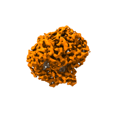 emd_11986.png emd_11986.png | 66.4 KB | ||
| Masks | emd_11986_msk_1.map | 8 MB | Mask map | |
| Others | emd_11986_additional_1.map.gzemd_11986_additional_2.map.gzemd_11986_half_map_1.map.gzemd_11986_half_map_2.map.gz | 6 MB 7.5 MB 6 MB 6 MB | ||
| Archive directory |  http://ftp.pdbj.org/pub/emdb/structures/EMD-11986ftp://ftp.pdbj.org/pub/emdb/structures/EMD-11986 http://ftp.pdbj.org/pub/emdb/structures/EMD-11986ftp://ftp.pdbj.org/pub/emdb/structures/EMD-11986 | HTTPS FTP |
-Related structure data
| Similar structure data |
|---|


-Links
| EMDB pages | EMDB (EBI/PDBe) / EMDataResource |
|---|
-Map
| File | Download / File: emd_11986.map.gz / Format: CCP4 / Size: 8 MB / Type: IMAGE STORED AS FLOATING POINT NUMBER (4 BYTES) | ||||||||||||||||||||||||||||||||||||||||||||||||||||||||||||
|---|---|---|---|---|---|---|---|---|---|---|---|---|---|---|---|---|---|---|---|---|---|---|---|---|---|---|---|---|---|---|---|---|---|---|---|---|---|---|---|---|---|---|---|---|---|---|---|---|---|---|---|---|---|---|---|---|---|---|---|---|---|
| Voxel size | X=Y=Z: 0.845 Å | ||||||||||||||||||||||||||||||||||||||||||||||||||||||||||||
| Density |
| ||||||||||||||||||||||||||||||||||||||||||||||||||||||||||||
| Symmetry | Space group: 1 | ||||||||||||||||||||||||||||||||||||||||||||||||||||||||||||
| Details | EMDB XML:
CCP4 map header:
| ||||||||||||||||||||||||||||||||||||||||||||||||||||||||||||
-Supplemental data
-Mask #1
| File | emd_11986_msk_1.map | ||||||||||||
|---|---|---|---|---|---|---|---|---|---|---|---|---|---|
| Projections & Slices |
| ||||||||||||
| Density Histograms |
 Z
Z Y
Y X
X


-Additional map: #1
| File | emd_11986_additional_1.map | ||||||||||||
|---|---|---|---|---|---|---|---|---|---|---|---|---|---|
| Projections & Slices |
| ||||||||||||
| Density Histograms |


-Additional map: #2
| File | emd_11986_additional_2.map | ||||||||||||
|---|---|---|---|---|---|---|---|---|---|---|---|---|---|
| Projections & Slices |
| ||||||||||||
| Density Histograms |


-Half map: Relion job648 halfmap2
| File | emd_11986_half_map_1.map | ||||||||||||
|---|---|---|---|---|---|---|---|---|---|---|---|---|---|
| Annotation | Relion job648 halfmap2 | ||||||||||||
| Projections & Slices |
| ||||||||||||
| Density Histograms |


-Half map: Relion job648 halfmap1
| File | emd_11986_half_map_2.map | ||||||||||||
|---|---|---|---|---|---|---|---|---|---|---|---|---|---|
| Annotation | Relion job648 halfmap1 | ||||||||||||
| Projections & Slices |
| ||||||||||||
| Density Histograms |


- Sample components
-Entire : Full-length exoglucanase Cel48S
| Entire | Name: Full-length exoglucanase Cel48S |
|---|---|
| Components |
|
-Supramolecule #1: Full-length exoglucanase Cel48S
| Supramolecule | Name: Full-length exoglucanase Cel48S / type: complex / ID: 1 / Parent: 0 / Macromolecule list: all |
|---|---|
| Source (natural) | Organism: Hungateiclostridium thermocellum DSM 1313 (bacteria) Location in cell: Extracellular |
| Molecular weight | Theoretical: 82 KDa |
-Macromolecule #1: exoglucanase Cel48S
| Macromolecule | Name: exoglucanase Cel48S / type: protein_or_peptide / ID: 1 / Enantiomer: LEVO / EC number: cellulose 1,4-beta-cellobiosidase (reducing end) |
|---|---|
| Source (natural) | Organism: Hungateiclostridium thermocellum DSM 1313 (bacteria) |
| Recombinant expression | Organism: Escherichia coli 'BL21-Gold(DE3)pLysS AG' (bacteria) |
| Sequence | String: MGPTKAPTKD GTSYKDLFLE LYGKIKDPKN GYFSPDEGIP YHSIETLIVE APDYGHVTTS EAFSYYVWLE AMYGNLTGNW SGVETAWKVM EDWIIPDSTE QPGMSSYNPN SPATYADEYE DPSYYPSELK FDTVRVGSDP VHNDLVSAYG PNMYLMHWLM DVDNWYGFGT ...String: MGPTKAPTKD GTSYKDLFLE LYGKIKDPKN GYFSPDEGIP YHSIETLIVE APDYGHVTTS EAFSYYVWLE AMYGNLTGNW SGVETAWKVM EDWIIPDSTE QPGMSSYNPN SPATYADEYE DPSYYPSELK FDTVRVGSDP VHNDLVSAYG PNMYLMHWLM DVDNWYGFGT GTRATFINTF QRGEQESTWE TIPHPSIEEF KYGGPNGFLD LFTKDRSYAK Q WRYTNAPD AEGRAIQAVY WANKWAKEQG KGSAVASVVS KAAKMGDFLR NDMFDKYFMK IGAQDKTPAT GYDSAHYLMA WYTAWGGGIG ASWAWKIGCS HAHFGYQNPF QGWVSATQSD FAPKSSNGKR DWTTSYKRQL EFYQWLQSAE GGIAGGATNS WNGRYEKYPA GTSTFYGMAY VPHPVYADPG SNQWFGFQAW SMQRVMEYYL ETGDSSVKNL IKKWVDWVMS EIKLYDDGTF AIPSDLEWSG QPDTWTGTYT GNPNLHVRVT SYGTDLGVAG SLANALATYA AATERWEGKL DTKARDMAAE LVNRAWYNFY CSEGKGVVTE EARADYKRFF EQEVYVPAGW SGTMPNGDKI QPGIKFIDIR TKYRQDPYYD IVYQAYLRGE APVLNYHRFW HEVDLAVAMG VLATYFPDMT YKVPGTPSTK LYGDVNDDGK VNSTDAVALK RYVLRSGISI NTDNADLNED GRVNSTDLGI LKRYILKEID TLPYKNHHHH HH |
-Experimental details
-Structure determination
| Method | cryo EM |
|---|---|
 Processing Processing | single particle reconstruction |
| Aggregation state | particle |
-Sample preparation
| Concentration | 0.35 mg/mL | ||||||||||||||||||
|---|---|---|---|---|---|---|---|---|---|---|---|---|---|---|---|---|---|---|---|
| Buffer | pH: 7.4 Component:
| ||||||||||||||||||
| Grid | Model: Quantifoil R0.6/1 / Material: GOLD / Mesh: 200 / Support film - Material: CARBON / Support film - topology: HOLEY / Pretreatment - Type: PLASMA CLEANING / Pretreatment - Time: 25 sec. / Pretreatment - Atmosphere: AIR | ||||||||||||||||||
| Vitrification | Cryogen name: ETHANE / Chamber humidity: 100 % / Chamber temperature: 277 K / Instrument: FEI VITROBOT MARK IV / Details: 4-5 s blotting time. |
- Electron microscopy
| Microscope | FEI TITAN KRIOS |
|---|---|
| Electron beam | Acceleration voltage: 300 kV / Electron source: FIELD EMISSION GUN |
| Electron optics | C2 aperture diameter: 50.0 µm / Calibrated defocus min: 0.5 µm / Illumination mode: FLOOD BEAM / Imaging mode: BRIGHT FIELDBright-field microscopy / Cs: 2.7 mm / Nominal defocus max: 1.5 µm / Nominal magnification: 58180 |
| Sample stage | Specimen holder model: FEI TITAN KRIOS AUTOGRID HOLDER / Cooling holder cryogen: NITROGEN |
| Image recording | Film or detector model: GATAN K2 SUMMIT (4k x 4k) / Detector mode: SUPER-RESOLUTION / Digitization - Frames/image: 1-60 / Number real images: 2000 / Average exposure time: 12.0 sec. / Average electron dose: 67.0 e/Å2 Details: Images were collected in movie-mode at 5 frames per second |
| Experimental equipment |  Model: Titan Krios / Image courtesy: FEI Company |
-Image processing
| Particle selection | Number selected: 1331906 |
|---|---|
| Startup model | Type of model: OTHER Details: Stochastic Gradient Descent (SGD) based initial model building of RELION 3.0 |
| Initial angle assignment | Type: MAXIMUM LIKELIHOOD / Software - Name: RELION (ver. 3.0) |
| Final 3D classification | Software - Name: RELION (ver. 3.0) |
| Final angle assignment | Type: MAXIMUM LIKELIHOOD |
| Final reconstruction | Number classes used: 1 / Applied symmetry - Point group: C1 (asymmetric) / Resolution.type: BY AUTHOR / Resolution: 3.4 Å / Resolution method: FSC 0.143 CUT-OFF / Software - Name: RELION (ver. 3.0) / Number images used: 55245 |
| FSC plot (resolution estimation) |  |
-Atomic model buiding 1
| Refinement | Space: REAL / Protocol: RIGID BODY FIT / Overall B value: 111 / Target criteria: 0.87 |
|---|
