 Movie
Movie Controller

[English] 日本語
 Yorodumi
- EMDB-11976: Actin filament structure from focal adhesions of mouse embryonic ... -
+ Open data

- Basic information
| Entry | Database: EMDB / ID: EMD-11976 | |||||||||
|---|---|---|---|---|---|---|---|---|---|---|
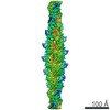
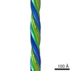
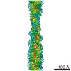
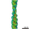
| Title | Actin filament structure from focal adhesions of mouse embryonic fibroblasts | |||||||||
 Map data Map data | Actin filament structure from focal adhesions of mouse embryonic fibroblasts | |||||||||
 Sample Sample |
| |||||||||
| Biological species |   Mus musculus (house mouse) Mus musculus (house mouse) | |||||||||
| Method | subtomogram averaging / cryo EM / Resolution: 17.0 Å | |||||||||
 Authors Authors | Martins B / Medalia O / Eibauer M | |||||||||
| Funding support |  Switzerland, European Union, 2 items Switzerland, European Union, 2 items
| |||||||||
 Citation Citation | Journal: Structure / Year: 2021 Title: Unveiling the polarity of actin filaments by cryo-electron tomography. Authors: Bruno Martins / Simona Sorrentino / Wen-Lu Chung / Meltem Tatli / Ohad Medalia / Matthias Eibauer / Abstract: The actin cytoskeleton plays a fundamental role in numerous cellular processes, such as cell motility, cytokinesis, and adhesion to the extracellular matrix. Revealing the polarity of individual ...The actin cytoskeleton plays a fundamental role in numerous cellular processes, such as cell motility, cytokinesis, and adhesion to the extracellular matrix. Revealing the polarity of individual actin filaments in intact cells would foster an unprecedented understanding of cytoskeletal processes and their associated mechanical forces. Cryo-electron tomography provides the means for high-resolution structural imaging of cells. However, the low signal-to-noise ratio of cryo-tomograms obscures the high frequencies, and therefore the polarity of actin filaments cannot be directly measured. Here, we developed a method that enables us to determine the polarity of actin filaments in cellular cryo-tomograms. We applied it to reveal the actin polarity distribution in focal adhesions, and show a linear relation between actin polarity and distance from the apical boundary of the adhesion site. | |||||||||
| History |
|
- Structure visualization
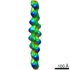
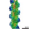
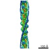
| Movie |
 Movie viewer Movie viewer |
|---|---|
| Structure viewer | EM map: SurfViewMolmilJmol/JSmol |
| Supplemental images |

- Downloads & links
-EMDB archive
| Map data | emd_11976.map.gz | 10.7 MB | EMDB map data format | |
|---|---|---|---|---|
| Header (meta data) | emd-11976-v30.xmlemd-11976.xml | 11 KB 11 KB | Display Display | EMDB header |
| Images | 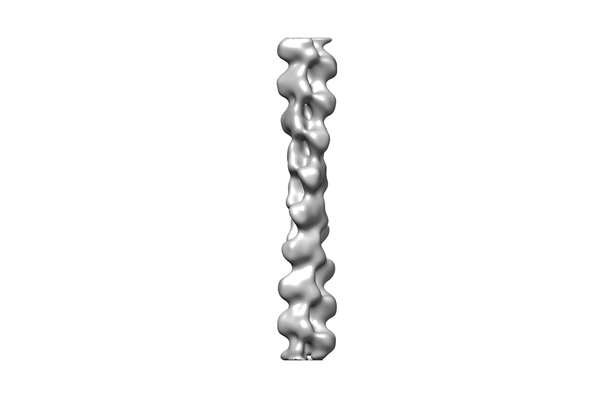 emd_11976.png emd_11976.png | 16.9 KB | ||
| Archive directory |  http://ftp.pdbj.org/pub/emdb/structures/EMD-11976ftp://ftp.pdbj.org/pub/emdb/structures/EMD-11976 http://ftp.pdbj.org/pub/emdb/structures/EMD-11976ftp://ftp.pdbj.org/pub/emdb/structures/EMD-11976 | HTTPS FTP |
-Related structure data
| Related structure data | |
|---|---|
| Similar structure data | |
| EM raw data | EMPIAR-10570 (Title: Actin filament structure from focal adhesions of mouse embryonic fibroblasts Data size: 314.1 Data #1: Actin filament structure from focal adhesions of mouse embryonic fibroblasts [tilt series]) |
-Links
| EMDB pages | EMDB (EBI/PDBe) / EMDataResource |
|---|
-Map
| File | Download / File: emd_11976.map.gz / Format: CCP4 / Size: 11.4 MB / Type: IMAGE STORED AS FLOATING POINT NUMBER (4 BYTES) | ||||||||||||||||||||||||||||||||||||||||||||||||||||||||||||
|---|---|---|---|---|---|---|---|---|---|---|---|---|---|---|---|---|---|---|---|---|---|---|---|---|---|---|---|---|---|---|---|---|---|---|---|---|---|---|---|---|---|---|---|---|---|---|---|---|---|---|---|---|---|---|---|---|---|---|---|---|---|
| Annotation | Actin filament structure from focal adhesions of mouse embryonic fibroblasts | ||||||||||||||||||||||||||||||||||||||||||||||||||||||||||||
| Voxel size | X=Y=Z: 3.443 Å | ||||||||||||||||||||||||||||||||||||||||||||||||||||||||||||
| Density |
| ||||||||||||||||||||||||||||||||||||||||||||||||||||||||||||
| Symmetry | Space group: 1 | ||||||||||||||||||||||||||||||||||||||||||||||||||||||||||||
| Details | EMDB XML:
CCP4 map header:
| ||||||||||||||||||||||||||||||||||||||||||||||||||||||||||||
-Supplemental data
- Sample components
-Entire : Actin filament from focal adhesions of mouse embryonic fibroblasts
| Entire | Name: Actin filament from focal adhesions of mouse embryonic fibroblastsMicrofilament |
|---|---|
| Components |
|
-Supramolecule #1: Actin filament from focal adhesions of mouse embryonic fibroblasts
| Supramolecule | Name: Actin filament from focal adhesions of mouse embryonic fibroblasts type: complex / ID: 1 / Parent: 0 / Macromolecule list: #1 |
|---|---|
| Source (natural) | Organism: Mus musculus (house mouse) |
-Experimental details
-Structure determination
| Method | cryo EM |
|---|---|
 Processing Processing | subtomogram averaging |
| Aggregation state | filament |
-Sample preparation
| Buffer | pH: 7.5 |
|---|---|
| Grid | Model: Quantifoil / Material: GOLD |
| Vitrification | Cryogen name: ETHANE / Instrument: HOMEMADE PLUNGER |
- Electron microscopy
| Microscope | FEI TITAN KRIOS |
|---|---|
| Electron beam | Acceleration voltage: 300 kV / Electron source: FIELD EMISSION GUN |
| Electron optics | Calibrated magnification: 42000 / Illumination mode: FLOOD BEAM / Imaging mode: BRIGHT FIELDBright-field microscopy / Cs: 2.7 mm / Nominal defocus max: 4.5 µm / Nominal defocus min: 3.5 µm / Nominal magnification: 14522 |
| Specialist optics | Energy filter - Name: GIF Quantum LS / Energy filter - Slit width: 20 eV |
| Sample stage | Specimen holder model: FEI TITAN KRIOS AUTOGRID HOLDER / Cooling holder cryogen: NITROGEN |
| Image recording | Film or detector model: GATAN K2 SUMMIT (4k x 4k) / Detector mode: SUPER-RESOLUTION / Average exposure time: 1.2 sec. / Average electron dose: 1.2 e/Å2 |
| Experimental equipment |  Model: Titan Krios / Image courtesy: FEI Company |
-Image processing
| Extraction | Number tomograms: 7 / Number images used: 43400 |
|---|---|
| CTF correction | Software - Name: TOM Toolbox |
| Final angle assignment | Type: NOT APPLICABLE |
| Final reconstruction | Applied symmetry - Helical parameters - Δz: 27.9 Å Applied symmetry - Helical parameters - Δ&Phi: -166.8 ° Applied symmetry - Helical parameters - Axial symmetry: C1 (asymmetric) Algorithm: FOURIER SPACE / Resolution.type: BY AUTHOR / Resolution: 17.0 Å / Resolution method: FSC 0.5 CUT-OFF / Software - Name: RELION (ver. 3.0.4) / Number subtomograms used: 9931 |
