 ムービー
ムービー コントローラー


+ データを開く

- 基本情報
基本情報
| 登録情報 | データベース: EMDB / ID: EMD-11870 | |||||||||
|---|---|---|---|---|---|---|---|---|---|---|
| タイトル | Tomogram of the actin network in an extracted and fixed mouse fibroblast lamellipodium. | |||||||||
 マップデータ マップデータ | Sample tomogram of an extracted and fixed mouse fibroblast lamellipodium. | |||||||||
 試料 試料 |
| |||||||||
| 生物種 |   Mus musculus (ハツカネズミ) Mus musculus (ハツカネズミ) | |||||||||
| 手法 | 電子線トモグラフィー法 / クライオ電子顕微鏡法 | |||||||||
 データ登録者 データ登録者 | Faessler F / Dimchev G / Hodirnau VV / Wan W / Schur FKM | |||||||||
| 資金援助 |  オーストリア, 1件 オーストリア, 1件
| |||||||||
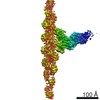
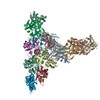
 引用 引用 | ジャーナル: Nat Commun / 年: 2020 タイトル: Cryo-electron tomography structure of Arp2/3 complex in cells reveals new insights into the branch junction. 著者: Florian Fäßler / Georgi Dimchev / Victor-Valentin Hodirnau / William Wan / Florian K M Schur /  要旨: The actin-related protein (Arp)2/3 complex nucleates branched actin filament networks pivotal for cell migration, endocytosis and pathogen infection. Its activation is tightly regulated and involves ...The actin-related protein (Arp)2/3 complex nucleates branched actin filament networks pivotal for cell migration, endocytosis and pathogen infection. Its activation is tightly regulated and involves complex structural rearrangements and actin filament binding, which are yet to be understood. Here, we report a 9.0 Å resolution structure of the actin filament Arp2/3 complex branch junction in cells using cryo-electron tomography and subtomogram averaging. This allows us to generate an accurate model of the active Arp2/3 complex in the branch junction and its interaction with actin filaments. Notably, our model reveals a previously undescribed set of interactions of the Arp2/3 complex with the mother filament, significantly different to the previous branch junction model. Our structure also indicates a central role for the ArpC3 subunit in stabilizing the active conformation. | |||||||||
| 履歴 |
|
- 構造の表示
構造の表示
| ムービー |
 ムービービューア ムービービューア |
|---|---|
| 添付画像 |

- ダウンロードとリンク
-EMDBアーカイブ
| マップデータ | emd_11870.map.gz | 33.3 MB | EMDBマップデータ形式 | |
|---|---|---|---|---|
| ヘッダ (付随情報) | emd-11870-v30.xmlemd-11870.xml | 12.5 KB 12.5 KB | 表示 表示 | EMDBヘッダ |
| 画像 | 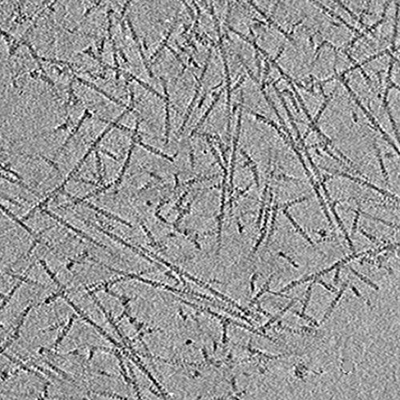 emd_11870.png emd_11870.png | 143.8 KB | ||
| アーカイブディレクトリ |  http://ftp.pdbj.org/pub/emdb/structures/EMD-11870ftp://ftp.pdbj.org/pub/emdb/structures/EMD-11870 http://ftp.pdbj.org/pub/emdb/structures/EMD-11870ftp://ftp.pdbj.org/pub/emdb/structures/EMD-11870 | HTTPS FTP |
-関連構造データ
-リンク
| EMDBのページ | EMDB (EBI/PDBe) / EMDataResource |
|---|
-マップ
| ファイル | ダウンロード / ファイル: emd_11870.map.gz / 形式: CCP4 / 大きさ: 52.7 MB / タイプ: IMAGE STORED AS SIGNED BYTE | ||||||||||||||||||||||||||||||||||||||||||||||||||||||||||||||||||||
|---|---|---|---|---|---|---|---|---|---|---|---|---|---|---|---|---|---|---|---|---|---|---|---|---|---|---|---|---|---|---|---|---|---|---|---|---|---|---|---|---|---|---|---|---|---|---|---|---|---|---|---|---|---|---|---|---|---|---|---|---|---|---|---|---|---|---|---|---|---|
| 注釈 | Sample tomogram of an extracted and fixed mouse fibroblast lamellipodium. | ||||||||||||||||||||||||||||||||||||||||||||||||||||||||||||||||||||
| ボクセルのサイズ | X=Y=Z: 17.096 Å | ||||||||||||||||||||||||||||||||||||||||||||||||||||||||||||||||||||
| 密度 |
| ||||||||||||||||||||||||||||||||||||||||||||||||||||||||||||||||||||
| 対称性 | 空間群: 1 | ||||||||||||||||||||||||||||||||||||||||||||||||||||||||||||||||||||
| 詳細 | EMDB XML:
CCP4マップ ヘッダ情報:
| ||||||||||||||||||||||||||||||||||||||||||||||||||||||||||||||||||||
-添付データ
- 試料の構成要素
-全体 : Actin network in a mouse fibroblast lamellipodium
| 全体 | 名称: Actin network in a mouse fibroblast lamellipodium |
|---|---|
| 要素 |
|
-超分子 #1: Actin network in a mouse fibroblast lamellipodium
| 超分子 | 名称: Actin network in a mouse fibroblast lamellipodium / タイプ: cell / ID: 1 / 親要素: 0 / 含まれる分子: #1-#8 詳細: Branched actin network of extracted and fixed mouse fibroblast lamellipodium |
|---|---|
| 由来(天然) | 生物種: Mus musculus (ハツカネズミ) |
-実験情報
-構造解析
| 手法 | クライオ電子顕微鏡法 |
|---|---|
 解析 解析 | 電子線トモグラフィー法 |
| 試料の集合状態 | cell |
-試料調製
| 緩衝液 | pH: 6.1 構成要素:
詳細: Adjust to pH 6.1 using NaOH | ||||||||||||
|---|---|---|---|---|---|---|---|---|---|---|---|---|---|
| グリッド | モデル: Quantifoil R2/2 / 材質: GOLD / メッシュ: 200 / 支持フィルム - 材質: CARBON / 支持フィルム - トポロジー: HOLEY / 前処理 - タイプ: GLOW DISCHARGE / 前処理 - 雰囲気: AIR 詳細: After glow discharging of the grid and prior to the seeding of cells, the grid was coated using 25ug/ml Fibronectin | ||||||||||||
| 凍結 | 凍結剤: ETHANE / チャンバー内湿度: 80 % / チャンバー内温度: 277 K 詳細: Leica GP2, 3,5sec back-blotting, sensor on, 0,1mm movement after contact, manually pre-blotted within the chamber prior to the application of fiducials. | ||||||||||||
| 切片作成 | その他: NO SECTIONING | ||||||||||||
| 位置合わせマーカー | Manufacturer: AURION / 直径: 10 nm |
- 電子顕微鏡法
| 顕微鏡 | FEI TITAN KRIOS |
|---|---|
| 電子線 | 加速電圧: 300 kV / 電子線源: FIELD EMISSION GUN |
| 電子光学系 | C2レンズ絞り径: 50.0 µm / 照射モード: FLOOD BEAM / 撮影モード: BRIGHT FIELDBright-field microscopy / Cs: 2.7 mm / 最大 デフォーカス(公称値): -0.0055 µm / 最小 デフォーカス(公称値): -0.00175 µm / 倍率(公称値): 42000 |
| 特殊光学系 | エネルギーフィルター - 名称: GIF Bioquantum / エネルギーフィルター - スリット幅: 20 eV |
| 試料ステージ | 試料ホルダーモデル: FEI TITAN KRIOS AUTOGRID HOLDER ホルダー冷却材: NITROGEN |
| 撮影 | フィルム・検出器のモデル: GATAN K3 BIOQUANTUM (6k x 4k) 実像数: 61 / 平均露光時間: 1.21 sec. / 平均電子線量: 2.79 e/Å2 詳細: Images were collected in movie-mode with 7 frames per tilt |
| 実験機器 |  モデル: Titan Krios / 画像提供: FEI Company |
-画像解析
| 最終 再構成 | アルゴリズム: BACK PROJECTION / ソフトウェア - 名称: IMOD (ver. 4.9.12) / 使用した粒子像数: 61 |
|---|
