Movie
Movie Controller

[English] 日本語
 Yorodumi
- EMDB-1149: Dodecameric structure of the small heat shock protein Acr1 from M... -
+ Open data

- Basic information
| Entry | Database: EMDB / ID: EMD-1149 | |||||||||
|---|---|---|---|---|---|---|---|---|---|---|
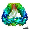
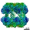
| Title | Dodecameric structure of the small heat shock protein Acr1 from Mycobacterium tuberculosis. | |||||||||
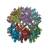
 Map data Map data | Negative stain EM map of dodecamer of M.tuberculosis Acr1 | |||||||||
 Sample Sample |
| |||||||||
| Function / homology |  Function and homology information Function and homology informationresponse to salt stress / response to hydrogen peroxide / protein homooligomerization / : / unfolded protein binding /  protein folding / protein complex oligomerization / response to heat / cytoplasm protein folding / protein complex oligomerization / response to heat / cytoplasmSimilarity search - Function | |||||||||
| Biological species |   Mycobacterium tuberculosis (bacteria) Mycobacterium tuberculosis (bacteria) | |||||||||
| Method | single particle reconstruction / negative staining / Resolution: 16.5 Å | |||||||||
 Authors Authors | Kennaway CK / Benesch JL / Gohlke U / Wang L / Robinson CV / Orlova EV / Saibil HR / Saibi HR / Keep NH | |||||||||
 Citation Citation | Journal: J Biol Chem / Year: 2005 Title: Dodecameric structure of the small heat shock protein Acr1 from Mycobacterium tuberculosis. Authors: Christopher K Kennaway / Justin L P Benesch / Ulrich Gohlke / Luchun Wang / Carol V Robinson / Elena V Orlova / Helen R Saibil / Nicholas H Keep /  Abstract: Small heat shock proteins are a ubiquitous and diverse family of stress proteins that have in common an alpha-crystallin domain. Mycobacterium tuberculosis has two small heat shock proteins, Acr1 ...Small heat shock proteins are a ubiquitous and diverse family of stress proteins that have in common an alpha-crystallin domain. Mycobacterium tuberculosis has two small heat shock proteins, Acr1 (alpha-crystallin-related protein 1, or Hsp16.3/16-kDa antigen) and Acr2 (HrpA), both of which are highly expressed under different stress conditions. Small heat shock proteins form large oligomeric assemblies and are commonly polydisperse. Nanoelectrospray mass spectrometry showed that Acr2 formed a range of oligomers composed of dimers and tetramers, whereas Acr1 was a dodecamer. Electron microscopy of Acr2 showed a variety of particle sizes. Using three-dimensional analysis of negative stain electron microscope images, we have shown that Acr1 forms a tetrahedral assembly with 12 polypeptide chains. The atomic structure of a related alpha-crystallin domain dimer was docked into the density to build a molecular structure of the dodecameric Acr1 complex. Along with the differential regulation of these two proteins, the differences in their quaternary structures demonstrated here supports their distinct functional roles. | |||||||||
| History |
|
- Structure visualization
| Movie |
Movie viewer |
|---|---|
| Structure viewer | EM map: SurfViewMolmilJmol/JSmol |
| Supplemental images |

- Downloads & links
-EMDB archive
| Map data | emd_1149.map.gz | 97.1 KB | EMDB map data format | |
|---|---|---|---|---|
| Header (meta data) | emd-1149-v30.xmlemd-1149.xml | 11 KB 11 KB | Display Display | EMDB header |
| Images |  1149.gif 1149.gif | 174.4 KB | ||
| Archive directory |  http://ftp.pdbj.org/pub/emdb/structures/EMD-1149ftp://ftp.pdbj.org/pub/emdb/structures/EMD-1149 http://ftp.pdbj.org/pub/emdb/structures/EMD-1149ftp://ftp.pdbj.org/pub/emdb/structures/EMD-1149 | HTTPS FTP |
-Related structure data
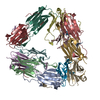
| Related structure data |  2byuMC M: atomic model generated by this map C: citing same article ( |
|---|---|
| Similar structure data |

-Links
| EMDB pages | EMDB (EBI/PDBe) / EMDataResource |
|---|---|
| Related items in Molecule of the Month |


-Map
| File | Download / File: emd_1149.map.gz / Format: CCP4 / Size: 1001 KB / Type: IMAGE STORED AS FLOATING POINT NUMBER (4 BYTES) | ||||||||||||||||||||||||||||||||||||||||||||||||||||||||||||||||||||
|---|---|---|---|---|---|---|---|---|---|---|---|---|---|---|---|---|---|---|---|---|---|---|---|---|---|---|---|---|---|---|---|---|---|---|---|---|---|---|---|---|---|---|---|---|---|---|---|---|---|---|---|---|---|---|---|---|---|---|---|---|---|---|---|---|---|---|---|---|---|
| Annotation | Negative stain EM map of dodecamer of M.tuberculosis Acr1 | ||||||||||||||||||||||||||||||||||||||||||||||||||||||||||||||||||||
| Voxel size | X=Y=Z: 3.3333 Å | ||||||||||||||||||||||||||||||||||||||||||||||||||||||||||||||||||||
| Density |
| ||||||||||||||||||||||||||||||||||||||||||||||||||||||||||||||||||||
| Symmetry | Space group: 1 | ||||||||||||||||||||||||||||||||||||||||||||||||||||||||||||||||||||
| Details | EMDB XML:
CCP4 map header:
| ||||||||||||||||||||||||||||||||||||||||||||||||||||||||||||||||||||
-Supplemental data
- Sample components
-Entire : Recombinant protein Acr1 From M.Tuberculosis made in E.coli.
| Entire | Name: Recombinant protein Acr1 From M.Tuberculosis made in E.coli. |
|---|---|
| Components |
|
-Supramolecule #1000: Recombinant protein Acr1 From M.Tuberculosis made in E.coli.
| Supramolecule | Name: Recombinant protein Acr1 From M.Tuberculosis made in E.coli. type: sample / ID: 1000 Details: Sample was monodisperse by mass spectrometry and light scattering Oligomeric state: dodecamer tetrahedral symmetry / Number unique components: 1 |
|---|---|
| Molecular weight | Experimental: 196 KDa / Theoretical: 196 KDa / Method: Mass spectrometry |
-Macromolecule #1: Acr1
| Macromolecule | Name: Acr1 / type: protein_or_peptide / ID: 1 / Name.synonym: Hsp16.3 / Details: monomer / Number of copies: 12 / Oligomeric state: dodecamer / Recombinant expression: Yes |
|---|---|
| Source (natural) | Organism: Mycobacterium tuberculosis (bacteria) / Strain: H37Rv / synonym: TB |
| Molecular weight | Experimental: 18.5 KDa / Theoretical: 18.5 KDa |
| Recombinant expression | Organism: Escherichia coli (E. coli) / Recombinant plasmid: pET15b |
| Sequence | InterPro: Alpha crystallin/Hsp20 domain |
-Experimental details
-Structure determination
| Method | negative staining |
|---|---|
 Processing Processing | single particle reconstruction |
| Aggregation state | particle |
-Sample preparation
| Concentration | 0.01 mg/mL |
|---|---|
| Buffer | pH: 7 / Details: 50 mM Tris pH 7 |
| Staining | Type: NEGATIVE / Details: Two 5 ul aliquots of 2% uranyl acetate. |
| Grid | Details: 400 mesh copper grid with carbon film |
| Vitrification | Cryogen name: NONE / Chamber temperature: 293 K |
- Electron microscopy
| Microscope | FEI TECNAI 12 |
|---|---|
| Electron beam | Acceleration voltage: 120 kV / Electron source: TUNGSTEN HAIRPIN |
| Electron optics | Illumination mode: FLOOD BEAM / Imaging mode: BRIGHT FIELDBright-field microscopy / Cs: 2.0 mm / Nominal defocus max: 0.6 µm / Nominal defocus min: 0.39 µm / Nominal magnification: 42000 |
| Sample stage | Specimen holder: side entry / Specimen holder model: OTHER |
| Temperature | Average: 293 K |
| Alignment procedure | Legacy - Astigmatism: corrected at 100k magnification |
| Date | Mar 26, 2004 |
| Image recording | Category: FILM / Film or detector model: KODAK SO-163 FILM / Digitization - Scanner: ZEISS SCAI / Digitization - Sampling interval: 14 µm / Number real images: 18 / Average electron dose: 10 e/Å2 / Od range: 1 / Bits/pixel: 8 |
-Image processing
| Final two d classification | Number classes: 400 |
|---|---|
| Final reconstruction | Applied symmetry - Point group: T (tetrahedral) / Algorithm: OTHER / Resolution.type: BY AUTHOR / Resolution: 16.5 Å / Resolution method: FSC 0.5 CUT-OFF / Software - Name: IMAGIC Details: Exact filtered back projection. Map made from 400 out of 600 class averages. Number images used: 6123 |
-Atomic model buiding 1
| Initial model | PDB ID: Chain - #0 - Chain ID: A / Chain - #1 - Chain ID: B |
|---|---|
| Software | Name: URO |
| Details | PDBEntryID_givenInChain. Protocol: rigid body. One dimer of 1gme A and B, residues 43-137 and 146-151, was fitted in URO and tetrahedral symmetry was used to generate the other five dimers. |
| Refinement | Space: RECIPROCAL / Protocol: RIGID BODY FIT / Target criteria: Density correlation |
| Output model | PDB-2byu: |