 Movie
Movie Controller

[English] 日本語
 Yorodumi
- EMDB-1117: Conservation of the capsid structure in tailed dsDNA bacteriophag... -
+ Open data

- Basic information
| Entry | Database: EMDB / ID: EMD-1117 | |||||||||
|---|---|---|---|---|---|---|---|---|---|---|
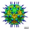
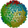
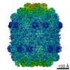
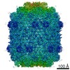
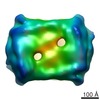
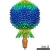
| Title | Conservation of the capsid structure in tailed dsDNA bacteriophages: the pseudoatomic structure of phi29. | |||||||||
 Map data Map data | This map is cryo-EM 3D reconstruction of a fiberless prohead particle from bacteriophage phi29 | |||||||||
 Sample Sample |
| |||||||||
| Biological species |   Bacillus phage phi29 (virus) Bacillus phage phi29 (virus) | |||||||||
| Method | single particle reconstruction / cryo EM / Resolution: 12.7 Å | |||||||||
 Authors Authors | Morais MC / Choi KH / Koti JS / Chipman PR / Anderson DL / Rossmann MG | |||||||||
 Citation Citation | Journal: Mol Cell / Year: 2005 Title: Conservation of the capsid structure in tailed dsDNA bacteriophages: the pseudoatomic structure of phi29. Authors: Marc C Morais / Kyung H Choi / Jaya S Koti / Paul R Chipman / Dwight L Anderson / Michael G Rossmann /  Abstract: Bacteriophage phi29 is one of the smallest and simplest known dsDNA phages, making it amenable to structural investigations. The three-dimensional structure of a fiberless, isometric variant has been ...Bacteriophage phi29 is one of the smallest and simplest known dsDNA phages, making it amenable to structural investigations. The three-dimensional structure of a fiberless, isometric variant has been determined to 7.9 A resolution by cryo-electron microscopy (cryo-EM), allowing the identification of alpha helices and beta sheets. Their arrangement indicates that the folds of the phi29 and bacteriophage HK97 capsid proteins are similar except for an additional immunoglobulin-like domain of the phi29 protein. An atomic model that incorporates these two domains fits well into the cryo-EM density of the T = 3, fiberless isometric phi29 particle, and cryo-EM structures of fibered isometric and fiberless prolate prohead phi29 particles at resolutions of 8.7 A and 12.7 A, respectively. Thus, phi29 joins the growing number of phages that utilize the HK97 capsid structure, suggesting that this protein fold may be as prevalent in capsids of dsDNA phages as the jelly roll fold is in eukaryotic viruses. | |||||||||
| History |
|
- Structure visualization
| Movie |
 Movie viewer Movie viewer |
|---|---|
| Structure viewer | EM map: SurfViewMolmilJmol/JSmol |
| Supplemental images |

- Downloads & links
-EMDB archive
| Map data | emd_1117.map.gz | 2.6 MB | EMDB map data format | |
|---|---|---|---|---|
| Header (meta data) | emd-1117-v30.xmlemd-1117.xml | 8.6 KB 8.6 KB | Display Display | EMDB header |
| Images |  1117.gif 1117.gif | 17.8 KB | ||
| Archive directory |  http://ftp.pdbj.org/pub/emdb/structures/EMD-1117ftp://ftp.pdbj.org/pub/emdb/structures/EMD-1117 http://ftp.pdbj.org/pub/emdb/structures/EMD-1117ftp://ftp.pdbj.org/pub/emdb/structures/EMD-1117 | HTTPS FTP |
-Validation report
| Summary document | emd_1117_validation.pdf.gz | 227 KB | Display | EMDB validaton report |
|---|---|---|---|---|
| Full document | emd_1117_full_validation.pdf.gz | 226.1 KB | Display | |
| Data in XML | emd_1117_validation.xml.gz | 5.9 KB | Display | |
| Arichive directory | https://ftp.pdbj.org/pub/emdb/validation_reports/EMD-1117ftp://ftp.pdbj.org/pub/emdb/validation_reports/EMD-1117 | HTTPS FTP |
-Related structure data


-Links
| EMDB pages | EMDB (EBI/PDBe) / EMDataResource |
|---|
-Map
| File | Download / File: emd_1117.map.gz / Format: CCP4 / Size: 15.3 MB / Type: IMAGE STORED AS FLOATING POINT NUMBER (4 BYTES) | ||||||||||||||||||||||||||||||||||||||||||||||||||||||||||||||||||||
|---|---|---|---|---|---|---|---|---|---|---|---|---|---|---|---|---|---|---|---|---|---|---|---|---|---|---|---|---|---|---|---|---|---|---|---|---|---|---|---|---|---|---|---|---|---|---|---|---|---|---|---|---|---|---|---|---|---|---|---|---|---|---|---|---|---|---|---|---|---|
| Annotation | This map is cryo-EM 3D reconstruction of a fiberless prohead particle from bacteriophage phi29 | ||||||||||||||||||||||||||||||||||||||||||||||||||||||||||||||||||||
| Voxel size | X=Y=Z: 4.24 Å | ||||||||||||||||||||||||||||||||||||||||||||||||||||||||||||||||||||
| Density |
| ||||||||||||||||||||||||||||||||||||||||||||||||||||||||||||||||||||
| Symmetry | Space group: 1 | ||||||||||||||||||||||||||||||||||||||||||||||||||||||||||||||||||||
| Details | EMDB XML:
CCP4 map header:
| ||||||||||||||||||||||||||||||||||||||||||||||||||||||||||||||||||||
-Supplemental data
- Sample components
-Entire : phi29 fiberless prohead particle
| Entire | Name: phi29 fiberless prohead particle |
|---|---|
| Components |
|
-Supramolecule #1000: phi29 fiberless prohead particle
| Supramolecule | Name: phi29 fiberless prohead particle / type: sample / ID: 1000 Oligomeric state: capsid protein forms t3 q5 prolate icosahedron Number unique components: 1 |
|---|---|
| Molecular weight | Theoretical: 12.4 MDa |
-Supramolecule #1: Bacillus phage phi29
| Supramolecule | Name: Bacillus phage phi29 / type: virus / ID: 1 / Name.synonym: phi29 / NCBI-ID: 10756 / Sci species name: Bacillus phage phi29 / Virus type: VIRUS-LIKE PARTICLE / Virus isolate: SPECIES / Virus enveloped: No / Virus empty: Yes / Syn species name: phi29 |
|---|---|
| Host (natural) | Organism:  |
| Molecular weight | Experimental: 12.4 MDa |
-Experimental details
-Structure determination
| Method | cryo EM |
|---|---|
 Processing Processing | single particle reconstruction |
| Aggregation state | particle |
-Sample preparation
| Buffer | pH: 7.8 / Details: 50 mM Tris-HCl pH7.8 10 mM MgCl2 100 mM NaCl |
|---|---|
| Grid | Details: holey carbon |
| Vitrification | Cryogen name: ETHANE / Instrument: HOMEMADE PLUNGER |
- Electron microscopy
| Microscope | FEI/PHILIPS CM300FEG/T |
|---|---|
| Image recording | Category: FILM / Film or detector model: KODAK SO-163 FILM / Digitization - Scanner: ZEISS SCAI / Digitization - Sampling interval: 7 µm / Number real images: 19 / Average electron dose: 20 e/Å2 / Details: after scanning, images binned by a factor of 2 / Bits/pixel: 8 |
| Electron beam | Acceleration voltage: 300 kV / Electron source:  FIELD EMISSION GUN FIELD EMISSION GUN |
| Electron optics | Illumination mode: FLOOD BEAM / Imaging mode: BRIGHT FIELD / Cs: 2.0 mm / Nominal defocus max: 3.781 µm / Nominal defocus min: 2.185 µm / Nominal magnification: 33000 |
| Sample stage | Specimen holder: Side entry liquid nitrogen-cooled cryo specimen holder Specimen holder model: GATAN LIQUID NITROGEN / Tilt angle min: 9 / Tilt angle max: 9 |
-Image processing
| CTF correction | Details: CTF correction included phase and amplitude correction for each micrograph |
|---|---|
| Final reconstruction | Applied symmetry - Point group: C5 (5 fold cyclic) / Algorithm: OTHER / Resolution.type: BY AUTHOR / Resolution: 12.7 Å / Resolution method: FSC 0.5 CUT-OFF / Software - Name: EMAN / Number images used: 12288 |
