 Movie
Movie Controller

[English] 日本語
 Yorodumi
- EMDB-11059: ATP-dependent partner switch links flagellar C-ring assembly with... -
+ Open data

- Basic information
| Entry | Database: EMDB / ID: EMD-11059 | |||||||||
|---|---|---|---|---|---|---|---|---|---|---|
| Title | ATP-dependent partner switch links flagellar C-ring assembly with gene expression | |||||||||
 Map data Map data | Flagellar motor of Shewanella putrefaciens in situ, wildtype | |||||||||
 Sample Sample |
| |||||||||
| Biological species |   Shewanella putrefaciens CN-32 (bacteria) Shewanella putrefaciens CN-32 (bacteria) | |||||||||
| Method | subtomogram averaging / cryo EM / Resolution: 82.0 Å | |||||||||
 Authors Authors | Blagotinsek V / Schwan M / Steinchen W / Mrusek D / Hook J / Rossmann FM / Freibert SA / Kressler D / Beeby M / Thormann KM / Bange G | |||||||||
| Funding support |  Germany, Germany,  United Kingdom, 2 items United Kingdom, 2 items
| |||||||||
 Citation Citation | Journal: Proc Natl Acad Sci U S A / Year: 2020 Title: An ATP-dependent partner switch links flagellar C-ring assembly with gene expression. Authors: Vitan Blagotinsek / Meike Schwan / Wieland Steinchen / Devid Mrusek / John C Hook / Florian Rossmann / Sven A Freibert / Hanna Kratzat / Guillaume Murat / Dieter Kressler / Roland Beckmann / ...Authors: Vitan Blagotinsek / Meike Schwan / Wieland Steinchen / Devid Mrusek / John C Hook / Florian Rossmann / Sven A Freibert / Hanna Kratzat / Guillaume Murat / Dieter Kressler / Roland Beckmann / Morgan Beeby / Kai M Thormann / Gert Bange /  Abstract: Bacterial flagella differ in their number and spatial arrangement. In many species, the MinD-type ATPase FlhG (also YlxH/FleN) is central to the numerical control of bacterial flagella, and its ...Bacterial flagella differ in their number and spatial arrangement. In many species, the MinD-type ATPase FlhG (also YlxH/FleN) is central to the numerical control of bacterial flagella, and its deletion in polarly flagellated bacteria typically leads to hyperflagellation. The molecular mechanism underlying this numerical control, however, remains enigmatic. Using the model species , we show that FlhG links assembly of the flagellar C ring with the action of the master transcriptional regulator FlrA (named FleQ in other species). While FlrA and the flagellar C-ring protein FliM have an overlapping binding site on FlhG, their binding depends on the ATP-dependent dimerization state of FlhG. FliM interacts with FlhG independent of nucleotide binding, while FlrA exclusively interacts with the ATP-dependent FlhG dimer and stimulates FlhG ATPase activity. Our in vivo analysis of FlhG partner switching between FliM and FlrA reveals its mechanism in the numerical restriction of flagella, in which the transcriptional activity of FlrA is down-regulated through a negative feedback loop. Our study demonstrates another level of regulatory complexity underlying the spationumerical regulation of flagellar biogenesis and implies that flagellar assembly transcriptionally regulates the production of more initial building blocks. | |||||||||
| History |
|
- Structure visualization
| Movie |
 Movie viewer Movie viewer |
|---|---|
| Structure viewer | EM map: SurfViewMolmilJmol/JSmol |
| Supplemental images |

- Downloads & links
-EMDB archive
| Map data | emd_11059.map.gz | 11.4 MB | EMDB map data format | |
|---|---|---|---|---|
| Header (meta data) | emd-11059-v30.xmlemd-11059.xml | 12.1 KB 12.1 KB | Display Display | EMDB header |
| Images | 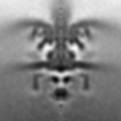 emd_11059.png emd_11059.png | 323.6 KB | ||
| Archive directory |  http://ftp.pdbj.org/pub/emdb/structures/EMD-11059ftp://ftp.pdbj.org/pub/emdb/structures/EMD-11059 http://ftp.pdbj.org/pub/emdb/structures/EMD-11059ftp://ftp.pdbj.org/pub/emdb/structures/EMD-11059 | HTTPS FTP |
-Related structure data
| Related structure data | C: citing same article ( |
|---|---|
| Similar structure data |


-Links
| EMDB pages | EMDB (EBI/PDBe) / EMDataResource |
|---|
-Map
| File | Download / File: emd_11059.map.gz / Format: CCP4 / Size: 12.9 MB / Type: IMAGE STORED AS FLOATING POINT NUMBER (4 BYTES) | ||||||||||||||||||||||||||||||||||||||||||||||||||||||||||||||||||||
|---|---|---|---|---|---|---|---|---|---|---|---|---|---|---|---|---|---|---|---|---|---|---|---|---|---|---|---|---|---|---|---|---|---|---|---|---|---|---|---|---|---|---|---|---|---|---|---|---|---|---|---|---|---|---|---|---|---|---|---|---|---|---|---|---|---|---|---|---|---|
| Annotation | Flagellar motor of Shewanella putrefaciens in situ, wildtype | ||||||||||||||||||||||||||||||||||||||||||||||||||||||||||||||||||||
| Voxel size | X=Y=Z: 8.28 Å | ||||||||||||||||||||||||||||||||||||||||||||||||||||||||||||||||||||
| Density |
| ||||||||||||||||||||||||||||||||||||||||||||||||||||||||||||||||||||
| Symmetry | Space group: 1 | ||||||||||||||||||||||||||||||||||||||||||||||||||||||||||||||||||||
| Details | EMDB XML:
CCP4 map header:
| ||||||||||||||||||||||||||||||||||||||||||||||||||||||||||||||||||||
-Supplemental data
- Sample components
-Entire : Flagellar motor of Shewanella putrefaciens in situ, wildtype
| Entire | Name: Flagellar motor of Shewanella putrefaciens in situ, wildtype |
|---|---|
| Components |
|
-Supramolecule #1: Flagellar motor of Shewanella putrefaciens in situ, wildtype
| Supramolecule | Name: Flagellar motor of Shewanella putrefaciens in situ, wildtype type: cell / ID: 1 / Parent: 0 |
|---|---|
| Source (natural) | Organism: Shewanella putrefaciens CN-32 (bacteria) |
-Experimental details
-Structure determination
| Method | cryo EM |
|---|---|
 Processing Processing | subtomogram averaging |
| Aggregation state | particle |
-Sample preparation
| Buffer | pH: 7 / Component - Name: LB medium |
|---|---|
| Grid | Model: UltrAuFoil / Material: GOLD / Mesh: 200 / Support film - Material: GOLD / Support film - topology: HOLEY / Support film - Film thickness: 50.0 nm / Pretreatment - Type: GLOW DISCHARGE / Pretreatment - Atmosphere: AIR / Pretreatment - Pressure: 0.02 kPa |
| Vitrification | Cryogen name: ETHANE-PROPANE / Chamber humidity: 100 % / Chamber temperature: 298 K / Instrument: FEI VITROBOT MARK IV Details: blot time 5 s, blot force 3, wait time 60s, drain time 0.5s,. |
- Electron microscopy
| Microscope | FEI TECNAI F20 |
|---|---|
| Electron beam | Acceleration voltage: 200 kV / Electron source: FIELD EMISSION GUN |
| Electron optics | C2 aperture diameter: 100.0 µm / Illumination mode: FLOOD BEAM / Imaging mode: BRIGHT FIELDBright-field microscopy / Cs: 2.0 mm / Nominal defocus max: 4.0 µm / Nominal defocus min: 3.0 µm / Nominal magnification: 25000 |
| Sample stage | Specimen holder model: GATAN 914 HIGH TILT LIQUID NITROGEN CRYO TRANSFER TOMOGRAPHY HOLDER Cooling holder cryogen: NITROGEN |
| Image recording | Film or detector model: FEI FALCON II (4k x 4k) / Detector mode: INTEGRATING / Digitization - Dimensions - Width: 4096 pixel / Digitization - Dimensions - Height: 4096 pixel / Number grids imaged: 5 / Average exposure time: 1.5 sec. / Average electron dose: 3.15 e/Å2 |
| Experimental equipment |  Model: Tecnai F20 / Image courtesy: FEI Company |
-Image processing
| Extraction | Number tomograms: 220 / Number images used: 224 / Reference model: sperical / Method: manual / Software - Name: PEET (ver. 1.10.1) |
|---|---|
| Final angle assignment | Type: ANGULAR RECONSTITUTION / Software - Name: PEET (ver. 1.10.1) |
| Final reconstruction | Applied symmetry - Point group: C100 (100 fold cyclic) / Algorithm: BACK PROJECTION / Resolution.type: BY AUTHOR / Resolution: 82.0 Å / Resolution method: FSC 0.5 CUT-OFF / Software - Name: PEET (ver. 1.10.1) / Number subtomograms used: 224 |
