 Movie
Movie Controller

[English] 日本語
 Yorodumi
- EMDB-1087: Three-dimensional structure of the bacterial multidrug transporte... -
+ Open data

- Basic information
| Entry | Database: EMDB / ID: EMD-1087 | |||||||||
|---|---|---|---|---|---|---|---|---|---|---|
| Title | Three-dimensional structure of the bacterial multidrug transporter EmrE shows it is an asymmetric homodimer. | |||||||||
 Map data Map data | Density map of the asymmetric dimer of EmrE,a multidrug transporter from Escherichia coli | |||||||||
 Sample Sample |
| |||||||||
| Function / homology |  Function and homology information Function and homology informationEmrE multidrug transporter complex / amino-acid betaine transmembrane transporter activity / choline transmembrane transporter activity / glycine betaine transport / choline transport / xenobiotic detoxification by transmembrane export across the plasma membrane / xenobiotic transport /  antiporter activity / response to osmotic stress / xenobiotic transmembrane transporter activity ...EmrE multidrug transporter complex / amino-acid betaine transmembrane transporter activity / choline transmembrane transporter activity / glycine betaine transport / choline transport / xenobiotic detoxification by transmembrane export across the plasma membrane / xenobiotic transport / antiporter activity / response to osmotic stress / xenobiotic transmembrane transporter activity / transmembrane transporter activity / xenobiotic metabolic process / transmembrane transport / cellular response to xenobiotic stimulus / membrane => GO:0016020 / response to xenobiotic stimulus / DNA damage response / membrane / identical protein binding / plasma membrane antiporter activity / response to osmotic stress / xenobiotic transmembrane transporter activity ...EmrE multidrug transporter complex / amino-acid betaine transmembrane transporter activity / choline transmembrane transporter activity / glycine betaine transport / choline transport / xenobiotic detoxification by transmembrane export across the plasma membrane / xenobiotic transport / antiporter activity / response to osmotic stress / xenobiotic transmembrane transporter activity / transmembrane transporter activity / xenobiotic metabolic process / transmembrane transport / cellular response to xenobiotic stimulus / membrane => GO:0016020 / response to xenobiotic stimulus / DNA damage response / membrane / identical protein binding / plasma membraneSimilarity search - Function | |||||||||
| Biological species |  Escherichia coli (E. coli) Escherichia coli (E. coli) | |||||||||
| Method | electron crystallography / cryo EM / negative staining / Resolution: 7.5 Å | |||||||||
 Authors Authors | Ubarretxena-Belandia I / Baldwin JM / Schuldiner S / Tate CG | |||||||||
 Citation Citation | Journal: EMBO J / Year: 2003 Title: Three-dimensional structure of the bacterial multidrug transporter EmrE shows it is an asymmetric homodimer. Authors: Iban Ubarretxena-Belandia / Joyce M Baldwin / Shimon Schuldiner / Christopher G Tate /  Abstract: The small multidrug resistance family of transporters is widespread in bacteria and is responsible for bacterial resistance to toxic aromatic cations by proton-linked efflux. We have determined the ...The small multidrug resistance family of transporters is widespread in bacteria and is responsible for bacterial resistance to toxic aromatic cations by proton-linked efflux. We have determined the three-dimensional (3D) structure of the Escherichia coli multidrug transporter EmrE by electron cryomicroscopy of 2D crystals, including data to 7.0 A resolution. The structure of EmrE consists of a bundle of eight transmembrane alpha-helices with one substrate molecule bound near the centre. The substrate binding chamber is formed from six helices and is accessible both from the aqueous phase and laterally from the lipid bilayer. The most remarkable feature of the structure of EmrE is that it is an asymmetric homodimer. The possible arrangement of the two polypeptides in the EmrE dimer is discussed based on the 3D density map. | |||||||||
| History |
|
- Structure visualization
| Movie |
Movie viewer |
|---|---|
| Structure viewer | EM map: SurfViewMolmilJmol/JSmol |
| Supplemental images |
- Downloads & links
-EMDB archive
| Map data | emd_1087.map.gz | 547.4 KB | EMDB map data format | |
|---|---|---|---|---|
| Header (meta data) | emd-1087-v30.xmlemd-1087.xml | 12.5 KB 12.5 KB | Display Display | EMDB header |
| Images |  1087.gif 1087.gif | 11.7 KB | ||
| Archive directory |  http://ftp.pdbj.org/pub/emdb/structures/EMD-1087ftp://ftp.pdbj.org/pub/emdb/structures/EMD-1087 http://ftp.pdbj.org/pub/emdb/structures/EMD-1087ftp://ftp.pdbj.org/pub/emdb/structures/EMD-1087 | HTTPS FTP |
-Related structure data
| Related structure data | 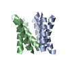 2i68M M: atomic model generated by this map |
|---|---|
| Similar structure data |
-Links
| EMDB pages | EMDB (EBI/PDBe) / EMDataResource |
|---|
-Map
| File | Download / File: emd_1087.map.gz / Format: CCP4 / Size: 579.1 KB / Type: IMAGE STORED AS FLOATING POINT NUMBER (4 BYTES) | ||||||||||||||||||||||||||||||||||||||||||||||||||||||||||||||||||||
|---|---|---|---|---|---|---|---|---|---|---|---|---|---|---|---|---|---|---|---|---|---|---|---|---|---|---|---|---|---|---|---|---|---|---|---|---|---|---|---|---|---|---|---|---|---|---|---|---|---|---|---|---|---|---|---|---|---|---|---|---|---|---|---|---|---|---|---|---|---|
| Annotation | Density map of the asymmetric dimer of EmrE,a multidrug transporter from Escherichia coli | ||||||||||||||||||||||||||||||||||||||||||||||||||||||||||||||||||||
| Voxel size | X: 0.48067 Å / Y: 0.57867 Å / Z: 2.5 Å | ||||||||||||||||||||||||||||||||||||||||||||||||||||||||||||||||||||
| Density |
| ||||||||||||||||||||||||||||||||||||||||||||||||||||||||||||||||||||
| Symmetry | Space group: 1 | ||||||||||||||||||||||||||||||||||||||||||||||||||||||||||||||||||||
| Details | EMDB XML:
CCP4 map header:
| ||||||||||||||||||||||||||||||||||||||||||||||||||||||||||||||||||||
-Supplemental data
- Sample components
-Entire : EmrE from E. coli containing the bound substrate tetraphenylphosp...
| Entire | Name: EmrE from E. coli containing the bound substrate tetraphenylphosphonium |
|---|---|
| Components |
|
-Supramolecule #1000: EmrE from E. coli containing the bound substrate tetraphenylphosp...
| Supramolecule | Name: EmrE from E. coli containing the bound substrate tetraphenylphosphonium type: sample / ID: 1000 Details: EmrE in the 2D crystals is in a functional state with the substrate (TPP+) bound in a binding pocket formed from 6 out of the 8 helices in the asymmetric dimer. Oligomeric state: Asymmetric homodimer / Number unique components: 1 |
|---|---|
| Molecular weight | Theoretical: 15.2 KDa |
-Macromolecule #1: EmrE
| Macromolecule | Name: EmrE / type: protein_or_peptide / ID: 1 / Name.synonym: MvrC, EB Details: 4-helix integral membrane protein; The engineered EmrE is fully functional as assessed by in vivo and in vitro transport assays Number of copies: 2 / Oligomeric state: dimer / Recombinant expression: Yes |
|---|---|
| Source (natural) | Organism: Escherichia coli (E. coli) / Strain: K12 / Location in cell: Inner membrane |
| Molecular weight | Experimental: 15.2 KDa / Theoretical: 15.2 KDa |
| Recombinant expression | Organism: Escherichia coli (E. coli) / Recombinant plasmid: pT7-7 |
| Sequence | UniProtKB: Multidrug transporter EmrE / GO: membrane => GO:0016020 / InterPro: Small drug/metabolite transporter protein family |
-Experimental details
-Structure determination
| Method | negative staining, cryo EM |
|---|---|
 Processing Processing | electron crystallography |
| Aggregation state | 2D array |
-Sample preparation
| Concentration | 1 mg/mL |
|---|---|
| Buffer | pH: 7.5 Details: 20 mM Sodium phosphate pH7.5, 100 mM NaCl, 2mM MgCl2, 1mM EDTA, 1 mM Na azide, 10uM tetraphenylphosphonium |
| Staining | Type: NEGATIVE Details: Crystals on carbon support were washed with 1% glucose pH 8 (aq ammonia) containing 25ug/ml bacitracin, 2 times 20ul, and blotted to dryness |
| Grid | Details: 300 mesh molybdenum grid |
| Vitrification | Cryogen name: NITROGEN / Chamber humidity: 45 % / Chamber temperature: 77 K / Instrument: HOMEMADE PLUNGER Details: Vitrification instrument: manual. Performed at room temperature Method: Blot to dryness for 10 seconds before freezing |
| Details | 2D crystals were grown in suspension by dialysis |
| Crystal formation | Details: 2D crystals were grown in suspension by dialysis |
- Electron microscopy
| Microscope | FEI TECNAI F30 |
|---|---|
| Electron beam | Acceleration voltage: 300 kV / Electron source: FIELD EMISSION GUN |
| Electron optics | Calibrated magnification: 57400 / Illumination mode: SPOT SCAN / Imaging mode: BRIGHT FIELDBright-field microscopy / Cs: 2.3 mm / Nominal defocus max: 1.6 µm / Nominal defocus min: 0.2 µm / Nominal magnification: 60000 |
| Sample stage | Specimen holder: Side entry liquid nitrogen-cooled cryo specimen holder Specimen holder model: GATAN LIQUID NITROGEN / Tilt angle max: 46 / Tilt series - Axis1 - Min angle: 0 ° / Tilt series - Axis1 - Max angle: 46 ° |
| Temperature | Min: 77 K / Max: 77 K / Average: 77 K |
| Alignment procedure | Legacy - Astigmatism: objective lens astigmatism was corrected at 200,000 times magnification |
| Image recording | Category: FILM / Film or detector model: KODAK SO-163 FILM / Digitization - Scanner: ZEISS SCAI / Digitization - Sampling interval: 7 µm / Number real images: 47 / Average electron dose: 15 e/Å2 |
| Tilt angle min | 0 |
| Experimental equipment |  Model: Tecnai F30 / Image courtesy: FEI Company |
-Image processing
| Crystal parameters | Unit cell - A: 72.1 Å / Unit cell - B: 86.8 Å / Unit cell - γ: 107.3 ° / Plane group: P 2 |
|---|---|
| CTF correction | Details: CTFFIND2 on each image |
| Final reconstruction | Algorithm: OTHER / Resolution.type: BY AUTHOR / Resolution: 7.5 Å / Resolution method: OTHER / Software - Name: MRC Details: Resolution in the membrane plane is 7.5 angstroms and 16 angstroms perpendicular to the membrane plane |
