 Movie
Movie Controller


+ Open data

- Basic information
| Entry | Database: EMDB / ID: EMD-10706 | |||||||||
|---|---|---|---|---|---|---|---|---|---|---|
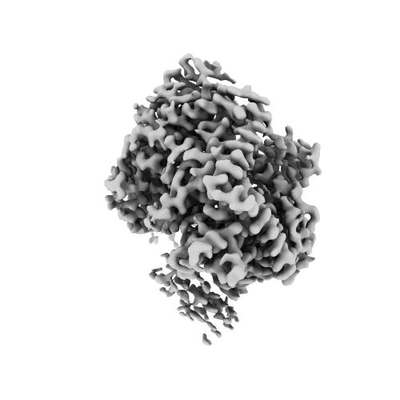
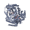
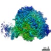
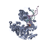
| Title | Cryo-EM structure of a Phenuiviridae L protein | |||||||||
 Map data Map data | ||||||||||
 Sample Sample |
| |||||||||
 Keywords Keywords |  Bunyavirus / Phenuiviridae / L protein / viral polymerase / cap-snatching / VIRAL PROTEIN Bunyavirus / Phenuiviridae / L protein / viral polymerase / cap-snatching / VIRAL PROTEIN | |||||||||
| Function / homology |  Function and homology information Function and homology informationhost cell endoplasmic reticulum / virion component / host cell endoplasmic reticulum-Golgi intermediate compartment / host cell Golgi apparatus / Hydrolases; Acting on ester bonds / hydrolase activity / RNA-directed RNA polymerase / viral RNA genome replication / RNA-dependent RNA polymerase activity / DNA-templated transcription / metal ion bindingSimilarity search - Function | |||||||||
| Biological species |  SFTS virus AH12 SFTS virus AH12 | |||||||||
| Method | single particle reconstruction / cryo EM / Resolution: 3.78 Å | |||||||||
 Authors Authors | Vogel D / Thorkelsson SR | |||||||||
| Funding support |  Germany, 2 items Germany, 2 items
| |||||||||
 Citation Citation | Journal: J Struct Biol / Year: 2012 Title: RELION: implementation of a Bayesian approach to cryo-EM structure determination. Authors: Sjors H W Scheres /  Abstract: RELION, for REgularized LIkelihood OptimizatioN, is an open-source computer program for the refinement of macromolecular structures by single-particle analysis of electron cryo-microscopy (cryo-EM) ...RELION, for REgularized LIkelihood OptimizatioN, is an open-source computer program for the refinement of macromolecular structures by single-particle analysis of electron cryo-microscopy (cryo-EM) data. Whereas alternative approaches often rely on user expertise for the tuning of parameters, RELION uses a Bayesian approach to infer parameters of a statistical model from the data. This paper describes developments that reduce the computational costs of the underlying maximum a posteriori (MAP) algorithm, as well as statistical considerations that yield new insights into the accuracy with which the relative orientations of individual particles may be determined. A so-called gold-standard Fourier shell correlation (FSC) procedure to prevent overfitting is also described. The resulting implementation yields high-quality reconstructions and reliable resolution estimates with minimal user intervention and at acceptable computational costs. | |||||||||
| History |
|
- Structure visualization
| Movie |
Movie viewer |
|---|---|
| Structure viewer | EM map: SurfViewMolmilJmol/JSmol |
| Supplemental images |
- Downloads & links
-EMDB archive
| Map data | emd_10706.map.gz | 28.6 MB | EMDB map data format | |
|---|---|---|---|---|
| Header (meta data) | emd-10706-v30.xmlemd-10706.xml | 19 KB 19 KB | Display Display | EMDB header |
| Images |  emd_10706.png emd_10706.png | 41.2 KB | ||
| Filedesc metadata | emd-10706.cif.gz | 7.6 KB | ||
| Archive directory |  http://ftp.pdbj.org/pub/emdb/structures/EMD-10706ftp://ftp.pdbj.org/pub/emdb/structures/EMD-10706 http://ftp.pdbj.org/pub/emdb/structures/EMD-10706ftp://ftp.pdbj.org/pub/emdb/structures/EMD-10706 | HTTPS FTP |
-Related structure data
| Related structure data |  6y6kMC  6xyaC M: atomic model generated by this map C: citing same article ( |
|---|---|
| Similar structure data |

-Links
| EMDB pages | EMDB (EBI/PDBe) / EMDataResource |
|---|
-Map
| File | Download / File: emd_10706.map.gz / Format: CCP4 / Size: 30.5 MB / Type: IMAGE STORED AS FLOATING POINT NUMBER (4 BYTES) | ||||||||||||||||||||||||||||||||||||||||||||||||||||||||||||
|---|---|---|---|---|---|---|---|---|---|---|---|---|---|---|---|---|---|---|---|---|---|---|---|---|---|---|---|---|---|---|---|---|---|---|---|---|---|---|---|---|---|---|---|---|---|---|---|---|---|---|---|---|---|---|---|---|---|---|---|---|---|
| Voxel size | X=Y=Z: 0.87 Å | ||||||||||||||||||||||||||||||||||||||||||||||||||||||||||||
| Density |
| ||||||||||||||||||||||||||||||||||||||||||||||||||||||||||||
| Symmetry | Space group: 1 | ||||||||||||||||||||||||||||||||||||||||||||||||||||||||||||
| Details | EMDB XML:
CCP4 map header:
| ||||||||||||||||||||||||||||||||||||||||||||||||||||||||||||
-Supplemental data
- Sample components
-Entire : Severe Fever with Thrombocytopenia Syndrome Virus L Protein
| Entire | Name: Severe Fever with Thrombocytopenia Syndrome Virus L Protein |
|---|---|
| Components |
|
-Supramolecule #1: Severe Fever with Thrombocytopenia Syndrome Virus L Protein
| Supramolecule | Name: Severe Fever with Thrombocytopenia Syndrome Virus L Protein type: complex / ID: 1 / Parent: 0 / Macromolecule list: #1 |
|---|---|
| Source (natural) | Organism: SFTS virus AH12 |
| Molecular weight | Theoretical: 238 KDa |
-Macromolecule #1: RNA-dependent RNA polymerase
| Macromolecule | Name: RNA-dependent RNA polymerase / type: protein_or_peptide / ID: 1 / Number of copies: 1 / Enantiomer: LEVO |
|---|---|
| Source (natural) | Organism: SFTS virus AH12 |
| Molecular weight | Theoretical: 235.742531 KDa |
| Recombinant expression | Organism:  Escherichia coli (E. coli) Escherichia coli (E. coli) |
| Sequence | String: MNLEVLCGRI NVENGLSLGE PGLYDQIYDR PGLPDLDVTV DATGVTVDIG AVPDSASQLG SSINAGLITI QLSEAYKINH DFTFSGLSK TTDRRLSEVF PITHDGSDGM TPDVIHTRLD GTIVVVEFST TRSHNIGGLE AAYRTKIEKY RDPISRRVDI M ENPRVFFG ...String: MNLEVLCGRI NVENGLSLGE PGLYDQIYDR PGLPDLDVTV DATGVTVDIG AVPDSASQLG SSINAGLITI QLSEAYKINH DFTFSGLSK TTDRRLSEVF PITHDGSDGM TPDVIHTRLD GTIVVVEFST TRSHNIGGLE AAYRTKIEKY RDPISRRVDI M ENPRVFFG VIVVSSGGVL SNMPLTQDEA EELMYRFCIA NEIYTKARSM DADIELQKSE EELEAISRAL SFFSLFEPNI ER VEGTFPN SEIKMLEQFL STPADVDFIT KTLKAKEVEA YADLCDSHYL KPEKTIQERL EINRCEAIDK TQDLLAGLHA RSN KQTSLN RGTVKLPPWL PKPSSESIDI KTDSGFGSLM DHGAYGELWA KCLLDVSLGN VEGVVSDPAK ELDIAISDDP EKDT PKEAK ITYRRFKPAL SSSARQEFSL QGVEGKKWKR MAANQKKEKE SHETLSPFLD VEDIGDFLTF NNLLTDSRYG DESIQ RAVS ILLEKASAMQ DTELTHALND SFKRNLSSNV VQWSLWVSCL AQELASALKQ HCRAGEFIIK KLKFWPIYVI IKPTKS SSH IFYSLGIRKA DVTRRLTGRV FSDTIDAGEW ELTEFKSLKT CKLTNLVNLP CTMLNSIAFW REKLGVAPWL VRKPCSE LR EQVGLTFLIS LEDKSKTEEI ITLTRYTQME GFVSPPMLPK PQKMLGKLDG PLRTKLQVYL LRKHLDCMVR IASQPFSL I PREGRVEWGG TFHAISGRST NLENMVNSWY IGYYKNKEES TELNALGEMY KKIVEMEEDK PSSPEFLGWG DTDSPKKHE FSRSFLRAAC SSLEREIAQR HGRQWKQNLE ERVLREIGTK NILDLASMKA TSNFSKDWEL YSEVQTKEYH RSKLLEKMAT LIEKGVMWY IDAVGQAWKA VLDDGCMRIC LFKKNQHGGL REIYVMDANA RLVQFGVETM ARCVCELSPH ETVANPRLKN S IIENHGLK SARSLGPGSI NINSSNDAKK WNQGHYTTKL ALVLCWFMPA KFHRFIWAAI SMFRRKKMMV DLRFLAHLSS KS ESRSSDP FREAMTDAFH GNRDVSWMDK GRTYIKTETG MMQGILHFTS SLLHSCVQSF YKSYFVSKLK EGYMGESISG VVD VIEGSD DSAIMISIRP KSDMDEVRSR FFVANLLHSV KFLNPLFGIY SSEKSTVNTV YCVEYNSEFH FHRHLVRPTL RWIA ASHQI SETEALASRQ EDYSNLLTQC LEGGASFSLT YLIQCAQLLH HYMLLGLCLH PLFGTFMGML ISDPDPALGF FLMDN PAFA GGAGFRFNLW RACKTTDLGR KYAYYFNEIQ GKTKGDEDYR ALDATSGGTL SHSVMVYWGD RKKYQALLNR MGLPED WVE QIDENPGVLY RRAANKKELL LKLAEKVHSP GVTSSLSKGH VVPRVVAAGV YLLSRHCFRF SSSIHGRGST QKASLIK LL MMSSISAMKH GGSLNPNQER MLFPQAQEYD RVCTLLEEVE HLTGKFVVRE RNIVRSRIDL FQEPVDLRCK AEDLVSEV W FGLKRTKLGP RLLKEEWDKL RASFAWLSTD PSETLRDGPF LSHVQFRNFI AHVDAKSRSV RLLGAPVKKS GGVTTISQV VRMNFFPGFS LEAEKSLDNQ ERLESISILK HVLFMVLNGP YTEEYKLEMI IEAFSTLVIP QPSEVIRKSR TMTLCLLSNY LSSRGGSIL DQIERAQSGT LGGFSKPQKT FVRPGGGVGY KGKGVWTGVM EDTHVQILID GDGTSNWLEE IRLSSDARLY D VIESIRRL CDDLGINNRV ASAYRGHCMV RLSGFKIKPA SRTDGCPVRI MERGFRIREL QNPDEVKMRV RGDILNLSVT IQ EGRVMNI LSYRPRDTDI SESAAAYLWS NRDLFSFGKK EPSCSWICLK TLDNWAWSHA SVLLANDRKT QGIDNRAMGN IFR DCLEGS LRKQGLMRSK LTEMVEKNVV PLTTQELVDI LEEDIDFSDV IAVELSEGSL DIESIFDGAP ILWSAEVEEF GEGV VAVSY SSKYYHLTLM DQAAITMCAI MGKEGCRGLL TEKRCMAAIR EQVRPFLIFL QIPEDSISWV SDQFCDSRGL DEEST IMWG UniProtKB: RNA-directed RNA polymerase L |
-Macromolecule #2: MAGNESIUM ION
| Macromolecule | Name: MAGNESIUM ION / type: ligand / ID: 2 / Number of copies: 1 / Formula: MG |
|---|---|
| Molecular weight | Theoretical: 24.305 Da |
-Experimental details
-Structure determination
| Method | cryo EM |
|---|---|
 Processing Processing | single particle reconstruction |
| Aggregation state | particle |
-Sample preparation
| Concentration | 0.15 mg/mL | ||||||||||||
|---|---|---|---|---|---|---|---|---|---|---|---|---|---|
| Buffer | pH: 7 Component:
| ||||||||||||
| Grid | Model: Quantifoil R2/1 / Pretreatment - Type: PLASMA CLEANING / Pretreatment - Time: 60 sec. / Details: Harrick Plasma cleaner | ||||||||||||
| Vitrification | Cryogen name: ETHANE-PROPANE / Chamber humidity: 100 % / Chamber temperature: 277.15 K / Instrument: FEI VITROBOT MARK IV / Details: Blot for 2s using blotting force -10. |
- Electron microscopy
| Microscope | TFS KRIOS |
|---|---|
| Electron beam | Acceleration voltage: 300 kV / Electron source: FIELD EMISSION GUN |
| Electron optics | C2 aperture diameter: 70.0 µm / Illumination mode: FLOOD BEAM / Imaging mode: BRIGHT FIELDBright-field microscopy / Cs: 2.7 mm / Nominal defocus max: 3.0 µm / Nominal defocus min: 0.5 µm / Nominal magnification: 105000 |
| Specialist optics | Energy filter - Name: GIF Bioquantum / Energy filter - Slit width: 20 eV |
| Sample stage | Specimen holder model: FEI TITAN KRIOS AUTOGRID HOLDER / Cooling holder cryogen: NITROGEN |
| Image recording | Film or detector model: GATAN K3 (6k x 4k) / Number grids imaged: 1 / Number real images: 2626 / Average exposure time: 3.0 sec. / Average electron dose: 59.45 e/Å2 |
| Experimental equipment |  Model: Titan Krios / Image courtesy: FEI Company |
-Image processing
| Particle selection | Number selected: 780000 Details: Particles were picked with Warp's pretrained agent BoxNet2Mask_20180918 |
|---|---|
| Startup model | Type of model: OTHER Details: An ab initio model generated by cisTEM from a screening dataset. |
| Initial angle assignment | Type: MAXIMUM LIKELIHOOD / Software - Name: RELION (ver. 3.0) |
| Final 3D classification | Number classes: 6 / Software - Name: RELION (ver. 3.0) |
| Final angle assignment | Type: MAXIMUM LIKELIHOOD / Software - Name: RELION (ver. 3.0) |
| Final reconstruction | Number classes used: 1 / Applied symmetry - Point group: C1 (asymmetric) / Algorithm: FOURIER SPACE / Resolution.type: BY AUTHOR / Resolution: 3.78 Å / Resolution method: FSC 0.143 CUT-OFF / Software - Name: RELION (ver. 3.0) / Number images used: 220000 |
-Atomic model buiding 1
| Refinement | Space: REAL / Protocol: AB INITIO MODEL / Overall B value: 225.217 |
|---|---|
| Output model | PDB-6y6k: |
