 ムービー
ムービー コントローラー


+ データを開く

- 基本情報
基本情報
| 登録情報 | データベース: EMDB / ID: EMD-1052 | |||||||||
|---|---|---|---|---|---|---|---|---|---|---|
| タイトル | Untangling desmosomal knots with electron tomography. | |||||||||
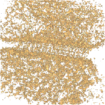
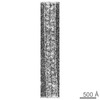
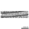
 マップデータ マップデータ | This is a 3D reconstruction map of an desmosome based on high pressure freezing/freeze-subsitution electron tomography | |||||||||
 試料 試料 |
| |||||||||
| 生物種 |   Mus musculus (ハツカネズミ) Mus musculus (ハツカネズミ) | |||||||||
| 手法 | 電子線トモグラフィー法 / クライオ電子顕微鏡法 / ネガティブ染色法 / 解像度: 30.0 Å | |||||||||
 データ登録者 データ登録者 | He W / Cowin P / Stokes DL | |||||||||
 引用 引用 | ジャーナル: Science / 年: 2003 タイトル: Untangling desmosomal knots with electron tomography. 著者: Wanzhong He / Pamela Cowin / David L Stokes /  要旨: Cell adhesion by adherens junctions and desmosomes relies on interactions between cadherin molecules. However, the molecular interfaces that define molecular specificity and that mediate adhesion ...Cell adhesion by adherens junctions and desmosomes relies on interactions between cadherin molecules. However, the molecular interfaces that define molecular specificity and that mediate adhesion remain controversial. We used electron tomography of plastic sections from neonatal mouse skin to visualize the organization of desmosomes in situ. The resulting three-dimensional maps reveal individual cadherin molecules forming discrete groups and interacting through their tips. Fitting of an x-ray crystal structure for C-cadherin to these maps is consistent with a flexible intermolecular interface mediated by an exchange of amino-terminal tryptophans. This flexibility suggests a novel mechanism for generating both cis and trans interactions and for propagating these adhesive interactions along the junction. | |||||||||
| 履歴 |
|
- 構造の表示
構造の表示
| ムービー |
 ムービービューア ムービービューア |
|---|---|
| 構造ビューア | EMマップ: SurfViewMolmilJmol/JSmol |
| 添付画像 |
- ダウンロードとリンク
-EMDBアーカイブ
| マップデータ | emd_1052.map.gz | 31.3 MB | EMDBマップデータ形式 | |
|---|---|---|---|---|
| ヘッダ (付随情報) | emd-1052-v30.xmlemd-1052.xml | 11.2 KB 11.2 KB | 表示 表示 | EMDBヘッダ |
| 画像 |  1052.gif 1052.gif | 106.6 KB | ||
| アーカイブディレクトリ |  http://ftp.pdbj.org/pub/emdb/structures/EMD-1052ftp://ftp.pdbj.org/pub/emdb/structures/EMD-1052 http://ftp.pdbj.org/pub/emdb/structures/EMD-1052ftp://ftp.pdbj.org/pub/emdb/structures/EMD-1052 | HTTPS FTP |
-関連構造データ
-リンク
| EMDBのページ | EMDB (EBI/PDBe) / EMDataResource |
|---|
-マップ
| ファイル | ダウンロード / ファイル: emd_1052.map.gz / 形式: CCP4 / 大きさ: 41.5 MB / タイプ: IMAGE STORED AS SIGNED INTEGER (2 BYTES) | ||||||||||||||||||||||||||||||||||||||||||||||||||||||||||||||||||||
|---|---|---|---|---|---|---|---|---|---|---|---|---|---|---|---|---|---|---|---|---|---|---|---|---|---|---|---|---|---|---|---|---|---|---|---|---|---|---|---|---|---|---|---|---|---|---|---|---|---|---|---|---|---|---|---|---|---|---|---|---|---|---|---|---|---|---|---|---|---|
| 注釈 | This is a 3D reconstruction map of an desmosome based on high pressure freezing/freeze-subsitution electron tomography | ||||||||||||||||||||||||||||||||||||||||||||||||||||||||||||||||||||
| ボクセルのサイズ | X=Y=Z: 7.266 Å | ||||||||||||||||||||||||||||||||||||||||||||||||||||||||||||||||||||
| 密度 |
| ||||||||||||||||||||||||||||||||||||||||||||||||||||||||||||||||||||
| 対称性 | 空間群: 1 | ||||||||||||||||||||||||||||||||||||||||||||||||||||||||||||||||||||
| 詳細 | EMDB XML:
CCP4マップ ヘッダ情報:
| ||||||||||||||||||||||||||||||||||||||||||||||||||||||||||||||||||||
-添付データ
- 試料の構成要素
-全体 : mouse skin
| 全体 | 名称: mouse skin皮膚 |
|---|---|
| 要素 |
|
-超分子 #1000: mouse skin
| 超分子 | 名称: mouse skin / タイプ: sample / ID: 1000 詳細: Skin from newborn mice frozen by high-pressure freezer followed by freeze-substitution, embedding in Epon resin and thin sectioning 集合状態: desmosome / Number unique components: 1 |
|---|
-超分子 #1: desmosome
| 超分子 | 名称: desmosome / タイプ: organelle_or_cellular_component / ID: 1 / Name.synonym: desmosome 詳細: in-situ desmosome from frozen skin; fresh skin from newborn mice frozen immediately with high pressure freezer. コピー数: 1 / 集合状態: unique / 組換発現: No / データベース: NCBI |
|---|---|
| 由来(天然) | 生物種: Mus musculus (ハツカネズミ) / 別称: mouse / 組織: mouse / 細胞: skin / Organelle: cell junction / 細胞中の位置: between two cells plasma membrane |
-実験情報
-構造解析
| 手法 | ネガティブ染色法, クライオ電子顕微鏡法 |
|---|---|
 解析 解析 | 電子線トモグラフィー法 |
-試料調製
| 緩衝液 | pH: 7.4 / 詳細: 0.5mM MgCl2 and 1-2mM CaCl2 in PBS |
|---|---|
| 染色 | タイプ: NEGATIVE 詳細: staining en bloc freeze-substitution with 1% OsO4/0.1% uranyl acetate in acetone, thin section stained with 3% uranyl acetate/SATO Pb post-stain |
| グリッド | 詳細: 200 mesh thin bar hexgonal cooper grid with formvar |
| 凍結 | 凍結剤: NITROGEN / チャンバー内湿度: 100 % / チャンバー内温度: 90 K / 装置: OTHER 詳細: Vitrification instrument: BalTec HPM 010. high pressure freezing at 2050 bar with liquid nitrigen Timed resolved state: 50msec / 手法: High pressure freezing |
- 電子顕微鏡法
| 顕微鏡 | FEI/PHILIPS CM200FEG |
|---|---|
| 電子線 | 加速電圧: 200 kV / 電子線源: FIELD EMISSION GUN |
| 電子光学系 | 照射モード: FLOOD BEAM / 撮影モード: BRIGHT FIELDBright-field microscopy / Cs: 2.0 mm / 最大 デフォーカス(公称値): 0.5 µm / 最小 デフォーカス(公称値): 0.3 µm / 倍率(公称値): 50000 |
| 試料ステージ | 試料ホルダー: high tilt / 試料ホルダーモデル: OTHER / Tilt series - Axis1 - Angle increment: 1 ° |
| 温度 | 最低: 293 K / 最高: 293 K / 平均: 293 K |
| アライメント法 | Legacy - 非点収差: objective lens astigmatism was corrected with thin carbon at 200-390kx Legacy - Electron beam tilt params: 0 |
| 詳細 | dose corresponds to the cumulative dose for the entire the dataset. |
| 日付 | 2002年11月23日 |
| 撮影 | カテゴリ: CCD / フィルム・検出器のモデル: GATAN MULTISCAN / 実像数: 300 / 詳細: imaging directly on the CCD |
-画像解析
| CTF補正 | 詳細: no CTF correction |
|---|---|
| 最終 再構成 | アルゴリズム: OTHER / 解像度のタイプ: BY AUTHOR / 解像度: 30.0 Å / 解像度の算出法: OTHER / ソフトウェア - 名称: IMOD 詳細: tomographic reconstruction from dual axis tilt series (tilt range:-78/+73; -68/+75,interval:1 degree). 使用した粒子像数: 150 |
| 詳細 | details 50nm thin section stained with 3% uranyl acetate and followed by SATO Lead stain, picked on formvar coated grids and both sides coated 5-10nm amorphous carbon. |
-原子モデル構築 1
| 初期モデル | PDB ID: |
|---|---|
| ソフトウェア | 名称: AmiraMol 3.0 |
| 詳細 | Protocol: Rigid Body. tomographic map used for fitting C-cadherin structure. #Results deposited in PDB database under codes 1Q55, 1Q5A, 1Q5B and 1Q5C. |
| 精密化 | 空間: REAL / プロトコル: RIGID BODY FIT |
| 得られたモデル | 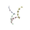 PDB-1q55:  PDB-1q5a:  PDB-1q5b:  PDB-1q5c: |

