 Movie
Movie Controller

[English] 日本語
 Yorodumi
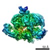
- EMDB-1043: Molecular architecture of the multiprotein splicing factor SF3b. -
+ Open data

- Basic information
| Entry | Database: EMDB / ID: EMD-1043 | |||||||||
|---|---|---|---|---|---|---|---|---|---|---|
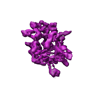
| Title | Molecular architecture of the multiprotein splicing factor SF3b. | |||||||||
 Map data Map data | 3D map of SF3b | |||||||||
 Sample Sample |
| |||||||||
| Biological species |   Homo sapiens (human) Homo sapiens (human) | |||||||||
| Method | single particle reconstruction / cryo EM / negative staining / Resolution: 9.7 Å | |||||||||
 Authors Authors | Golas MM / Sander B / Will CL / Luhrmann R / Stark H | |||||||||
 Citation Citation | Journal: Science / Year: 2003 Title: Molecular architecture of the multiprotein splicing factor SF3b. Authors: Monika M Golas / Bjoern Sander / Cindy L Will / Reinhard Lührmann / Holger Stark /  Abstract: The splicing factor SF3b is a multiprotein complex essential for the accurate excision of introns from pre-messenger RNA. As an integral component of the U2 small nuclear ribonucleoprotein (snRNP) ...The splicing factor SF3b is a multiprotein complex essential for the accurate excision of introns from pre-messenger RNA. As an integral component of the U2 small nuclear ribonucleoprotein (snRNP) and the U11/U12 di-snRNP, SF3b is involved in the recognition of the pre-messenger RNA's branch site within the major and minor spliceosomes. We have determined the three-dimensional structure of the human SF3b complex by single-particle electron cryomicroscopy at a resolution of less than 10 angstroms, allowing identification of protein domains with known structural folds. The best fit of a modeled RNA-recognition motif indicates that the protein p14 is located in the central cavity of the complex. The 22 tandem helical repeats of the protein SF3b155 are located in the outer shell of the complex enclosing p14. | |||||||||
| History |
|
- Structure visualization
| Movie |
 Movie viewer Movie viewer |
|---|---|
| Structure viewer | EM map: SurfViewMolmilJmol/JSmol |
| Supplemental images |
- Downloads & links
-EMDB archive
| Map data | emd_1043.map.gz | 1.1 MB | EMDB map data format | |
|---|---|---|---|---|
| Header (meta data) | emd-1043-v30.xmlemd-1043.xml | 9.5 KB 9.5 KB | Display Display | EMDB header |
| Images |  1043.gif 1043.gif | 21.5 KB | ||
| Archive directory |  http://ftp.pdbj.org/pub/emdb/structures/EMD-1043ftp://ftp.pdbj.org/pub/emdb/structures/EMD-1043 http://ftp.pdbj.org/pub/emdb/structures/EMD-1043ftp://ftp.pdbj.org/pub/emdb/structures/EMD-1043 | HTTPS FTP |
-Related structure data
| Similar structure data |
|---|


-Links
| EMDB pages | EMDB (EBI/PDBe) / EMDataResource |
|---|
-Map
| File | Download / File: emd_1043.map.gz / Format: CCP4 / Size: 7.8 MB / Type: IMAGE STORED AS FLOATING POINT NUMBER (4 BYTES) | ||||||||||||||||||||||||||||||||||||||||||||||||||||||||||||
|---|---|---|---|---|---|---|---|---|---|---|---|---|---|---|---|---|---|---|---|---|---|---|---|---|---|---|---|---|---|---|---|---|---|---|---|---|---|---|---|---|---|---|---|---|---|---|---|---|---|---|---|---|---|---|---|---|---|---|---|---|---|
| Annotation | 3D map of SF3b | ||||||||||||||||||||||||||||||||||||||||||||||||||||||||||||
| Voxel size | X=Y=Z: 1.6 Å | ||||||||||||||||||||||||||||||||||||||||||||||||||||||||||||
| Density |
| ||||||||||||||||||||||||||||||||||||||||||||||||||||||||||||
| Symmetry | Space group: 1 | ||||||||||||||||||||||||||||||||||||||||||||||||||||||||||||
| Details | EMDB XML:
CCP4 map header:
| ||||||||||||||||||||||||||||||||||||||||||||||||||||||||||||
-Supplemental data
- Sample components
-Entire : SF3b, multiprotein splicing factor
| Entire | Name: SF3b, multiprotein splicing factor |
|---|---|
| Components |
|
-Supramolecule #1000: SF3b, multiprotein splicing factor
| Supramolecule | Name: SF3b, multiprotein splicing factor / type: sample / ID: 1000 / Oligomeric state: monomer / Number unique components: 1 |
|---|---|
| Molecular weight | Experimental: 445 KDa / Theoretical: 450 KDa / Method: protein sequences |
-Macromolecule #1: splicing related proteins
| Macromolecule | Name: splicing related proteins / type: protein_or_peptide / ID: 1 / Number of copies: 1 / Oligomeric state: Monomer / Recombinant expression: No |
|---|---|
| Source (natural) | Organism: Homo sapiens (human) / synonym: Human / Cell: Hela / Organelle: Nucleus / Location in cell: Nucleus |
| Molecular weight | Experimental: 450 KDa / Theoretical: 445 KDa |
-Experimental details
-Structure determination
| Method | negative staining, cryo EM |
|---|---|
 Processing Processing | single particle reconstruction |
| Aggregation state | particle |
-Sample preparation
| Concentration | 0.05 mg/mL |
|---|---|
| Buffer | pH: 7.9 Details: 20 mM Hepes, 600 mM KCl, 1.5 mM Mg2 Cl2, 5% glycerol, 0.5 mM DTT, 0.5 mM PMSF |
| Staining | Type: NEGATIVE Details: The SF3b complexes were sandwiched in 2% uranyl formate between two layers of thin carbon using a perforated carbon grid as support. After 2 min at RT, the sample was frozen in liquid nitrogen. |
| Grid | Details: 200 mesh copper grid |
| Vitrification | Cryogen name: NITROGEN |
- Electron microscopy
| Microscope | FEI/PHILIPS CM200FEG |
|---|---|
| Electron beam | Acceleration voltage: 200 kV / Electron source: FIELD EMISSION GUN |
| Electron optics | Calibrated magnification: 49500 / Illumination mode: FLOOD BEAM / Imaging mode: BRIGHT FIELDBright-field microscopy / Cs: 2 mm / Nominal defocus max: 1.5 µm / Nominal defocus min: 0.15 µm / Nominal magnification: 50000 |
| Sample stage | Specimen holder: side entry cryo holder / Specimen holder model: GATAN LIQUID NITROGEN |
| Temperature | Average: 93 K |
| Alignment procedure | Legacy - Astigmatism: 175,000 times magnification |
| Image recording | Category: FILM / Film or detector model: KODAK SO-163 FILM / Digitization - Scanner: PRIMESCAN / Digitization - Sampling interval: 4 µm / Number real images: 37 / Average electron dose: 20 e/Å2 / Bits/pixel: 8 |
-Image processing
| CTF correction | Details: Each particle |
|---|---|
| Final reconstruction | Applied symmetry - Point group: C1 (asymmetric) / Algorithm: OTHER / Resolution.type: BY AUTHOR / Resolution: 9.7 Å / Resolution method: FSC 0.5 CUT-OFF / Software - Name: Imagic / Number images used: 31000 |
| Details | interactively |
-Atomic model buiding 1
| Software | Name: Situs, Amira |
|---|---|
| Refinement | Overall B value: 28 |
