 Movie
Movie Controller

[English] 日本語
 Yorodumi
- EMDB-10263: Structure of Coxsackievirus A10 complexed with its receptor KREMEN1 -
+ Open data

- Basic information
| Entry | Database: EMDB / ID: EMD-10263 | ||||||||||||
|---|---|---|---|---|---|---|---|---|---|---|---|---|---|
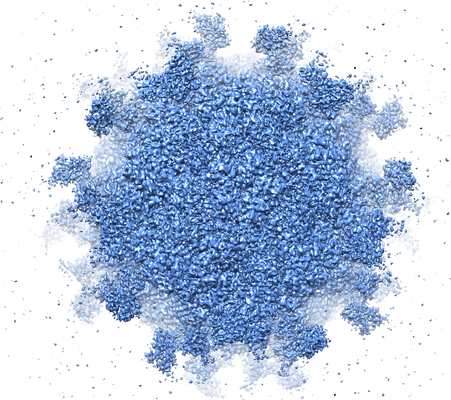
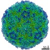
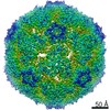
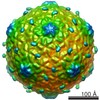
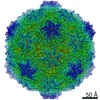
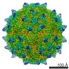
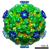
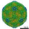
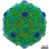
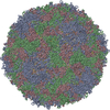
| Title | Structure of Coxsackievirus A10 complexed with its receptor KREMEN1 | ||||||||||||
 Map data Map data | |||||||||||||
 Sample Sample |
| ||||||||||||
| Function / homology |  Function and homology information Function and homology informationSignaling by LRP5 mutants / Negative regulation of TCF-dependent signaling by WNT ligand antagonists / negative regulation of axon regeneration /  cell communication / negative regulation of ossification / regulation of canonical Wnt signaling pathway / limb development / symbiont-mediated suppression of host cytoplasmic pattern recognition receptor signaling pathway via inhibition of MDA-5 activity / picornain 2A / symbiont-mediated suppression of host mRNA export from nucleus ...Signaling by LRP5 mutants / Negative regulation of TCF-dependent signaling by WNT ligand antagonists / negative regulation of axon regeneration / cell communication / negative regulation of ossification / regulation of canonical Wnt signaling pathway / limb development / symbiont-mediated suppression of host cytoplasmic pattern recognition receptor signaling pathway via inhibition of MDA-5 activity / picornain 2A / symbiont-mediated suppression of host mRNA export from nucleus / symbiont genome entry into host cell via pore formation in plasma membrane / picornain 3C / T=pseudo3 icosahedral viral capsid / TCF dependent signaling in response to WNT / host cell cytoplasmic vesicle membrane / negative regulation of canonical Wnt signaling pathway / cytoplasmic vesicle membrane / endocytosis involved in viral entry into host cell / Wnt signaling pathway / : / nucleoside-triphosphate phosphatase / protein complex oligomerization / monoatomic ion channel activity / RNA helicase activity / induction by virus of host autophagy / RNA-directed RNA polymerase / symbiont-mediated suppression of host gene expression / viral RNA genome replication / cysteine-type endopeptidase activity / RNA-dependent RNA polymerase activity / neuronal cell body / DNA-templated transcription / apoptotic process / virion attachment to host cell / structural molecule activity / ATP hydrolysis activity / proteolysis / RNA binding / ATP binding / membrane / metal ion binding / plasma membrane cell communication / negative regulation of ossification / regulation of canonical Wnt signaling pathway / limb development / symbiont-mediated suppression of host cytoplasmic pattern recognition receptor signaling pathway via inhibition of MDA-5 activity / picornain 2A / symbiont-mediated suppression of host mRNA export from nucleus ...Signaling by LRP5 mutants / Negative regulation of TCF-dependent signaling by WNT ligand antagonists / negative regulation of axon regeneration / cell communication / negative regulation of ossification / regulation of canonical Wnt signaling pathway / limb development / symbiont-mediated suppression of host cytoplasmic pattern recognition receptor signaling pathway via inhibition of MDA-5 activity / picornain 2A / symbiont-mediated suppression of host mRNA export from nucleus / symbiont genome entry into host cell via pore formation in plasma membrane / picornain 3C / T=pseudo3 icosahedral viral capsid / TCF dependent signaling in response to WNT / host cell cytoplasmic vesicle membrane / negative regulation of canonical Wnt signaling pathway / cytoplasmic vesicle membrane / endocytosis involved in viral entry into host cell / Wnt signaling pathway / : / nucleoside-triphosphate phosphatase / protein complex oligomerization / monoatomic ion channel activity / RNA helicase activity / induction by virus of host autophagy / RNA-directed RNA polymerase / symbiont-mediated suppression of host gene expression / viral RNA genome replication / cysteine-type endopeptidase activity / RNA-dependent RNA polymerase activity / neuronal cell body / DNA-templated transcription / apoptotic process / virion attachment to host cell / structural molecule activity / ATP hydrolysis activity / proteolysis / RNA binding / ATP binding / membrane / metal ion binding / plasma membraneSimilarity search - Function | ||||||||||||
| Biological species |  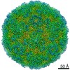 Coxsackievirus A10 / Coxsackievirus A10 /  Homo sapiens (human) Homo sapiens (human) | ||||||||||||
| Method | single particle reconstruction / cryo EM / Resolution: 3.9 Å | ||||||||||||
 Authors Authors | Zhao Y / Zhou D / Ni T / Karia D / Kotecha A / Wang X / Rao Z / Jones EY / Fry EE / Ren J / Stuart DI | ||||||||||||
| Funding support |  United Kingdom, 3 items United Kingdom, 3 items
| ||||||||||||
 Citation Citation | Journal: Nat Commun / Year: 2020 Title: Hand-foot-and-mouth disease virus receptor KREMEN1 binds the canyon of Coxsackie Virus A10. Authors: Yuguang Zhao / Daming Zhou / Tao Ni / Dimple Karia / Abhay Kotecha / Xiangxi Wang / Zihe Rao / E Yvonne Jones / Elizabeth E Fry / Jingshan Ren / David I Stuart /   Abstract: Coxsackievirus A10 (CV-A10) is responsible for an escalating number of severe infections in children, but no prophylactics or therapeutics are currently available. KREMEN1 (KRM1) is the entry ...Coxsackievirus A10 (CV-A10) is responsible for an escalating number of severe infections in children, but no prophylactics or therapeutics are currently available. KREMEN1 (KRM1) is the entry receptor for the largest receptor-group of hand-foot-and-mouth disease causing viruses, which includes CV-A10. We report here structures of CV-A10 mature virus alone and in complex with KRM1 as well as of the CV-A10 A-particle. The receptor spans the viral canyon with a large footprint on the virus surface. The footprint has some overlap with that seen for the neonatal Fc receptor complexed with enterovirus E6 but is larger and distinct from that of another enterovirus receptor SCARB2. Reduced occupancy of a particle-stabilising pocket factor in the complexed virus and the presence of both unbound and expanded virus particles suggests receptor binding initiates a cascade of conformational changes that produces expanded particles primed for viral uncoating. | ||||||||||||
| History |
|
- Structure visualization
| Movie |
Movie viewer |
|---|---|
| Structure viewer | EM map: SurfViewMolmilJmol/JSmol |
| Supplemental images |
- Downloads & links
-EMDB archive
| Map data | emd_10263.map.gz | 47.3 MB | EMDB map data format | |
|---|---|---|---|---|
| Header (meta data) | emd-10263-v30.xmlemd-10263.xml | 16.7 KB 16.7 KB | Display Display | EMDB header |
| Images |  emd_10263.png emd_10263.png | 299.9 KB | ||
| Archive directory |  http://ftp.pdbj.org/pub/emdb/structures/EMD-10263ftp://ftp.pdbj.org/pub/emdb/structures/EMD-10263 http://ftp.pdbj.org/pub/emdb/structures/EMD-10263ftp://ftp.pdbj.org/pub/emdb/structures/EMD-10263 | HTTPS FTP |
-Related structure data
| Related structure data | 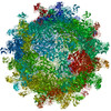 6snwMC 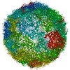 6smgC 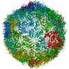 6snbC M: atomic model generated by this map C: citing same article ( |
|---|---|
| Similar structure data |
-Links
| EMDB pages | EMDB (EBI/PDBe) / EMDataResource |
|---|---|
| Related items in Molecule of the Month |


-Map
| File | Download / File: emd_10263.map.gz / Format: CCP4 / Size: 244.1 MB / Type: IMAGE STORED AS FLOATING POINT NUMBER (4 BYTES) | ||||||||||||||||||||||||||||||||||||||||||||||||||||||||||||
|---|---|---|---|---|---|---|---|---|---|---|---|---|---|---|---|---|---|---|---|---|---|---|---|---|---|---|---|---|---|---|---|---|---|---|---|---|---|---|---|---|---|---|---|---|---|---|---|---|---|---|---|---|---|---|---|---|---|---|---|---|---|
| Voxel size | X=Y=Z: 1.35 Å | ||||||||||||||||||||||||||||||||||||||||||||||||||||||||||||
| Density |
| ||||||||||||||||||||||||||||||||||||||||||||||||||||||||||||
| Symmetry | Space group: 1 | ||||||||||||||||||||||||||||||||||||||||||||||||||||||||||||
| Details | EMDB XML:
CCP4 map header:
| ||||||||||||||||||||||||||||||||||||||||||||||||||||||||||||
-Supplemental data
- Sample components
+Entire : Coxsackievirus A10
+Supramolecule #1: Coxsackievirus A10
+Supramolecule #2: Coxsackievirus A10
+Supramolecule #3: KREMEN1
+Macromolecule #1: Capsid protein VP1
+Macromolecule #2: Coxsackievirus VP2
+Macromolecule #3: Capsid protein VP3
+Macromolecule #4: Coxsackievirus VP4
+Macromolecule #5: Kremen protein 1
+Macromolecule #6: SPHINGOSINE

+Macromolecule #7: 2-acetamido-2-deoxy-beta-D-glucopyranose

-Experimental details
-Structure determination
| Method | cryo EM |
|---|---|
 Processing Processing | single particle reconstruction |
| Aggregation state | particle |
-Sample preparation
| Buffer | pH: 8 |
|---|---|
| Vitrification | Cryogen name: ETHANE |
- Electron microscopy
| Microscope | FEI POLARA 300 |
|---|---|
| Electron beam | Acceleration voltage: 300 kV / Electron source: FIELD EMISSION GUN |
| Electron optics | Illumination mode: FLOOD BEAM / Imaging mode: BRIGHT FIELDBright-field microscopy |
| Image recording | Film or detector model: GATAN K2 SUMMIT (4k x 4k) / Number grids imaged: 1 / Average electron dose: 35.0 e/Å2 |
| Experimental equipment |  Model: Tecnai Polara / Image courtesy: FEI Company |
-Image processing
| Startup model | Type of model: PDB ENTRY PDB model - PDB ID: |
|---|---|
| Initial angle assignment | Type: MAXIMUM LIKELIHOOD |
| Final angle assignment | Type: MAXIMUM LIKELIHOOD |
| Final reconstruction | Resolution.type: BY AUTHOR / Resolution: 3.9 Å / Resolution method: FSC 0.143 CUT-OFF / Number images used: 1597 |
-Atomic model buiding 1
| Refinement | Space: REAL / Protocol: RIGID BODY FIT |
|---|---|
| Output model | PDB-6snw: |
