 Movie
Movie Controller


+ Open data

- Basic information
| Entry | Database: EMDB / ID: EMD-0790 | |||||||||
|---|---|---|---|---|---|---|---|---|---|---|
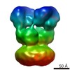
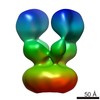
| Title | GluK3 receptor complex with kainate | |||||||||
 Map data Map data | Refined map | |||||||||
 Sample Sample |
| |||||||||
| Function / homology |  Function and homology information Function and homology informationPresynaptic function of Kainate receptors / regulation of presynaptic membrane potential / adenylate cyclase inhibiting G protein-coupled glutamate receptor activity / G protein-coupled glutamate receptor signaling pathway / kainate selective glutamate receptor complex / Activation of Ca-permeable Kainate Receptor / negative regulation of synaptic transmission, glutamatergic / glutamate receptor signaling pathway /  glutamate receptor activity / kainate selective glutamate receptor activity ...Presynaptic function of Kainate receptors / regulation of presynaptic membrane potential / adenylate cyclase inhibiting G protein-coupled glutamate receptor activity / G protein-coupled glutamate receptor signaling pathway / kainate selective glutamate receptor complex / Activation of Ca-permeable Kainate Receptor / negative regulation of synaptic transmission, glutamatergic / glutamate receptor signaling pathway / glutamate receptor activity / kainate selective glutamate receptor activity / glutamate-gated receptor activity / ligand-gated monoatomic ion channel activity involved in regulation of presynaptic membrane potential / dendrite cytoplasm / regulation of membrane potential / monoatomic ion transmembrane transport / transmitter-gated monoatomic ion channel activity involved in regulation of postsynaptic membrane potential / synaptic transmission, glutamatergic / postsynaptic density membrane / modulation of chemical synaptic transmission / terminal bouton / presynaptic membrane / perikaryon / chemical synaptic transmission / postsynaptic membrane / axon / dendrite / glutamatergic synapse / plasma membrane glutamate receptor activity / kainate selective glutamate receptor activity ...Presynaptic function of Kainate receptors / regulation of presynaptic membrane potential / adenylate cyclase inhibiting G protein-coupled glutamate receptor activity / G protein-coupled glutamate receptor signaling pathway / kainate selective glutamate receptor complex / Activation of Ca-permeable Kainate Receptor / negative regulation of synaptic transmission, glutamatergic / glutamate receptor signaling pathway / glutamate receptor activity / kainate selective glutamate receptor activity / glutamate-gated receptor activity / ligand-gated monoatomic ion channel activity involved in regulation of presynaptic membrane potential / dendrite cytoplasm / regulation of membrane potential / monoatomic ion transmembrane transport / transmitter-gated monoatomic ion channel activity involved in regulation of postsynaptic membrane potential / synaptic transmission, glutamatergic / postsynaptic density membrane / modulation of chemical synaptic transmission / terminal bouton / presynaptic membrane / perikaryon / chemical synaptic transmission / postsynaptic membrane / axon / dendrite / glutamatergic synapse / plasma membraneSimilarity search - Function | |||||||||
| Biological species |  Rattus norvegicus (Norway rat) Rattus norvegicus (Norway rat) | |||||||||
| Method | single particle reconstruction / cryo EM / Resolution: 9.6 Å | |||||||||
 Authors Authors | Kumar J / Kumari J / Burada AP | |||||||||
| Funding support |  India, 1 items India, 1 items
| |||||||||
 Citation Citation | Journal: Int J Biol Macromol / Year: 2020 Title: Structural dynamics of the GluK3-kainate receptor neurotransmitter binding domains revealed by cryo-EM. Authors: Jyoti Kumari / Ameya D Bendre / Sumedha Bhosale / Rajesh Vinnakota / Ananth P Burada / Giancarlo Tria / Raimond B G Ravelli / Peter J Peters / Manali Joshi / Janesh Kumar /  Abstract: Kainate receptors belong to the ionotropic glutamate receptor family and play critical roles in the regulation of synaptic networks. The kainate receptor subunit GluK3 has unique functional ...Kainate receptors belong to the ionotropic glutamate receptor family and play critical roles in the regulation of synaptic networks. The kainate receptor subunit GluK3 has unique functional properties and contributes to presynaptic facilitation at the hippocampal mossy fiber synapses along with roles at the post-synapses. To gain structural insights into the unique functional properties and dynamics of GluK3 receptor, we imaged them via electron microscopy in the apo-state and in complex with either agonist kainate or antagonist UBP301. Our analysis of all the GluK3 full-length structures not only provides insights into the receptor transitions between desensitized and closed states but also reveals a "non-classical" conformation of neurotransmitter binding domain in the closed-state distinct from that observed in AMPA and other kainate receptor structures. We show by molecular dynamics simulations that Asp759 influences the stability of the LBD dimers and hence could be responsible for the observed conformational variability and dynamics of the GluK3 via electron microscopy. Lower dimer stability could explain faster desensitization and low agonist sensitivity of GluK3. In overview, our work helps to associate biochemistry and physiology of GluK3 receptors with their structural biology and offers structural insights into the unique functional properties of these atypical receptors. | |||||||||
| History |
|
- Structure visualization
| Movie |
Movie viewer |
|---|---|
| Structure viewer | EM map: SurfViewMolmilJmol/JSmol |
| Supplemental images |

- Downloads & links
-EMDB archive
| Map data | emd_0790.map.gz | 325.2 MB | EMDB map data format | |
|---|---|---|---|---|
| Header (meta data) | emd-0790-v30.xmlemd-0790.xml | 16.7 KB 16.7 KB | Display Display | EMDB header |
| Images | 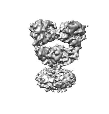 emd_0790.png emd_0790.png | 35.9 KB | ||
| Others | emd_0790_half_map_1.map.gzemd_0790_half_map_2.map.gz | 323 MB 323 MB | ||
| Archive directory |  http://ftp.pdbj.org/pub/emdb/structures/EMD-0790ftp://ftp.pdbj.org/pub/emdb/structures/EMD-0790 http://ftp.pdbj.org/pub/emdb/structures/EMD-0790ftp://ftp.pdbj.org/pub/emdb/structures/EMD-0790 | HTTPS FTP |
-Related structure data
| Related structure data |  6kzmMC  0839C  6l6fC M: atomic model generated by this map C: citing same article ( |
|---|---|
| Similar structure data |


-Links
| EMDB pages | EMDB (EBI/PDBe) / EMDataResource |
|---|---|
| Related items in Molecule of the Month |


-Map
| File | Download / File: emd_0790.map.gz / Format: CCP4 / Size: 347.6 MB / Type: IMAGE STORED AS FLOATING POINT NUMBER (4 BYTES) | ||||||||||||||||||||||||||||||||||||||||||||||||||||||||||||||||||||
|---|---|---|---|---|---|---|---|---|---|---|---|---|---|---|---|---|---|---|---|---|---|---|---|---|---|---|---|---|---|---|---|---|---|---|---|---|---|---|---|---|---|---|---|---|---|---|---|---|---|---|---|---|---|---|---|---|---|---|---|---|---|---|---|---|---|---|---|---|---|
| Annotation | Refined map | ||||||||||||||||||||||||||||||||||||||||||||||||||||||||||||||||||||
| Voxel size | X=Y=Z: 0.935 Å | ||||||||||||||||||||||||||||||||||||||||||||||||||||||||||||||||||||
| Density |
| ||||||||||||||||||||||||||||||||||||||||||||||||||||||||||||||||||||
| Symmetry | Space group: 1 | ||||||||||||||||||||||||||||||||||||||||||||||||||||||||||||||||||||
| Details | EMDB XML:
CCP4 map header:
| ||||||||||||||||||||||||||||||||||||||||||||||||||||||||||||||||||||
-Supplemental data
-Half map: HALF MAP A
| File | emd_0790_half_map_1.map | ||||||||||||
|---|---|---|---|---|---|---|---|---|---|---|---|---|---|
| Annotation | HALF MAP A | ||||||||||||
| Projections & Slices |
| ||||||||||||
| Density Histograms |
 Z
Z Y
Y X
X


-Half map: Half map B
| File | emd_0790_half_map_2.map | ||||||||||||
|---|---|---|---|---|---|---|---|---|---|---|---|---|---|
| Annotation | Half map B | ||||||||||||
| Projections & Slices |
| ||||||||||||
| Density Histograms |


- Sample components
-Entire : GluK3 complex with agonist Kainate
| Entire | Name: GluK3 complex with agonist Kainate |
|---|---|
| Components |
|
-Supramolecule #1: GluK3 complex with agonist Kainate
| Supramolecule | Name: GluK3 complex with agonist Kainate / type: complex / ID: 1 / Parent: 0 / Macromolecule list: all |
|---|---|
| Source (natural) | Organism: Rattus norvegicus (Norway rat) |
| Recombinant expression | Organism:  Homo sapiens (human) / Recombinant cell: HEK293 GnTi- / Recombinant plasmid: pEGBacMam Homo sapiens (human) / Recombinant cell: HEK293 GnTi- / Recombinant plasmid: pEGBacMam |
-Macromolecule #1: Glutamate receptor ionotropic, kainate 3
| Macromolecule | Name: Glutamate receptor ionotropic, kainate 3 / type: protein_or_peptide / ID: 1 / Number of copies: 4 / Enantiomer: LEVO |
|---|---|
| Source (natural) | Organism: Rattus norvegicus (Norway rat) |
| Molecular weight | Theoretical: 93.699562 KDa |
| Recombinant expression | Organism: Homo sapiens (human) |
| Sequence | String: HVIRIGGIFE YADGPNAQVM NAEEHAFRFS ANIINRNRTL LPNTTLTYDI QRIHFHDSFE ATKKACDQLA LGVVAIFGPS QGSTTNAVQ SICNALEVPH IQLRWKHHPL DNKDTFYVNL YPDYASLSHA ILDLVQSLKW RSATVVYDDS TGLIRLQELI M APSRYNIR ...String: HVIRIGGIFE YADGPNAQVM NAEEHAFRFS ANIINRNRTL LPNTTLTYDI QRIHFHDSFE ATKKACDQLA LGVVAIFGPS QGSTTNAVQ SICNALEVPH IQLRWKHHPL DNKDTFYVNL YPDYASLSHA ILDLVQSLKW RSATVVYDDS TGLIRLQELI M APSRYNIR LKIRQLPIDS DDSRPLLKEM KRGREFRIIF DCSHTMAAQI LKQAMAMGMM TEYYHFIFTT LDLYALDLEP YR YSGVNLT GFRILNVDNP HVSAIVEKWS MERLQAAPRA ESGLLDGVMM TDAALLYDAV HIVSVTYQRA PQMTVNSLQC HRH KAWRFG GRFMNFIKEA QWEGLTGRIV FNKTSGLRTD FDLDIISLKE DGLEKVGVWS PADGLNITEV AKGRGPNVTD SLTN RSLIV TTLLEEPFVM FRKSDRTLYG NDRFEGYCID LLKELAHILG FSYEIRLVED GKYGAQDDKG QWNGMVKELI DHKAD LAVA PLTITHVREK AIDFSKPFMT LGVSILYRKP NGTNPSVFSF LNPLSPDIWM YVLLAYLGVS VVLFVIARFS PYEWYD AHP CNPGSEVVEN NFTLLNSFWF GMGSLMQQGS ELMPKALSTR IIGGIWWFFT LIIISSYTAN LAAFLTVERM ESPIDSA DD LAKQTKIEYG AVKDGATMTF FKKSKISTFE KMWAFMSSKP SALVKNNEEG IQRTLTADYA LLMESTTIEY ITQRNCNL T QIGGLIDSKG YGIGTPMGSP YRDKITIAIL QLQEEDKLHI MKEKWWRGSG CPEEENKEAS ALGIQKIGGI FIVLAAGLV LSVLVAVGEF IYKLRKTAER EQRSGLVPR |
-Experimental details
-Structure determination
| Method | cryo EM |
|---|---|
 Processing Processing | single particle reconstruction |
| Aggregation state | particle |
-Sample preparation
| Concentration | 1.7 mg/mL |
|---|---|
| Buffer | pH: 8 / Component - Concentration: 1.7 1.7 / Component - Formula: NaClSodium chloride / Component - Name: Sodium Chloride / Details: 20 mM Tris pH 8.0, 150 mM NaCl |
| Grid | Model: Quantifoil R1.2/1.3 / Material: GOLD / Mesh: 300 / Support film - Material: CARBON / Support film - topology: HOLEY / Pretreatment - Type: GLOW DISCHARGE |
| Vitrification | Cryogen name: ETHANE / Details: Multiple application, blotted for 3 seconds. |
- Electron microscopy
| Microscope | FEI TECNAI ARCTICA |
|---|---|
| Electron beam | Acceleration voltage: 200 kV / Electron source: FIELD EMISSION GUN |
| Electron optics | Illumination mode: SPOT SCAN / Imaging mode: BRIGHT FIELDBright-field microscopy |
| Image recording | Film or detector model: FEI FALCON III (4k x 4k) / Number real images: 2845 / Average electron dose: 16.73 e/Å2 |
| Experimental equipment |  Model: Talos Arctica / Image courtesy: FEI Company |
-Image processing
| Particle selection | Number selected: 46106 |
|---|---|
| CTF correction | Software - Name: Gctf (ver. v1.06) |
| Startup model | Type of model: PDB ENTRY PDB model - PDB ID: |
| Initial angle assignment | Type: NOT APPLICABLE |
| Final angle assignment | Type: NOT APPLICABLE |
| Final reconstruction | Applied symmetry - Point group: C1 (asymmetric) / Resolution.type: BY AUTHOR / Resolution: 9.6 Å / Resolution method: FSC 0.143 CUT-OFF / Software - Name: cryoSPARC (ver. v1) / Details: Gold-standard refinement of independent half maps / Number images used: 46106 |

