 Movie
Movie Controller

[English] 日本語
 Yorodumi
- EMDB-0529: Purified ribosome - A complete data processing workflow for CryoE... -
+ Open data

- Basic information
| Entry | Database: EMDB / ID: EMD-0529 | |||||||||
|---|---|---|---|---|---|---|---|---|---|---|
| Title | Purified ribosome - A complete data processing workflow for CryoET and subtomogram averaging | |||||||||
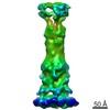
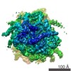
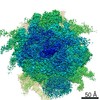
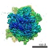
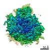
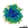
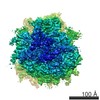
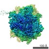
 Map data Map data | Averaged structure of purified ribosome from EMPIAR-10064 dataset, after subtilt refinement in EMAN2. | |||||||||
 Sample Sample |
| |||||||||
| Biological species |  | |||||||||
| Method | electron tomography / cryo EM / Resolution: 8.4 Å | |||||||||
 Authors Authors | Chen M / Bell JM / Ludtke SJ | |||||||||
| Funding support |  United States, 1 items United States, 1 items
| |||||||||
 Citation Citation | Journal: Nat Methods / Year: 2019 Title: A complete data processing workflow for cryo-ET and subtomogram averaging. Authors: Muyuan Chen / James M Bell / Xiaodong Shi / Stella Y Sun / Zhao Wang / Steven J Ludtke /  Abstract: Electron cryotomography is currently the only method capable of visualizing cells in three dimensions at nanometer resolutions. While modern instruments produce massive amounts of tomography data ...Electron cryotomography is currently the only method capable of visualizing cells in three dimensions at nanometer resolutions. While modern instruments produce massive amounts of tomography data containing extremely rich structural information, data processing is very labor intensive and the results are often limited by the skills of the personnel rather than the data. We present an integrated workflow that covers the entire tomography data processing pipeline, from automated tilt series alignment to subnanometer resolution subtomogram averaging. Resolution enhancement is made possible through the use of per-particle per-tilt contrast transfer function correction and alignment. The workflow greatly reduces human bias, increases throughput and more closely approaches data-limited resolution for subtomogram averaging in both purified macromolecules and cells. | |||||||||
| History |
|
- Structure visualization
| Movie |
 Movie viewer Movie viewer |
|---|---|
| Structure viewer | EM map: SurfViewMolmilJmol/JSmol |
| Supplemental images |

- Downloads & links
-EMDB archive
| Map data | emd_0529.map.gz | 4.4 MB | EMDB map data format | |
|---|---|---|---|---|
| Header (meta data) | emd-0529-v30.xmlemd-0529.xml | 10.6 KB 10.6 KB | Display Display | EMDB header |
| FSC (resolution estimation) | emd_0529_fsc.xml | 6.6 KB | Display | FSC data file |
| Images |  emd_0529.png emd_0529.png | 85.9 KB | ||
| Others | emd_0529_additional.map.gz | 21 MB | ||
| Archive directory |  http://ftp.pdbj.org/pub/emdb/structures/EMD-0529ftp://ftp.pdbj.org/pub/emdb/structures/EMD-0529 http://ftp.pdbj.org/pub/emdb/structures/EMD-0529ftp://ftp.pdbj.org/pub/emdb/structures/EMD-0529 | HTTPS FTP |
-Validation report
| Summary document | emd_0529_validation.pdf.gz | 79 KB | Display | EMDB validaton report |
|---|---|---|---|---|
| Full document | emd_0529_full_validation.pdf.gz | 78.1 KB | Display | |
| Data in XML | emd_0529_validation.xml.gz | 493 B | Display | |
| Arichive directory | https://ftp.pdbj.org/pub/emdb/validation_reports/EMD-0529ftp://ftp.pdbj.org/pub/emdb/validation_reports/EMD-0529 | HTTPS FTP |
-Related structure data

-Links
| EMDB pages | EMDB (EBI/PDBe) / EMDataResource |
|---|---|
| Related items in Molecule of the Month |

-Map
| File | Download / File: emd_0529.map.gz / Format: CCP4 / Size: 22.2 MB / Type: IMAGE STORED AS FLOATING POINT NUMBER (4 BYTES) | ||||||||||||||||||||||||||||||||||||||||||||||||||||||||||||
|---|---|---|---|---|---|---|---|---|---|---|---|---|---|---|---|---|---|---|---|---|---|---|---|---|---|---|---|---|---|---|---|---|---|---|---|---|---|---|---|---|---|---|---|---|---|---|---|---|---|---|---|---|---|---|---|---|---|---|---|---|---|
| Annotation | Averaged structure of purified ribosome from EMPIAR-10064 dataset, after subtilt refinement in EMAN2. | ||||||||||||||||||||||||||||||||||||||||||||||||||||||||||||
| Voxel size | X=Y=Z: 2.62 Å | ||||||||||||||||||||||||||||||||||||||||||||||||||||||||||||
| Density |
| ||||||||||||||||||||||||||||||||||||||||||||||||||||||||||||
| Symmetry | Space group: 1 | ||||||||||||||||||||||||||||||||||||||||||||||||||||||||||||
| Details | EMDB XML:
CCP4 map header:
| ||||||||||||||||||||||||||||||||||||||||||||||||||||||||||||
-Supplemental data
-Additional map: Averaged structure after subtomogram refinement but before subtilt...
| File | emd_0529_additional.map | ||||||||||||
|---|---|---|---|---|---|---|---|---|---|---|---|---|---|
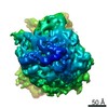
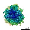
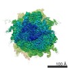
| Annotation | Averaged structure after subtomogram refinement but before subtilt refinement | ||||||||||||
| Projections & Slices |
| ||||||||||||
| Density Histograms |
 Z
Z Y
Y X
X


- Sample components
-Entire : Ribosome from EMPIAR-10064, MixedCTEM dataset
| Entire | Name: Ribosome from EMPIAR-10064, MixedCTEM dataset |
|---|---|
| Components |
|
-Supramolecule #1: Ribosome from EMPIAR-10064, MixedCTEM dataset
| Supramolecule | Name: Ribosome from EMPIAR-10064, MixedCTEM dataset / type: complex / ID: 1 / Parent: 0 |
|---|---|
| Source (natural) | Organism: |
-Experimental details
-Structure determination
| Method | cryo EM |
|---|---|
 Processing Processing | electron tomography |
| Aggregation state | particle |
-Sample preparation
| Buffer | pH: 7 |
|---|---|
| Grid | Details: unspecified |
| Vitrification | Cryogen name: ETHANE |
| Sectioning | Other: NO SECTIONING |
- Electron microscopy
| Microscope | FEI TITAN KRIOS |
|---|---|
| Image recording | Film or detector model: GATAN K2 SUMMIT (4k x 4k) / Average electron dose: 30.0 e/Å2 |
| Electron beam | Acceleration voltage: 300 kV / Electron source:  FIELD EMISSION GUN FIELD EMISSION GUN |
| Electron optics | Illumination mode: FLOOD BEAM / Imaging mode: BRIGHT FIELD |
| Experimental equipment |  Model: Titan Krios / Image courtesy: FEI Company |
-Image processing
| Final reconstruction | Algorithm: FOURIER SPACE / Resolution.type: BY AUTHOR / Resolution: 8.4 Å / Resolution method: FSC 0.143 CUT-OFF / Software - Name: EMAN2 (ver. 2.3) / Number images used: 3239 |
|---|---|
| CTF correction | Software - Name: EMAN2 (ver. 2.3) |
| FSC plot (resolution estimation) |  |
