 Movie
Movie Controller


+ Open data

- Basic information
| Entry | Database: EMDB / ID: EMD-0069 | |||||||||||||||
|---|---|---|---|---|---|---|---|---|---|---|---|---|---|---|---|---|
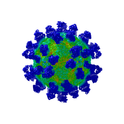
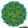
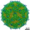
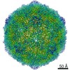
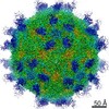
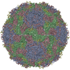
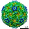
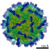
| Title | High-resolution Cryo-EM of Fab-labeled human parechovirus 3 | |||||||||||||||
 Map data Map data | Human parechovirus type 3 in complex with neutralizing human monoclonal antibody Fabs Post-processed map, B factor -70 | |||||||||||||||
 Sample Sample |
| |||||||||||||||
| Function / homology |  Function and homology information Function and homology informationRNA-protein covalent cross-linking / : / T=pseudo3 icosahedral viral capsid / host cell cytoplasmic vesicle membrane / cytoplasmic vesicle membrane /  viral capsid / : / protein complex oligomerization / monoatomic ion channel activity / RNA helicase activity ...RNA-protein covalent cross-linking / : / T=pseudo3 icosahedral viral capsid / host cell cytoplasmic vesicle membrane / cytoplasmic vesicle membrane / viral capsid / : / protein complex oligomerization / monoatomic ion channel activity / RNA helicase activity / symbiont entry into host cell / viral RNA genome replication / cysteine-type endopeptidase activity / RNA-dependent RNA polymerase activity / DNA-templated transcription / virion attachment to host cell / structural molecule activity / RNA binding / ATP binding viral capsid / : / protein complex oligomerization / monoatomic ion channel activity / RNA helicase activity ...RNA-protein covalent cross-linking / : / T=pseudo3 icosahedral viral capsid / host cell cytoplasmic vesicle membrane / cytoplasmic vesicle membrane / viral capsid / : / protein complex oligomerization / monoatomic ion channel activity / RNA helicase activity / symbiont entry into host cell / viral RNA genome replication / cysteine-type endopeptidase activity / RNA-dependent RNA polymerase activity / DNA-templated transcription / virion attachment to host cell / structural molecule activity / RNA binding / ATP bindingSimilarity search - Function | |||||||||||||||
| Biological species |  Homo sapiens (human) / Homo sapiens (human) /  Human parechovirus 3 Human parechovirus 3 | |||||||||||||||
| Method | single particle reconstruction / cryo EM / Resolution: 2.8 Å | |||||||||||||||
 Authors Authors | Domanska A / Flatt JW / Jukonen JJJ / Geraets JA / Butcher SJ | |||||||||||||||
| Funding support |  Finland, 4 items Finland, 4 items
| |||||||||||||||
 Citation Citation | Journal: J Virol / Year: 2019 Title: A 2.8-Angstrom-Resolution Cryo-Electron Microscopy Structure of Human Parechovirus 3 in Complex with Fab from a Neutralizing Antibody. Authors: Aušra Domanska / Justin W Flatt / Joonas J J Jukonen / James A Geraets / Sarah J Butcher / Abstract: Human parechovirus 3 (HPeV3) infection is associated with sepsis characterized by significant immune activation and subsequent tissue damage in neonates. Strategies to limit infection have been ...Human parechovirus 3 (HPeV3) infection is associated with sepsis characterized by significant immune activation and subsequent tissue damage in neonates. Strategies to limit infection have been unsuccessful due to inadequate molecular diagnostic tools for early detection and the lack of a vaccine or specific antiviral therapy. Toward the latter, we present a 2.8-Å-resolution structure of HPeV3 in complex with fragments from a neutralizing human monoclonal antibody, AT12-015, using cryo-electron microscopy (cryo-EM) and image reconstruction. Modeling revealed that the epitope extends across neighboring asymmetric units with contributions from capsid proteins VP0, VP1, and VP3. Antibody decoration was found to block binding of HPeV3 to cultured cells. Additionally, at high resolution, it was possible to model a stretch of RNA inside the virion and, from this, identify the key features that drive and stabilize protein-RNA association during assembly. Human parechovirus 3 (HPeV3) is receiving increasing attention as a prevalent cause of sepsis-like symptoms in neonates, for which, despite the severity of disease, there are no effective treatments available. Structural and molecular insights into virus neutralization are urgently needed, especially as clinical cases are on the rise. Toward this goal, we present the first structure of HPeV3 in complex with fragments from a neutralizing monoclonal antibody. At high resolution, it was possible to precisely define the epitope that, when targeted, prevents virions from binding to cells. Such an atomic-level description is useful for understanding host-pathogen interactions and viral pathogenesis mechanisms and for finding potential cures for infection and disease. | |||||||||||||||
| History |
|
- Structure visualization
| Movie |
Movie viewer |
|---|---|
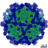
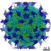
| Structure viewer | EM map: SurfViewMolmilJmol/JSmol |
| Supplemental images |
- Downloads & links
-EMDB archive
| Map data | emd_0069.map.gz | 391.7 MB | EMDB map data format | |
|---|---|---|---|---|
| Header (meta data) | emd-0069-v30.xmlemd-0069.xml | 21.7 KB 21.7 KB | Display Display | EMDB header |
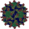
| Images |  emd_0069.png emd_0069.png | 111.2 KB | ||
| Archive directory |  http://ftp.pdbj.org/pub/emdb/structures/EMD-0069ftp://ftp.pdbj.org/pub/emdb/structures/EMD-0069 http://ftp.pdbj.org/pub/emdb/structures/EMD-0069ftp://ftp.pdbj.org/pub/emdb/structures/EMD-0069 | HTTPS FTP |
-Related structure data
| Related structure data |  6gv4MC M: atomic model generated by this map C: citing same article ( |
|---|---|
| Similar structure data | |
| EM raw data | EMPIAR-10983 (Title: High-resolution Cryo-EM of Fab-labeled human parechovirus 3 Data size: 3.3 TB Data #1: Unaligned multi-frame micrographs of HPeV3-fab complex [micrographs - multiframe]) |
-Links
| EMDB pages | EMDB (EBI/PDBe) / EMDataResource |
|---|---|
| Related items in Molecule of the Month |


-Map
| File | Download / File: emd_0069.map.gz / Format: CCP4 / Size: 421.9 MB / Type: IMAGE STORED AS FLOATING POINT NUMBER (4 BYTES) | ||||||||||||||||||||||||||||||||||||||||||||||||||||||||||||
|---|---|---|---|---|---|---|---|---|---|---|---|---|---|---|---|---|---|---|---|---|---|---|---|---|---|---|---|---|---|---|---|---|---|---|---|---|---|---|---|---|---|---|---|---|---|---|---|---|---|---|---|---|---|---|---|---|---|---|---|---|---|
| Annotation | Human parechovirus type 3 in complex with neutralizing human monoclonal antibody Fabs Post-processed map, B factor -70 | ||||||||||||||||||||||||||||||||||||||||||||||||||||||||||||
| Voxel size | X=Y=Z: 1.06 Å | ||||||||||||||||||||||||||||||||||||||||||||||||||||||||||||
| Density |
| ||||||||||||||||||||||||||||||||||||||||||||||||||||||||||||
| Symmetry | Space group: 1 | ||||||||||||||||||||||||||||||||||||||||||||||||||||||||||||
| Details | EMDB XML:
CCP4 map header:
| ||||||||||||||||||||||||||||||||||||||||||||||||||||||||||||
-Supplemental data
- Sample components
-Entire : Human parechovirus type 3 in complex with fabs from AT12-015
| Entire | Name: Human parechovirus type 3 in complex with fabs from AT12-015 |
|---|---|
| Components |
|
-Supramolecule #1: Human parechovirus type 3 in complex with fabs from AT12-015
| Supramolecule | Name: Human parechovirus type 3 in complex with fabs from AT12-015 type: complex / ID: 1 / Parent: 0 / Macromolecule list: all |
|---|---|
| Molecular weight | Theoretical: 7.7 MDa |
-Supramolecule #3: fabs from AT12-015
| Supramolecule | Name: fabs from AT12-015 / type: complex / ID: 3 / Parent: 1 / Macromolecule list: #5-#6 |
|---|---|
| Source (natural) | Organism: Homo sapiens (human) |
| Recombinant expression | Organism: Homo sapiens (human) |
-Supramolecule #2: Human parechovirus 3
| Supramolecule | Name: Human parechovirus 3 / type: virus / ID: 2 / Parent: 1 / Macromolecule list: #1-#4 / NCBI-ID: 195055 / Sci species name: Human parechovirus 3 / Sci species strain: A308/99 / Virus type: VIRION / Virus isolate: STRAIN / Virus enveloped: No / Virus empty: No |
|---|
-Macromolecule #1: RNA (5'-R(*UP*GP*GP*UP*AP*UP*UP*U)-3')
| Macromolecule | Name: RNA (5'-R(*UP*GP*GP*UP*AP*UP*UP*U)-3') / type: rna / ID: 1 / Details: RNA / Number of copies: 1 |
|---|---|
| Source (natural) | Organism: Human parechovirus 3 / Organ: colon adenocarcinoma |
| Molecular weight | Theoretical: 2.505489 KDa |
| Sequence | String: UGGUAUUU |
-Macromolecule #2: VP0
| Macromolecule | Name: VP0 / type: protein_or_peptide / ID: 2 / Number of copies: 1 / Enantiomer: LEVO |
|---|---|
| Source (natural) | Organism: Human parechovirus 3 / Organ: colon adenocarcinoma |
| Molecular weight | Theoretical: 31.770135 KDa |
| Sequence | String: MESIKDLVNV ATGAMDTLSL SNVETEVNNI ISGNEVGGEI ITKVADDASN LLGPNSFATT AQPENKDVVQ ATTTVNTTNL TQHPSAPTI PFTPDFKNVD NFHSMAYDIT TGDKNPSKLI RLDTASWQTS YSRQYEITTV ELPKSFWDDT RKPAYGQAKY F AAVRCGFH ...String: MESIKDLVNV ATGAMDTLSL SNVETEVNNI ISGNEVGGEI ITKVADDASN LLGPNSFATT AQPENKDVVQ ATTTVNTTNL TQHPSAPTI PFTPDFKNVD NFHSMAYDIT TGDKNPSKLI RLDTASWQTS YSRQYEITTV ELPKSFWDDT RKPAYGQAKY F AAVRCGFH FQVQVNVNQG TAGSALVVYE PKPVIDSRQY LEFGSLTNLP HVLMNLAETT QADLCIPYVA DTNYVKTDSS DL GQLRVYV WTPLSVPTGA SNEVDVTVMG SLLQLDFQNP RPYGEDVEIY DN |
-Macromolecule #3: VP1
| Macromolecule | Name: VP1 / type: protein_or_peptide / ID: 3 / Number of copies: 1 / Enantiomer: LEVO |
|---|---|
| Source (natural) | Organism: Human parechovirus 3 / Organ: colon adenocarcinoma |
| Molecular weight | Theoretical: 25.913127 KDa |
| Sequence | String: NSWGSQMDLT DPLCIEDNME NCKQSISPNE LGLTSAQDDG PLGNEKPNYF LNFRTMNVDI FTVSHTKVDN IFGRAWYVTS HDFNNGDTW RQKLTFPKEG HGMLSQFFAY FTGEINIHIL YMAKQGFLRV AHTYDTEDNR KTFLSSNGVI TIPAGEQMTL S VPFYSNKP ...String: NSWGSQMDLT DPLCIEDNME NCKQSISPNE LGLTSAQDDG PLGNEKPNYF LNFRTMNVDI FTVSHTKVDN IFGRAWYVTS HDFNNGDTW RQKLTFPKEG HGMLSQFFAY FTGEINIHIL YMAKQGFLRV AHTYDTEDNR KTFLSSNGVI TIPAGEQMTL S VPFYSNKP LRTVRHDSAL GFLMCRPSMH GTTRTTVEVY VSLRCPNFFF PVPAPKPTGS RATALSDESP Y |
-Macromolecule #4: VP3
| Macromolecule | Name: VP3 / type: protein_or_peptide / ID: 4 / Details: polypeptide chain / Number of copies: 1 / Enantiomer: LEVO |
|---|---|
| Source (natural) | Organism: Human parechovirus 3 / Organ: colon adenocarcinoma |
| Molecular weight | Theoretical: 28.757551 KDa |
| Sequence | String: GPNKANVSKF NKRKFLTAST KYKWTRTKVD IAEGPGTMNM ANVLSTTGAQ SVALVGERAF YDPRTAGSKS RFDDMIKIAQ LFSVMSDNT TPSSSSGIDK YGYFDWAATV APQNMVHRNV VTLDQFPNLN LFMNTYSYFR GSLIIRLSIY ASTFNRGRLR M GFFPNCTH ...String: GPNKANVSKF NKRKFLTAST KYKWTRTKVD IAEGPGTMNM ANVLSTTGAQ SVALVGERAF YDPRTAGSKS RFDDMIKIAQ LFSVMSDNT TPSSSSGIDK YGYFDWAATV APQNMVHRNV VTLDQFPNLN LFMNTYSYFR GSLIIRLSIY ASTFNRGRLR M GFFPNCTH DTQLELDNAI YTICDIGSDN SFELTIPYSF STWMRKTHGH QLGLFQVEVL NRLTYNSSSP NKVHCIVQGR LG DDAKFFC PTGSLVSFQ |
-Macromolecule #5: AT12-015 antibody variable heavy
| Macromolecule | Name: AT12-015 antibody variable heavy / type: protein_or_peptide / ID: 5 / Details: polypeptide chain / Number of copies: 1 / Enantiomer: LEVO |
|---|---|
| Source (natural) | Organism: Homo sapiens (human) |
| Molecular weight | Theoretical: 13.128613 KDa |
| Recombinant expression | Organism: Homo sapiens (human) |
| Sequence | String: EVQLLESGGG LVQPGGSLRL SCAASGFTFS SYAMSWVRQA PGKGLEWVSA ISGGGDSRYY ADSVKGRFTI SRDNSKNTLY LQMNSLGAE DTALYYCAKR LGRVAEYYFD YWGQGTLVTV SP |
-Macromolecule #6: AT12-015 antibody variable light
| Macromolecule | Name: AT12-015 antibody variable light / type: protein_or_peptide / ID: 6 / Details: polypeptide chain / Number of copies: 1 / Enantiomer: LEVO |
|---|---|
| Source (natural) | Organism: Homo sapiens (human) |
| Molecular weight | Theoretical: 12.339752 KDa |
| Recombinant expression | Organism: Homo sapiens (human) |
| Sequence | String: DIQMTQSPST LSASVGDRVT ITCRTSQSIS NWLAWYQQKP GKAPKLLIYQ ASTLENGVPS RFTGSGSGTE FSLTISSLQP DDFATYYCQ QYNNYMALTF GGGTKVEIKR TVAA |
-Experimental details
-Structure determination
| Method | cryo EM |
|---|---|
 Processing Processing | single particle reconstruction |
| Aggregation state | particle |
-Sample preparation
| Concentration | 0.1 mg/mL | ||||||||||||
|---|---|---|---|---|---|---|---|---|---|---|---|---|---|
| Buffer | pH: 7.5 Component:
| ||||||||||||
| Grid | Material: COPPER / Mesh: 400 / Support film - Material: CARBON / Support film - topology: LACEY / Pretreatment - Type: GLOW DISCHARGE Details: ultrathin carbon-coated lacey 400-mesh copper grids (Ted Pella product #01824) | ||||||||||||
| Vitrification | Cryogen name: ETHANE / Chamber temperature: 295 K / Instrument: HOMEMADE PLUNGER Details: We could not control humidity during plunging. It was ambient humidity. Blot for 1 s before plunging.. |
- Electron microscopy
| Microscope | FEI TITAN KRIOS |
|---|---|
| Electron beam | Acceleration voltage: 300 kV / Electron source: FIELD EMISSION GUN |
| Electron optics | Illumination mode: FLOOD BEAM / Imaging mode: BRIGHT FIELDBright-field microscopy / Cs: 2.7 mm / Nominal defocus max: 2.5 µm / Nominal defocus min: 0.5 µm / Nominal magnification: 75000 |
| Sample stage | Specimen holder model: FEI TITAN KRIOS AUTOGRID HOLDER / Cooling holder cryogen: NITROGEN |
| Image recording | Film or detector model: FEI FALCON II (4k x 4k) / Detector mode: INTEGRATING / Digitization - Frames/image: 2-17 / Number grids imaged: 2 / Number real images: 6541 / Average exposure time: 1.0 sec. / Average electron dose: 48.0 e/Å2 |
| Experimental equipment |  Model: Titan Krios / Image courtesy: FEI Company |
-Image processing
| Particle selection | Number selected: 217212 Details: automatic particle selection in RELION using template generated from manually selected particles |
|---|---|
| CTF correction | Details: GCTF was used to estimate ctf |
| Startup model | Type of model: OTHER Details: Low resolution cryo-EM map done in AUTO3DEM from images of the same sample taken with FEI Tecnai TF20 on CCD camera |
| Initial angle assignment | Type: ANGULAR RECONSTITUTION / Software - Name: RELION (ver. 2.0) |
| Final 3D classification | Number classes: 10 / Software - Name: RELION (ver. 2.1) |
| Final angle assignment | Type: MAXIMUM LIKELIHOOD / Software - Name: RELION (ver. 2.0) |
| Final reconstruction | Number classes used: 3 / Applied symmetry - Point group: I (icosahedral) / Resolution.type: BY AUTHOR / Resolution: 2.8 Å / Resolution method: FSC 0.143 CUT-OFF / Software - Name: RELION (ver. 2.1) / Number images used: 74927 |
-Atomic model buiding 1
| Details | Initial model was generated in I-TASSER and SWISSMODEL using 4z92 and 4udf as reference. Initial rigid fit of the model to the map was done in UCSF Chimera. Model refinement was done in Coot and MDFF. |
|---|---|
| Refinement | Space: REAL / Protocol: OTHER |
| Output model | PDB-6gv4: |
