 Movie
Movie Controller
[English] 日本語
 Yorodumi
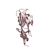
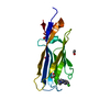
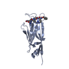
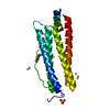
- SASDDD9: Cardiac myosin binding protein C: tri-helix bundle from the motif... -
+ Open data

- Basic information
| Entry | Database: SASBDB / ID: SASDDD9 |
|---|---|
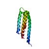
 Sample Sample | Cardiac myosin binding protein C: tri-helix bundle from the motif with C2 domain
|
| Function / homology |  Function and homology information Function and homology informationC zone / regulation of muscle filament sliding / striated muscle myosin thick filament / cardiac myofibril / regulation of striated muscle contraction / positive regulation of ATP-dependent activity / Striated Muscle Contraction / A band / structural constituent of muscle / ventricular cardiac muscle tissue morphogenesis ...C zone / regulation of muscle filament sliding / striated muscle myosin thick filament / cardiac myofibril / regulation of striated muscle contraction / positive regulation of ATP-dependent activity / Striated Muscle Contraction / A band / structural constituent of muscle / ventricular cardiac muscle tissue morphogenesis / sarcomere organization /  myosin binding / myosin heavy chain binding / ATPase activator activity / heart morphogenesis / cardiac muscle contraction / titin binding / sarcomere / actin binding / cell adhesion / identical protein binding / metal ion binding / cytosol myosin binding / myosin heavy chain binding / ATPase activator activity / heart morphogenesis / cardiac muscle contraction / titin binding / sarcomere / actin binding / cell adhesion / identical protein binding / metal ion binding / cytosolSimilarity search - Function |
| Biological species | human sequence obtained using E. coli expression |
 Citation Citation | Journal: Structure / Year: 2016 Title: A Highly Conserved Yet Flexible Linker Is Part of a Polymorphic Protein-Binding Domain in Myosin-Binding Protein C. Authors: Katharine A Michie / Ann H Kwan / Chang-Shung Tung / J Mitchell Guss / Jill Trewhella /   Abstract: The nuclear magnetic resonance (NMR) structure of the tri-helix bundle (THB) of the m-domain plus C2 (ΔmC2) of myosin-binding protein C (MyBP-C) has revealed a highly flexible seven-residue linker ...The nuclear magnetic resonance (NMR) structure of the tri-helix bundle (THB) of the m-domain plus C2 (ΔmC2) of myosin-binding protein C (MyBP-C) has revealed a highly flexible seven-residue linker between the structured THB and C2. Bioinformatics shows significant patterns of conservation across the THB-linker sequence, with the linker containing a strictly conserved serine in all MyBP-C isoforms. Clinically linked mutations further support the functional significance of the THB-linker region. NMR, small-angle X-ray scattering, and binding studies show the THB-linker plus the first ten residues of C2 undergo dramatic changes when ΔmC2 binds Ca-calmodulin, with the linker and C2 N-terminal residues contributing significantly to the affinity. Modeling of all available experimental data indicates that the THB tertiary structure must be disrupted to form the complex. These results are discussed in the context of the THB-linker and the N-terminal residues of C2 forming a polymorphic binding domain that could accommodate multiple binding partners in the dynamic sarcomere. |
 Contact author Contact author |
|
- Structure visualization
| Structure viewer | Molecule: MolmilJmol/JSmol |
|---|
- Downloads & links
-Data source
| SASBDB page |  SASDDD9 SASDDD9 |
|---|
-Related structure data


-External links
| Related items in Molecule of the Month |
|---|


-Models
| Model #2155 |   Type: atomic / Comment: one state of 2-state MultiFoXS model, weight 0.753 / Chi-square value: 3.56671154143 / P-value: 0.820000  Search similar-shape structures of this assembly by Omokage search (details) Search similar-shape structures of this assembly by Omokage search (details) |
|---|---|
| Model #2156 |  Type: atomic / Comment: one state of 2-state MultiFoXS model, weight 0.247 / Chi-square value: 3.56671154143 / P-value: 0.820000 Search similar-shape structures of this assembly by Omokage search (details) |
-Sample
| Sample | Name: Cardiac myosin binding protein C: tri-helix bundle from the motif with C2 domain Specimen concentration: 0.98-4.89 |
|---|---|
| Buffer | Name: 150 mM NaCl, 10 mM MES, 2 mM TCEP, 1 mM NaN3 at 4°C / pH: 6.5 |
| Entity #1178 | Name: tri-helix bundle-C2 / Type: protein Description: cardiac myosin binding protein C: tri-helix bundle-C2 Formula weight: 15.474 / Num. of mol.: 1 / Source: human sequence obtained using E. coli expression / References: UniProt: Q14896 Sequence: GPGSEDVWEI LRQAPPSEYE RIAFQYGVTD LRGMLKRLKG MRRDEKKSTA FQKKLEPAYQ VSKGHKIRLT VELADHDAEV KWLKNGQEIQ MSGSKYIFES IGAKRTLTIS QCSLADDAAY QCVVGGEKCS TELFVKE |
-Experimental information
| Beam | Instrument name: Australian Synchrotron SAXS/WAXS / City: Melbourne / 国: Australia / Shape: Point / Type of source: X-ray synchrotronSynchrotron / Wavelength: 0.10332 Å / Dist. spec. to detc.: 2.68024 mm | ||||||||||||||||||||||||||||||||||||
|---|---|---|---|---|---|---|---|---|---|---|---|---|---|---|---|---|---|---|---|---|---|---|---|---|---|---|---|---|---|---|---|---|---|---|---|---|---|
| Detector | Name: Pilatus 1M / Pixsize x: 172 mm | ||||||||||||||||||||||||||||||||||||
| Scan |  Measurement date: Apr 18, 2015 / Cell temperature: 22 °C / Exposure time: 1 sec. / Number of frames: 26 / Unit: 1/A /
| ||||||||||||||||||||||||||||||||||||
| Distance distribution function P(R) | 
| ||||||||||||||||||||||||||||||||||||
| Result |  Comments: The concentration series SAXS data and EOM models are provided in the full entry zip archive.
|
