 Movie
Movie Controller
[English] 日本語
 Yorodumi
- SASDBH7: Dps1 truncated, DNA binding protein under starvation conditions (... -
+ Open data

- Basic information
| Entry | Database: SASBDB / ID: SASDBH7 |
|---|---|
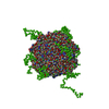
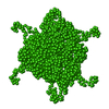
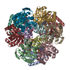
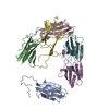
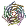
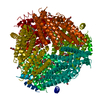
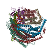
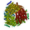
 Sample Sample | Dps1 truncated, DNA binding protein under starvation conditions (SEC-SAXS)
|
| Function / homology |  Function and homology information Function and homology information Oxidoreductases; Oxidizing metal ions / oxidoreductase activity, acting on metal ions / nucleoid / ferric iron binding / intracellular iron ion homeostasis / DNA binding / cytoplasm Oxidoreductases; Oxidizing metal ions / oxidoreductase activity, acting on metal ions / nucleoid / ferric iron binding / intracellular iron ion homeostasis / DNA binding / cytoplasmSimilarity search - Function |
| Biological species |  Deinococcus radiodurans R1 (radioresistant) Deinococcus radiodurans R1 (radioresistant) |
 Citation Citation | Journal: J Mol Biol / Year: 2017 Title: SAXS Structural Studies of Dps from Deinococcus radiodurans Highlights the Conformation of the Mobile N-Terminal Extensions. Authors: Sandra P Santos / Maxime G Cuypers / Adam Round / Stephanie Finet / Theyencheri Narayanan / Edward P Mitchell / Célia V Romão /    Abstract: The radiation-resistant bacterium Deinococcus radiodurans contains two DNA-binding proteins from starved cells (Dps): Dps1 (DR2263) and Dps2 (DRB0092). These are suggested to play a role in DNA ...The radiation-resistant bacterium Deinococcus radiodurans contains two DNA-binding proteins from starved cells (Dps): Dps1 (DR2263) and Dps2 (DRB0092). These are suggested to play a role in DNA interaction and manganese and iron storage. The proteins assemble as a conserved dodecameric structure with structurally uncharacterised N-terminal extensions. In the case of DrDps1, these extensions have been proposed to be involved in DNA interactions, while in DrDps2, their function has yet to be established. The reported data reveal the relative position of the N-terminal extensions to the dodecameric sphere in solution for both Dps. The low-resolution small angle X-ray scattering (SAXS) results show that the N-terminal extensions protrude from the spherical shell of both proteins. The SAXS envelope of a truncated form of DrDps1 without the N-terminal extensions appears as a dodecameric sphere, contrasting strongly with the protrusions observed in the full-length models. The effect of iron incorporation into DrDps2 was investigated by static and stopped-flow SAXS measurements, revealing dynamic structural changes upon iron binding and core formation, as reflected by a quick alteration of its radius of gyration. The truncated and full-length versions of DrDps were also compared on the basis of their interaction with DNA to analyse functional roles of the N-terminal extensions. DrDps1 N-terminal protrusions appear to be directly involved with DNA, whilst those from DrDps2 are indirectly associated with DNA binding. Furthermore, detection of DrDps2 in the D. radiodurans membrane fraction suggests that the N-terminus of the protein interacts with the membrane. |
 Contact author Contact author |
|
- Structure visualization
| Structure viewer | Molecule: MolmilJmol/JSmol |
|---|
- Downloads & links
-Data source
| SASBDB page |  SASDBH7 SASDBH7 |
|---|
-Related structure data
| Related structure data | C: citing same article ( |
|---|---|
| Similar structure data |
-External links
| Related items in Molecule of the Month |
|---|


-Models
| Model #889 |  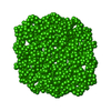 Type: dummy / Software: (2.2i) / Radius of dummy atoms: 1.90 A / Symmetry : P23 / Chi-square value: 10.5625 Search similar-shape structures of this assembly by Omokage search (details) Search similar-shape structures of this assembly by Omokage search (details) |
|---|
-Sample
| Sample | Name: Dps1 truncated, DNA binding protein under starvation conditions (SEC-SAXS) Specimen concentration: 10 mg/ml |
|---|---|
| Buffer | Name: 20 mM Tris-HCl, 150 mM NaCl / pH: 7.5 |
| Entity #422 | Name: Dps1t / Type: protein Description: N-terminal truncated DNA protection during starvation protein 1 Formula weight: 18.037 / Num. of mol.: 12 / Source: Deinococcus radiodurans R1 / References: UniProt: Q9RS64 Sequence: HYLEEKEFQT VAETLQRNLA TTISLYLKFK KYHWDIRGRF FRDLHLAYDE FIAEIFPSID EQAERLVALG GSPLAAPADL ARYSTVQVPQ ETVRDARTQV ADLVQDLSRV GKGYRDDSQA CDEANDPVTA DMYNGYAATI DKIRWMLQAI MDDERLD |
-Experimental information
| Beam | Instrument name: ESRF BM29 / City: Grenoble / 国: France / Type of source: X-ray synchrotronSynchrotron / Wavelength: 0.09919 Å / Dist. spec. to detc.: 2.81 mm | |||||||||||||||||||||||||||||||||
|---|---|---|---|---|---|---|---|---|---|---|---|---|---|---|---|---|---|---|---|---|---|---|---|---|---|---|---|---|---|---|---|---|---|---|
| Detector | Name: Pilatus 1M | |||||||||||||||||||||||||||||||||
| Scan | 
| |||||||||||||||||||||||||||||||||
| Distance distribution function P(R) | 
| |||||||||||||||||||||||||||||||||
| Result | 
|
