 Movie
Movie Controller
[English] 日本語
 Yorodumi
- SASDAN4: Calmodulin:peptide complex (Calmodulin + C-terminal region of hum... -
+ Open data

- Basic information
| Entry | Database: SASBDB / ID: SASDAN4 |
|---|---|
 Sample Sample | Calmodulin:peptide complex
|
| Function / homology |  Function and homology information Function and homology information: / establishment of protein localization to mitochondrial membrane / type 3 metabotropic glutamate receptor binding / regulation of synaptic vesicle endocytosis / negative regulation of high voltage-gated calcium channel activity / regulation of synaptic vesicle exocytosis / negative regulation of calcium ion export across plasma membrane / regulation of cardiac muscle cell action potential / response to corticosterone / positive regulation of ryanodine-sensitive calcium-release channel activity ...: / establishment of protein localization to mitochondrial membrane / type 3 metabotropic glutamate receptor binding / regulation of synaptic vesicle endocytosis / negative regulation of high voltage-gated calcium channel activity / regulation of synaptic vesicle exocytosis / negative regulation of calcium ion export across plasma membrane / regulation of cardiac muscle cell action potential / response to corticosterone / positive regulation of ryanodine-sensitive calcium-release channel activity / regulation of cell communication by electrical coupling involved in cardiac conduction / negative regulation of peptidyl-threonine phosphorylation /  nitric-oxide synthase binding / protein phosphatase activator activity / positive regulation of cyclic-nucleotide phosphodiesterase activity / positive regulation of phosphoprotein phosphatase activity / adenylate cyclase binding / catalytic complex / detection of calcium ion / negative regulation of ryanodine-sensitive calcium-release channel activity / regulation of cardiac muscle contraction / positive regulation of DNA binding / regulation of cardiac muscle contraction by regulation of the release of sequestered calcium ion / phosphatidylinositol 3-kinase binding / enzyme regulator activity / regulation of release of sequestered calcium ion into cytosol by sarcoplasmic reticulum / regulation of calcium-mediated signaling / positive regulation of protein dephosphorylation / voltage-gated potassium channel complex / titin binding / positive regulation of protein autophosphorylation / calcium channel complex / activation of adenylate cyclase activity / response to amphetamine / substantia nigra development / adenylate cyclase activator activity / nitric-oxide synthase regulator activity / regulation of heart rate / sarcomere / protein serine/threonine kinase activator activity / positive regulation of peptidyl-threonine phosphorylation / regulation of cytokinesis / positive regulation of nitric-oxide synthase activity / mitochondrial membrane / positive regulation of protein serine/threonine kinase activity / spindle microtubule / synaptic vesicle membrane / spindle pole / response to calcium ion / calcium-dependent protein binding / G2/M transition of mitotic cell cycle / disordered domain specific binding / myelin sheath / growth cone / vesicle / transmembrane transporter binding / G protein-coupled receptor signaling pathway / protein domain specific binding / centrosome / calcium ion binding / protein kinase binding / protein-containing complex / nucleus / plasma membrane / cytoplasm nitric-oxide synthase binding / protein phosphatase activator activity / positive regulation of cyclic-nucleotide phosphodiesterase activity / positive regulation of phosphoprotein phosphatase activity / adenylate cyclase binding / catalytic complex / detection of calcium ion / negative regulation of ryanodine-sensitive calcium-release channel activity / regulation of cardiac muscle contraction / positive regulation of DNA binding / regulation of cardiac muscle contraction by regulation of the release of sequestered calcium ion / phosphatidylinositol 3-kinase binding / enzyme regulator activity / regulation of release of sequestered calcium ion into cytosol by sarcoplasmic reticulum / regulation of calcium-mediated signaling / positive regulation of protein dephosphorylation / voltage-gated potassium channel complex / titin binding / positive regulation of protein autophosphorylation / calcium channel complex / activation of adenylate cyclase activity / response to amphetamine / substantia nigra development / adenylate cyclase activator activity / nitric-oxide synthase regulator activity / regulation of heart rate / sarcomere / protein serine/threonine kinase activator activity / positive regulation of peptidyl-threonine phosphorylation / regulation of cytokinesis / positive regulation of nitric-oxide synthase activity / mitochondrial membrane / positive regulation of protein serine/threonine kinase activity / spindle microtubule / synaptic vesicle membrane / spindle pole / response to calcium ion / calcium-dependent protein binding / G2/M transition of mitotic cell cycle / disordered domain specific binding / myelin sheath / growth cone / vesicle / transmembrane transporter binding / G protein-coupled receptor signaling pathway / protein domain specific binding / centrosome / calcium ion binding / protein kinase binding / protein-containing complex / nucleus / plasma membrane / cytoplasmSimilarity search - Function |
| Biological species |  Homo sapiens (human) Homo sapiens (human) |
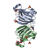
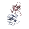
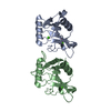
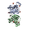
 Citation Citation | Journal: BMC Struct Biol / Year: 2008 Title: Interaction between the C-terminal region of human myelin basic protein and calmodulin: analysis of complex formation and solution structure. Authors: Viivi Majava / Maxim V Petoukhov / Nobuhiro Hayashi / Päivi Pirilä / Dmitri I Svergun / Petri Kursula /  Abstract: BACKGROUND: The myelin sheath is a multilamellar membrane structure wrapped around the axon, enabling the saltatory conduction of nerve impulses in vertebrates. Myelin basic protein, one of the most ...BACKGROUND: The myelin sheath is a multilamellar membrane structure wrapped around the axon, enabling the saltatory conduction of nerve impulses in vertebrates. Myelin basic protein, one of the most abundant myelin-specific proteins, is an intrinsically disordered protein that has been shown to bind calmodulin. In this study, we focus on a 19-mer synthetic peptide from the predicted calmodulin-binding segment near the C-terminus of human myelin basic protein. RESULTS: The interaction of native human myelin basic protein with calmodulin was confirmed by affinity chromatography. The binding of the myelin basic protein peptide to calmodulin was tested with ...RESULTS: The interaction of native human myelin basic protein with calmodulin was confirmed by affinity chromatography. The binding of the myelin basic protein peptide to calmodulin was tested with isothermal titration calorimetry (ITC) in different temperatures, and Kd was observed to be in the low muM range, as previously observed for full-length myelin basic protein. Surface plasmon resonance showed that the peptide bound to calmodulin, and binding was accompanied by a conformational change; furthermore, gel filtration chromatography indicated a decrease in the hydrodynamic radius of calmodulin in the presence of the peptide. NMR spectroscopy was used to map the binding area to reside mainly within the hydrophobic pocket of the C-terminal lobe of calmodulin. The solution structure obtained by small-angle X-ray scattering indicates binding of the myelin basic protein peptide into the interlobal groove of calmodulin, while calmodulin remains in an extended conformation. CONCLUSION: Taken together, our results give a detailed structural insight into the interaction of calmodulin with a C-terminal segment of a major myelin protein, the myelin basic protein. The used ...CONCLUSION: Taken together, our results give a detailed structural insight into the interaction of calmodulin with a C-terminal segment of a major myelin protein, the myelin basic protein. The used 19-mer peptide interacts mainly with the C-terminal lobe of calmodulin, and a conformational change accompanies binding, suggesting a novel mode of calmodulin-target protein interaction. Calmodulin does not collapse and wrap around the peptide tightly; instead, it remains in an extended conformation in the solution structure. The observed affinity can be physiologically relevant, given the high abundance of both binding partners in the nervous system. |
 Contact author Contact author |
|
- Structure visualization
| Structure viewer | Molecule: MolmilJmol/JSmol |
|---|
- Downloads & links
-Data source
| SASBDB page |  SASDAN4 SASDAN4 |
|---|
-Related structure data
| Similar structure data |
|---|


-External links
| Related items in Molecule of the Month |
|---|


-Models
| Model #63 |   Type: atomic / Software: Sasref / Chi-square value: 4.7524  Search similar-shape structures of this assembly by Omokage search (details) Search similar-shape structures of this assembly by Omokage search (details) |
|---|
-Sample
| Sample | Name: Calmodulin:peptide complex / Sample MW: 18.84 kDa / Specimen concentration: 5.30-20.00 / Entity id: 58 / 59 |
|---|---|
| Buffer | Name: Tris75 / Concentration: 25.00 mM / pH: 7.5 / Composition: NaCl 200.000 mM |
| Entity #58 | Type: protein / Description: Calmodulin / Formula weight: 16.84 / Num. of mol.: 1 / Source: Homo sapiens / References: UniProt: P62158 Sequence: MADQLTEEQI AEFKEAFSLF DKDGDGTITT KELGTVMRSL GQNPTEAELQ DMINEVDADG NGTIDFPEFL TMMARKMKDT DSEEEIREAF RVFDKDGNGY ISAAELRHVM TNLGEKLTDE EVDEMIREAD IDGDGQVNYE EFVQMMTAK |
| Entity #59 | Name: peptide / Type: protein Description: C-terminal region of human myelin basic protein Formula weight: 2 / Num. of mol.: 1 / Source: Homo sapiens Sequence: HKGFKGVDAQ GTLSKIFKL |
-Experimental information
| Beam | Instrument name: DORIS III X33  / City: Hamburg / 国: Germany / Type of source: X-ray synchrotronSynchrotron / City: Hamburg / 国: Germany / Type of source: X-ray synchrotronSynchrotron | ||||||||||||||||||
|---|---|---|---|---|---|---|---|---|---|---|---|---|---|---|---|---|---|---|---|
| Detector | Name: MAR 345 Image Plate | ||||||||||||||||||
| Scan | 
| ||||||||||||||||||
| Distance distribution function P(R) | 
| ||||||||||||||||||
| Result | 
|
