 Movie
Movie Controller

+ Open data

- Basic information
| Entry | Database: SASBDB / ID: SASDAM8 |
|---|---|
 Sample Sample | MHV-68 LANA
|
| Function / homology | : / Protein LANA1-like, DNA-binding domain / Epstein Barr virus nuclear antigen-1, DNA-binding domain superfamily / host cell nucleus /  DNA binding / identical protein binding / 73 protein DNA binding / identical protein binding / 73 protein Function and homology information Function and homology information |
| Biological species |  Murid herpesvirus 4 (Murine herpesvirus 68) Murid herpesvirus 4 (Murine herpesvirus 68) |
 Citation Citation | Journal: Nucleic Acids Res / Year: 2015 Title: KSHV but not MHV-68 LANA induces a strong bend upon binding to terminal repeat viral DNA. Authors: Rajesh Ponnusamy / Maxim V Petoukhov / Bruno Correia / Tania F Custodio / Franceline Juillard / Min Tan / Marta Pires de Miranda / Maria A Carrondo / J Pedro Simas / Kenneth M Kaye / Dmitri ...Authors: Rajesh Ponnusamy / Maxim V Petoukhov / Bruno Correia / Tania F Custodio / Franceline Juillard / Min Tan / Marta Pires de Miranda / Maria A Carrondo / J Pedro Simas / Kenneth M Kaye / Dmitri I Svergun / Colin E McVey /    Abstract: Latency-associated nuclear antigen (LANA) is central to episomal tethering, replication and transcriptional regulation of γ2-herpesviruses. LANA binds cooperatively to the terminal repeat (TR) ...Latency-associated nuclear antigen (LANA) is central to episomal tethering, replication and transcriptional regulation of γ2-herpesviruses. LANA binds cooperatively to the terminal repeat (TR) region of the viral episome via adjacent LANA binding sites (LBS), but the molecular mechanism by which LANA assembles on the TR remains elusive. We show that KSHV LANA and MHV-68 LANA proteins bind LBS DNA using strikingly different modes. Solution structure of LANA complexes revealed that while kLANA tetramer is intrinsically bent both in the free and bound state to LBS1-2 DNA, mLANA oligomers instead adopt a rigid linear conformation. In addition, we report a novel non-ring kLANA structure that displays more flexibility at its assembly interface than previously demonstrated. We identified a hydrophobic pivot point located at the dimer-dimer assembly interface, which gives rotational freedom for kLANA to adopt variable conformations to accommodate both LBS1-2 and LBS2-1-3 DNA. Alterations in the arrangement of LBS within TR or at the tetramer assembly interface have a drastic effect on the ability of kLANA binding. We also show kLANA and mLANA DNA binding functions can be reciprocated. Although KSHV and MHV-68 are closely related, the findings provide new insights into how the structure, oligomerization, and DNA binding of LANA have evolved differently to assemble on the TR DNA. |
 Contact author Contact author |
|
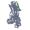
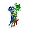
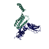
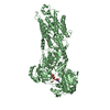
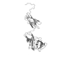
- Structure visualization
| Structure viewer | Molecule: MolmilJmol/JSmol |
|---|
- Downloads & links
 SASDAM8


-Models
| Model #326 |  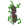 Type: mix / Software: EOM / Radius of dummy atoms: 1.90 A / Chi-square value: 1.69  Search similar-shape structures of this assembly by Omokage search (details) Search similar-shape structures of this assembly by Omokage search (details) |
|---|---|
| Model #327 | 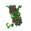 Type: mix / Software: EOM / Radius of dummy atoms: 1.90 A / Chi-square value: 1.69 Search similar-shape structures of this assembly by Omokage search (details) |
| Model #328 | 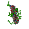 Type: mix / Software: EOM / Radius of dummy atoms: 1.90 A / Chi-square value: 1.69 Search similar-shape structures of this assembly by Omokage search (details) |
-Sample
| Sample | Name: MHV-68 LANA / Specimen concentration: 5 mg/ml |
|---|---|
| Buffer | Name: 25 mM Na/K Phosphate / pH: 7.5 |
| Entity #193 | Name: mLANA / Type: protein / Description: Latency-associated nuclear antigen / Formula weight: 21.721 / Num. of mol.: 4 / Source: Murid herpesvirus 4 / References: UniProt: O41974 Sequence: MPTSPPTTRN TTSGKTRSGC KRRCFNKPAA MPPKRRRAPK RPAPPPPPGC QGDEESSQGT QTPNPPSPPV PPSSPTLPSS PVPPSSPVHE PPSPSPPPAP PSPDVDVEGL DVGETDDPGP PPPKRYSRYQ KPHNPSDPLP KKYQGMRRHL QVTAPRLFDP EGHPPTHFKS ...Sequence: MPTSPPTTRN TTSGKTRSGC KRRCFNKPAA MPPKRRRAPK RPAPPPPPGC QGDEESSQGT QTPNPPSPPV PPSSPTLPSS PVPPSSPVHE PPSPSPPPAP PSPDVDVEGL DVGETDDPGP PPPKRYSRYQ KPHNPSDPLP KKYQGMRRHL QVTAPRLFDP EGHPPTHFKS AVMFSSTHPY TLNKLHKCIQ SKHVLSTPVS CLPLVPGTTQ QCVTYYLLSF VEDKKQAKKL KRVVLAYCEK YHSSVEGTIV KAKPYFPLPE PPTEPPTDPE QPSTSTQASG TQHGPTASLD AGAEQGATGS PGSSPGQQGQ GSQT |
-Experimental information
| Beam | Instrument name: PETRA III P12 / City: Hamburg / 国: Germany / Type of source: X-ray synchrotronSynchrotron / Wavelength: 0.12 Å / Dist. spec. to detc.: 3.1 mm | ||||||||||||||||||
|---|---|---|---|---|---|---|---|---|---|---|---|---|---|---|---|---|---|---|---|
| Detector | Name: Pilatus 2M | ||||||||||||||||||
| Scan | 
| ||||||||||||||||||
| Distance distribution function P(R) | 
| ||||||||||||||||||
| Result |  Comments: Study of oligomeric states of mLANA and kLANA and their complexes with DNA
|
