 Movie
Movie Controller
[English] 日本語
 Yorodumi
- SASDAB8: Protein Interacting with C-kinase 1 (PICK1) LKV, dimer contributi... -
+ Open data

- Basic information
| Entry | Database: SASBDB / ID: SASDAB8 |
|---|---|
 Sample Sample | Protein Interacting with C-kinase 1 (PICK1) LKV, dimer contribution (data decomposition).
|
| Function / homology |  Function and homology information Function and homology informationG protein-coupled glutamate receptor binding / membrane curvature sensor activity / postsynaptic endocytic zone / postsynaptic early endosome / glial cell development / cellular response to decreased oxygen levels /  Arp2/3 complex binding / postsynaptic specialization / regulation of Arp2/3 complex-mediated actin nucleation / negative regulation of Arp2/3 complex-mediated actin nucleation ...G protein-coupled glutamate receptor binding / membrane curvature sensor activity / postsynaptic endocytic zone / postsynaptic early endosome / glial cell development / cellular response to decreased oxygen levels / Arp2/3 complex binding / postsynaptic specialization / regulation of Arp2/3 complex-mediated actin nucleation / negative regulation of Arp2/3 complex-mediated actin nucleation / dopamine transport / SNAP receptor activity / monoamine transport / protein kinase C-activating G protein-coupled receptor signaling pathway / dendritic spine organization / regulation of postsynaptic neurotransmitter receptor internalization / long-term synaptic depression / dendritic spine maintenance / receptor clustering / Trafficking of GluR2-containing AMPA receptors / regulation of insulin secretion / positive regulation of receptor internalization / protein targeting / cellular response to glucose starvation / ionotropic glutamate receptor binding / cytoskeletal protein binding / ephrin receptor binding / trans-Golgi network membrane / G protein-coupled receptor binding / protein kinase C binding / intracellular protein transport / phospholipid binding / receptor tyrosine kinase binding / actin filament binding / synaptic vesicle / GTPase binding / presynaptic membrane / dendritic spine / postsynaptic density / cytoskeleton / neuron projection / protein domain specific binding / protein phosphorylation / negative regulation of gene expression / signaling receptor binding / glutamatergic synapse / synapse / dendrite / perinuclear region of cytoplasm / Golgi apparatus / protein-containing complex / mitochondrion / identical protein binding / metal ion binding / plasma membrane / cytoplasm Arp2/3 complex binding / postsynaptic specialization / regulation of Arp2/3 complex-mediated actin nucleation / negative regulation of Arp2/3 complex-mediated actin nucleation ...G protein-coupled glutamate receptor binding / membrane curvature sensor activity / postsynaptic endocytic zone / postsynaptic early endosome / glial cell development / cellular response to decreased oxygen levels / Arp2/3 complex binding / postsynaptic specialization / regulation of Arp2/3 complex-mediated actin nucleation / negative regulation of Arp2/3 complex-mediated actin nucleation / dopamine transport / SNAP receptor activity / monoamine transport / protein kinase C-activating G protein-coupled receptor signaling pathway / dendritic spine organization / regulation of postsynaptic neurotransmitter receptor internalization / long-term synaptic depression / dendritic spine maintenance / receptor clustering / Trafficking of GluR2-containing AMPA receptors / regulation of insulin secretion / positive regulation of receptor internalization / protein targeting / cellular response to glucose starvation / ionotropic glutamate receptor binding / cytoskeletal protein binding / ephrin receptor binding / trans-Golgi network membrane / G protein-coupled receptor binding / protein kinase C binding / intracellular protein transport / phospholipid binding / receptor tyrosine kinase binding / actin filament binding / synaptic vesicle / GTPase binding / presynaptic membrane / dendritic spine / postsynaptic density / cytoskeleton / neuron projection / protein domain specific binding / protein phosphorylation / negative regulation of gene expression / signaling receptor binding / glutamatergic synapse / synapse / dendrite / perinuclear region of cytoplasm / Golgi apparatus / protein-containing complex / mitochondrion / identical protein binding / metal ion binding / plasma membrane / cytoplasmSimilarity search - Function |
| Biological species |  Rattus norvegicus (Norway rat) Rattus norvegicus (Norway rat) |
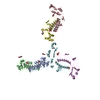
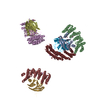
 Citation Citation | Journal: Structure / Year: 2015 Title: Structure of Dimeric and Tetrameric Complexes of the BAR Domain Protein PICK1 Determined by Small-Angle X-Ray Scattering. Authors: Morten L Karlsen / Thor S Thorsen / Niklaus Johner / Ina Ammendrup-Johnsen / Simon Erlendsson / Xinsheng Tian / Jens B Simonsen / Rasmus Høiberg-Nielsen / Nikolaj M Christensen / George ...Authors: Morten L Karlsen / Thor S Thorsen / Niklaus Johner / Ina Ammendrup-Johnsen / Simon Erlendsson / Xinsheng Tian / Jens B Simonsen / Rasmus Høiberg-Nielsen / Nikolaj M Christensen / George Khelashvili / Werner Streicher / Kaare Teilum / Bente Vestergaard / Harel Weinstein / Ulrik Gether / Lise Arleth / Kenneth L Madsen /   Abstract: PICK1 is a neuronal scaffolding protein containing a PDZ domain and an auto-inhibited BAR domain. BAR domains are membrane-sculpting protein modules generating membrane curvature and promoting ...PICK1 is a neuronal scaffolding protein containing a PDZ domain and an auto-inhibited BAR domain. BAR domains are membrane-sculpting protein modules generating membrane curvature and promoting membrane fission. Previous data suggest that BAR domains are organized in lattice-like arrangements when stabilizing membranes but little is known about structural organization of BAR domains in solution. Through a small-angle X-ray scattering (SAXS) analysis, we determine the structure of dimeric and tetrameric complexes of PICK1 in solution. SAXS and biochemical data reveal a strong propensity of PICK1 to form higher-order structures, and SAXS analysis suggests an offset, parallel mode of BAR-BAR oligomerization. Furthermore, unlike accessory domains in other BAR domain proteins, the positioning of the PDZ domains is flexible, enabling PICK1 to perform long-range, dynamic scaffolding of membrane-associated proteins. Together with functional data, these structural findings are compatible with a model in which oligomerization governs auto-inhibition of BAR domain function. |
 Contact author Contact author |
|
- Structure visualization
| Structure viewer | Molecule: MolmilJmol/JSmol |
|---|
- Downloads & links
-Data source
| SASBDB page |  SASDAB8 SASDAB8 |
|---|
-Related structure data
| Similar structure data |
|---|
-External links
| Related items in Molecule of the Month |
|---|


-Models
| Model #318 |  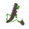 Type: mix / Software: EOM / Radius of dummy atoms: 1.90 A / Chi-square value: 3.818116  Search similar-shape structures of this assembly by Omokage search (details) Search similar-shape structures of this assembly by Omokage search (details) |
|---|---|
| Model #319 | 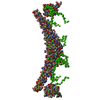 Type: mix / Software: EOM / Radius of dummy atoms: 1.90 A / Chi-square value: 3.818116 Search similar-shape structures of this assembly by Omokage search (details) |
| Model #322 | 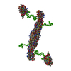 Type: mix / Software: EOM / Radius of dummy atoms: 1.90 A / Chi-square value: 3.818116 Search similar-shape structures of this assembly by Omokage search (details) |
-Sample
| Sample | Name: Protein Interacting with C-kinase 1 (PICK1) LKV, dimer contribution (data decomposition). Specimen concentration: 4.20-8.80 |
|---|---|
| Buffer | Name: 50 mM Tris 125 mM NaCl 0.01 vol% reduced TX-100 / Concentration: 50.00 mM / pH: 7.4 / Composition: 125 mM NaCl, 0.01 vol% reduced TX-100 |
| Entity #184 | Type: protein / Description: PRKCA-binding protein / Formula weight: 46.506 / Num. of mol.: 2 / Source: Rattus norvegicus / References: UniProt: Q9EP80 Sequence: MFADLDYDIE EDKLGIPTVP GKVTLQKDAQ NLIGISIGGG AQYCPCLYIV QVFDNTPAAL DGTVAAGDEI TGVNGRSIKG KTKVEVAKMI QEVKGEVTIH YNKLQADPKQ GMSLDIVLKK VKHRLVENMS SGTADALGLS RAILCNDGLV KRLEELERTA ELYKGMTEHT ...Sequence: MFADLDYDIE EDKLGIPTVP GKVTLQKDAQ NLIGISIGGG AQYCPCLYIV QVFDNTPAAL DGTVAAGDEI TGVNGRSIKG KTKVEVAKMI QEVKGEVTIH YNKLQADPKQ GMSLDIVLKK VKHRLVENMS SGTADALGLS RAILCNDGLV KRLEELERTA ELYKGMTEHT KNLLRAFYEL SQTHRAFGDV FSVIGVREPQ PAASEAFVKF ADAHRSIEKF GIRLLKTIKP MLTDLNTYLN KAIPDTRLTI KKYLDVKFEY LSYCLKVKEM DDEEYSCIAL GEPLYRVSGN YEYRLILRCR QEARARFSQM RKDVLEKMEL LDQKHVQDIV FQLQRLVSTM SKYYNDCYAV LRDADVFPIE VDLAHTTLAY GPNQGGFTDG EDEEEEEEDG AAREVSKDAR GATGPTDKGG SWLKV |
-Experimental information
| Beam | Instrument name: DORIS III X33  / City: Hamburg / 国: Germany / Type of source: X-ray synchrotronSynchrotron / City: Hamburg / 国: Germany / Type of source: X-ray synchrotronSynchrotron | ||||||||||||||||||||||||||||||||||||
|---|---|---|---|---|---|---|---|---|---|---|---|---|---|---|---|---|---|---|---|---|---|---|---|---|---|---|---|---|---|---|---|---|---|---|---|---|---|
| Detector | Name: MAR 345 Image Plate | ||||||||||||||||||||||||||||||||||||
| Scan | 
| ||||||||||||||||||||||||||||||||||||
| Distance distribution function P(R) | 
| ||||||||||||||||||||||||||||||||||||
| Result |  Comments: Structure and flexibility of PICK1 LKV mutant dimer was characterized by SAXS using data obtained by decomposing scattering from two polydisperse samples into dimer (and tetramer) ...Comments: Structure and flexibility of PICK1 LKV mutant dimer was characterized by SAXS using data obtained by decomposing scattering from two polydisperse samples into dimer (and tetramer) contributions. To obtain the dimer scattering, two samples were measured at 4.4 and 8,8 mg/ml, and decomposed as described in the reference. Models were fitted to data using a combination of rigid body modelling and EOM, based on structural components defined by NMR and MD simulations. PDB files represent structures found in the optimal ensemble. All data, in addition to that displayed for this entry, are available in the .zip archive.
|
