 Movie
Movie Controller


+ Open data

- Basic information
| Entry | Database: PDB / ID: 9eq3 | ||||||||||||
|---|---|---|---|---|---|---|---|---|---|---|---|---|---|
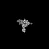
| Title | Structure of IgE HMM5 bound to FceRIa cryo-EM class 8 | ||||||||||||
 Components Components |
| ||||||||||||
 Keywords Keywords |  IMMUNE SYSTEM / IgE / Fc receptor / allergy / antibody IMMUNE SYSTEM / IgE / Fc receptor / allergy / antibody | ||||||||||||
| Function / homology |  Function and homology information Function and homology informationhigh-affinity IgE receptor activity / type I hypersensitivity / eosinophil degranulation / IgE binding / type 2 immune response / Fc epsilon receptor (FCERI) signaling / mast cell degranulation / regulation of immune response / Role of LAT2/NTAL/LAB on calcium mobilization / FCERI mediated Ca+2 mobilization ...high-affinity IgE receptor activity / type I hypersensitivity / eosinophil degranulation / IgE binding / type 2 immune response / Fc epsilon receptor (FCERI) signaling / mast cell degranulation / regulation of immune response / Role of LAT2/NTAL/LAB on calcium mobilization / FCERI mediated Ca+2 mobilization / FCERI mediated MAPK activation / FCERI mediated NF-kB activation / transmembrane signaling receptor activity / cell surface receptor signaling pathway / cell surface / plasma membraneSimilarity search - Function | ||||||||||||
| Biological species |  Homo sapiens (human) Homo sapiens (human) | ||||||||||||
| Method | ELECTRON MICROSCOPY / single particle reconstruction / cryo EM / Resolution: 6.9 Å | ||||||||||||
 Authors Authors | Andersen, G.R. / Jensen, R.K. | ||||||||||||
| Funding support |  Denmark, 3items Denmark, 3items
| ||||||||||||
 Citation Citation | Journal: To be published Title: Structure of IgE HMM5 bound to FceRIa cryo-EM class 8 Authors: Andersen, G.R. / Jensen, R.K. | ||||||||||||
| History |
|
- Structure visualization
| Structure viewer | Molecule: MolmilJmol/JSmol |
|---|
- Downloads & links
-Download
| PDBx/mmCIF format | 9eq3.cif.gz | 309.4 KB | Display | PDBx/mmCIF format |
|---|---|---|---|---|
| PDB format | pdb9eq3.ent.gz | Display | PDB format | |
| PDBx/mmJSON format | 9eq3.json.gz | Tree view | PDBx/mmJSON format | |
| Others |  Other downloads Other downloads |
-Validation report
| Arichive directory | https://data.pdbj.org/pub/pdb/validation_reports/eq/9eq3ftp://data.pdbj.org/pub/pdb/validation_reports/eq/9eq3 | HTTPS FTP |
|---|
-Related structure data
| Related structure data | 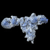 19895MC M: map data used to model this data C: citing same article ( |
|---|---|
| Similar structure data |
-Links
 PDBj
PDBj


- Assembly
| Deposited unit | 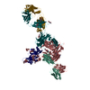
|
|---|---|
| 1 |
|
-Components
-Protein , 2 types, 3 molecules HXR
| #1: Protein | Mass: 60203.508 Da / Num. of mol.: 2 Source method: isolated from a genetically manipulated source Source: (gene. exp.) Homo sapiens (human) / Production host: Homo sapiens (human)#3: Protein | | Mass: 19880.070 Da / Num. of mol.: 1 Source method: isolated from a genetically manipulated source Source: (gene. exp.) Homo sapiens (human) / Gene: FCER1A, FCE1A / Production host: Homo sapiens (human) / References: UniProt: P12319 |
|---|
-Antibody , 1 types, 2 molecules LY
| #2: Antibody | Mass: 23384.795 Da / Num. of mol.: 2 Source method: isolated from a genetically manipulated source Source: (gene. exp.) Homo sapiens (human) / Production host: Homo sapiens (human) |
|---|
-Sugars , 5 types, 17 molecules 
| #4: Polysaccharide | / Mass: 910.823 Da / Num. of mol.: 3 Source method: isolated from a genetically manipulated source #5: Polysaccharide | alpha-D-mannopyranose-(1-3)-[alpha-D-mannopyranose-(1-6)]alpha-D-mannopyranose-(1-4)-2-acetamido-2- ...alpha-D-mannopyranose-(1-3)-[alpha-D-mannopyranose-(1-6)]alpha-D-mannopyranose-(1-4)-2-acetamido-2-deoxy-beta-D-glucopyranose-(1-4)-2-acetamido-2-deoxy-beta-D-glucopyranose | / Mass: 910.823 Da / Num. of mol.: 1Source method: isolated from a genetically manipulated source #6: Polysaccharide | beta-D-mannopyranose-(1-4)-2-acetamido-2-deoxy-beta-D-glucopyranose-(1-4)-2-acetamido-2-deoxy-beta- ...beta-D-mannopyranose-(1-4)-2-acetamido-2-deoxy-beta-D-glucopyranose-(1-4)-2-acetamido-2-deoxy-beta-D-glucopyranose / Mass: 586.542 Da / Num. of mol.: 4Source method: isolated from a genetically manipulated source #7: Polysaccharide | 2-acetamido-2-deoxy-beta-D-glucopyranose-(1-4)-2-acetamido-2-deoxy-beta-D-glucopyranose | / Mass: 424.401 Da / Num. of mol.: 1Source method: isolated from a genetically manipulated source #8: Sugar | ChemComp-NAG / N-Acetylglucosamine Type: D-saccharide, beta linking / Mass: 221.208 Da / Num. of mol.: 8 / Source method: obtained synthetically / Formula: C8H15NO6 / Feature type: SUBJECT OF INVESTIGATION Type: D-saccharide, beta linking / Mass: 221.208 Da / Num. of mol.: 8 / Source method: obtained synthetically / Formula: C8H15NO6 / Feature type: SUBJECT OF INVESTIGATION |
|---|
-Details
| Has ligand of interest | Y |
|---|
-Experimental details
-Experiment
| Experiment | Method: ELECTRON MICROSCOPY |
|---|---|
| EM experiment | Aggregation state: PARTICLE / 3D reconstruction method: single particle reconstruction |
- Sample preparation
| Component | Name: complex of IgE HMM5 and the ectodomain of FceRIa / Type: COMPLEX / Entity ID: #1-#3 / Source: RECOMBINANT |
|---|---|
| Molecular weight | Value: 0.21 MDa / Experimental value: NO |
| Source (natural) | Organism: Homo sapiens (human) |
| Source (recombinant) | Organism: Homo sapiens (human) |
| Buffer solution | pH: 7.5 |
| Specimen | Embedding applied: NO / Shadowing applied: NO / Staining applied: NO / Vitrification applied: YES |
| Vitrification | Cryogen name: ETHANE |
- Electron microscopy imaging
| Experimental equipment |  Model: Titan Krios / Image courtesy: FEI Company |
|---|---|
| Microscopy | Model: FEI TITAN KRIOS |
| Electron gun | Electron source: FIELD EMISSION GUN / Accelerating voltage: 300 kV / Illumination mode: FLOOD BEAM |
| Electron lens | Mode: BRIGHT FIELDBright-field microscopy / Nominal defocus max: 1800 nm / Nominal defocus min: 800 nm / Cs: 2.7 mm / C2 aperture diameter: 70 µm |
| Image recording | Electron dose: 59.16 e/Å2 / Film or detector model: GATAN K3 (6k x 4k) |
- Processing
| CTF correction | Type: PHASE FLIPPING AND AMPLITUDE CORRECTION |
|---|---|
| 3D reconstruction | Resolution: 6.9 Å / Resolution method: FSC 0.143 CUT-OFF / Num. of particles: 19161 / Symmetry type: POINT |
