 ムービー
ムービー コントローラー


+ データを開く

- 基本情報
基本情報
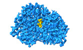
| 登録情報 | データベース: PDB / ID: 8ybe | ||||||
|---|---|---|---|---|---|---|---|
| タイトル | Cryo-EM structure of Maltose Binding Protein | ||||||
 要素 要素 | Maltose/maltodextrin-binding periplasmic protein | ||||||
 キーワード キーワード | SUGAR BINDING PROTEIN /  maltose binding protein / Cryo-EM (低温電子顕微鏡法) / sub-50kDa / atomic resolution. maltose binding protein / Cryo-EM (低温電子顕微鏡法) / sub-50kDa / atomic resolution. | ||||||
| 機能・相同性 |  機能・相同性情報 機能・相同性情報detection of maltose stimulus / maltose transport complex / maltose binding / maltose transport / maltodextrin transmembrane transport / carbohydrate transport / carbohydrate transmembrane transporter activity / ATP-binding cassette (ABC) transporter complex, substrate-binding subunit-containing / ATP-binding cassette (ABC) transporter complex / cell chemotaxis ...detection of maltose stimulus / maltose transport complex / maltose binding / maltose transport / maltodextrin transmembrane transport / carbohydrate transport / carbohydrate transmembrane transporter activity / ATP-binding cassette (ABC) transporter complex, substrate-binding subunit-containing / ATP-binding cassette (ABC) transporter complex / cell chemotaxis / outer membrane-bounded periplasmic space / ペリプラズム / DNA damage response / 生体膜類似検索 - 分子機能 | ||||||
| 生物種 |  Escherichia coli (大腸菌) Escherichia coli (大腸菌) | ||||||
| 手法 | 電子顕微鏡法 / 単粒子再構成法 / クライオ電子顕微鏡法 / 解像度: 2.3 Å | ||||||
 データ登録者 データ登録者 | Yoo, Y. / Park, K. / Kim, H. | ||||||
| 資金援助 | 1件
| ||||||
 引用 引用 | ジャーナル: To Be Published タイトル: Atomic resolution structure of MBP using Cryo-EM 著者: Yoo, Y. / Park, K. / Kim, H. #1: ジャーナル: J Mol Biol / 年: 2001タイトル: Crystal structures of the maltodextrin/maltose-binding protein complexed with reduced oligosaccharides: flexibility of tertiary structure and ligand binding. 著者: X Duan / J A Hall / H Nikaido / F A Quiocho /  要旨: The structure of the maltodextrin or maltose-binding protein, an initial receptor for bacterial ABC-type active transport and chemotaxis, consists of two globular domains that are separated by a ...The structure of the maltodextrin or maltose-binding protein, an initial receptor for bacterial ABC-type active transport and chemotaxis, consists of two globular domains that are separated by a groove wherein the ligand is bound and enclosed by an inter-domain rotation. Here, we report the determination of the crystal structures of the protein complexed with reduced maltooligosaccharides (maltotriitol and maltotetraitol) in both the "closed" and "open" forms. Although these modified sugars bind to the receptor, they are not transported by the wild-type transporter. In the closed structures, the reduced sugars are buried in the groove and bound by both domains, one domain mainly by hydrogen-bonding interactions and the other domain primarily by non-polar interactions with aromatic side-chains. In the open structures, which abrogate both cellular activities of active transport and chemotaxis because of the large separation between the two domains, the sugars are bound almost exclusively to the domain rich in aromatic residues. The binding site for the open chain glucitol residue extends to a subsite that is distinct from those for the glucose residues that were uncovered in prior structural studies of the binding of active linear maltooligosaccharides. Occupation of this subsite may also account for the inability of the reduced oligosaccharides to be transported. The structures reported here, combined with those previously determined for several other complexes with active oligosaccharides in the closed form and with cyclodextrin in the open form, revealed at least four distinct modes of ligand binding but with only one being functionally active. This versatility reflects the flexibility of the protein, from very large motions of interdomain rotation to more localized side-chain conformational changes, and adaptation by the oligosaccharides as well. | ||||||
| 履歴 |
|
- 構造の表示
構造の表示
| 構造ビューア | 分子: MolmilJmol/JSmol |
|---|
- ダウンロードとリンク
-ダウンロード
| PDBx/mmCIF形式 | 8ybe.cif.gz | 89.2 KB | 表示 | PDBx/mmCIF形式 |
|---|---|---|---|---|
| PDB形式 | pdb8ybe.ent.gz | 63.7 KB | 表示 | PDB形式 |
| PDBx/mmJSON形式 | 8ybe.json.gz | ツリー表示 | PDBx/mmJSON形式 | |
| その他 |  その他のダウンロード その他のダウンロード |
-検証レポート
| アーカイブディレクトリ | https://data.pdbj.org/pub/pdb/validation_reports/yb/8ybeftp://data.pdbj.org/pub/pdb/validation_reports/yb/8ybe | HTTPS FTP |
|---|
-関連構造データ
-リンク
 PDBj
PDBj


- 集合体
集合体
| 登録構造単位 | 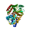
|
|---|---|
| 1 |
|
-要素
| #1: タンパク質 | 分子量: 40912.398 Da / 分子数: 1 / 変異: A338V / 由来タイプ: 組換発現 / 由来: (組換発現) Escherichia coli (大腸菌) / 遺伝子: malE, b4034, JW3994 / 発現宿主: Escherichia coli (大腸菌) / 参照: UniProt: P0AEX9 |
|---|---|
| #2: 多糖 | alpha-D-glucopyranose-(1-4)-alpha-D-glucopyranose  オリゴ糖, Oligosaccharideオリゴ糖 オリゴ糖, Oligosaccharideオリゴ糖クラス: 栄養素 栄養素 / 分子量: 342.297 Da / 分子数: 1 / 由来タイプ: 組換発現 / 詳細: oligosaccharide / 参照: alpha-maltose |
| #3: 水 | ChemComp-HOH / 水 分子量: 18.015 Da / 分子数: 197 / 由来タイプ: 天然 / 式: H2O 分子量: 18.015 Da / 分子数: 197 / 由来タイプ: 天然 / 式: H2O |
| 研究の焦点であるリガンドがあるか | N |
-実験情報
-実験
| 実験 | 手法: 電子顕微鏡法 |
|---|---|
| EM実験 | 試料の集合状態: PARTICLE / 3次元再構成法: 単粒子再構成法 |
- 試料調製
試料調製
| 構成要素 | 名称: Maltose binding protein monomer / タイプ: COMPLEX / Entity ID: #1 / 由来: RECOMBINANT |
|---|---|
| 分子量 | 実験値: NO |
| 由来(天然) | 生物種: Escherichia coli (大腸菌) |
| 由来(組換発現) | 生物種: Escherichia coli (大腸菌) |
| 緩衝液 | pH: 7.5 |
| 試料 | 濃度: 2 mg/ml / 包埋: NO / シャドウイング: NO / 染色: NO / 凍結: YES |
| 試料支持 | グリッドの材料: GOLD / グリッドのサイズ: 300 divisions/in. / グリッドのタイプ: UltrAuFoil R1.2/1.3 |
| 急速凍結 | 凍結剤: ETHANE |
- 電子顕微鏡撮影
| 実験機器 |  モデル: Titan Krios / 画像提供: FEI Company |
|---|---|
| 顕微鏡 | モデル: FEI TITAN KRIOS |
| 電子銃 | 電子線源: FIELD EMISSION GUN / 加速電圧: 300 kV / 照射モード: FLOOD BEAM |
| 電子レンズ | モード: BRIGHT FIELDBright-field microscopy / 最大 デフォーカス(公称値): 1800 nm / 最小 デフォーカス(公称値): 400 nm |
| 撮影 | 電子線照射量: 60 e/Å2 フィルム・検出器のモデル: FEI FALCON IV (4k x 4k) |
- 解析
解析
| EMソフトウェア |
| ||||||||||||||||||
|---|---|---|---|---|---|---|---|---|---|---|---|---|---|---|---|---|---|---|---|
| CTF補正 | タイプ: PHASE FLIPPING AND AMPLITUDE CORRECTION | ||||||||||||||||||
| 対称性 | 点対称性: C1 (非対称) | ||||||||||||||||||
| 3次元再構成 | 解像度: 2.3 Å / 解像度の算出法: FSC 0.143 CUT-OFF / 粒子像の数: 517853 / 対称性のタイプ: POINT |
