[English] 日本語
 Yorodumi
- PDB-8ti2: Cryo-EM structure of a SUR1/Kir6.2-Q52R ATP-sensitive potassium c... -
+ Open data
- Basic information
| Entry | Database: PDB / ID: 8ti2 | |||||||||
|---|---|---|---|---|---|---|---|---|---|---|
| Title | Cryo-EM structure of a SUR1/Kir6.2-Q52R ATP-sensitive potassium channel in the presence of PIP2 in the open conformation | |||||||||
 Components Components |
| |||||||||
 Keywords Keywords |  TRANSPORT PROTEIN / TRANSPORT PROTEIN /  ATP-sensitive potassium channel / ATP-sensitive potassium channel /  KATP channel / KATP channel /  SUR1 / Kir6.2-Q52R / potassium transport / metabolic sensor / SUR1 / Kir6.2-Q52R / potassium transport / metabolic sensor /  diabetes / diabetes /  phospholipid binding / phospholipid binding /  PIP2 PIP2 | |||||||||
| Function / homology |  Function and homology information Function and homology informationRegulation of insulin secretion /  ATP sensitive Potassium channels / ABC-family proteins mediated transport / response to resveratrol / ventricular cardiac muscle tissue development / ATP-activated inward rectifier potassium channel activity / cell body fiber / inward rectifying potassium channel / ATP sensitive Potassium channels / ABC-family proteins mediated transport / response to resveratrol / ventricular cardiac muscle tissue development / ATP-activated inward rectifier potassium channel activity / cell body fiber / inward rectifying potassium channel /  sulfonylurea receptor activity / CAMKK-AMPK signaling cascade ...Regulation of insulin secretion / sulfonylurea receptor activity / CAMKK-AMPK signaling cascade ...Regulation of insulin secretion /  ATP sensitive Potassium channels / ABC-family proteins mediated transport / response to resveratrol / ventricular cardiac muscle tissue development / ATP-activated inward rectifier potassium channel activity / cell body fiber / inward rectifying potassium channel / ATP sensitive Potassium channels / ABC-family proteins mediated transport / response to resveratrol / ventricular cardiac muscle tissue development / ATP-activated inward rectifier potassium channel activity / cell body fiber / inward rectifying potassium channel /  sulfonylurea receptor activity / CAMKK-AMPK signaling cascade / voltage-gated monoatomic ion channel activity involved in regulation of presynaptic membrane potential / sulfonylurea receptor activity / CAMKK-AMPK signaling cascade / voltage-gated monoatomic ion channel activity involved in regulation of presynaptic membrane potential /  inward rectifier potassium channel activity / ATPase-coupled monoatomic cation transmembrane transporter activity / inward rectifier potassium channel activity / ATPase-coupled monoatomic cation transmembrane transporter activity /  nervous system process / regulation of monoatomic ion transmembrane transport / inorganic cation transmembrane transport / nervous system process / regulation of monoatomic ion transmembrane transport / inorganic cation transmembrane transport /  action potential / action potential /  ankyrin binding / Ion homeostasis / response to ATP / response to testosterone / potassium ion import across plasma membrane / ankyrin binding / Ion homeostasis / response to ATP / response to testosterone / potassium ion import across plasma membrane /  voltage-gated potassium channel activity / regulation of insulin secretion / voltage-gated potassium channel activity / regulation of insulin secretion /  axolemma / axolemma /  intercalated disc / negative regulation of insulin secretion / ABC-type transporter activity / potassium ion transmembrane transport / intercalated disc / negative regulation of insulin secretion / ABC-type transporter activity / potassium ion transmembrane transport /  T-tubule / T-tubule /  heat shock protein binding / heat shock protein binding /  regulation of membrane potential / acrosomal vesicle / response to ischemia / determination of adult lifespan / cellular response to glucose stimulus / positive regulation of protein localization to plasma membrane / regulation of membrane potential / acrosomal vesicle / response to ischemia / determination of adult lifespan / cellular response to glucose stimulus / positive regulation of protein localization to plasma membrane /  sarcolemma / potassium ion transport / cellular response to nicotine / glucose metabolic process / response to estradiol / sarcolemma / potassium ion transport / cellular response to nicotine / glucose metabolic process / response to estradiol /  presynaptic membrane / presynaptic membrane /  nuclear envelope / cellular response to tumor necrosis factor / transmembrane transporter binding / response to hypoxia / nuclear envelope / cellular response to tumor necrosis factor / transmembrane transporter binding / response to hypoxia /  endosome / response to xenobiotic stimulus / neuronal cell body / glutamatergic synapse / apoptotic process / endosome / response to xenobiotic stimulus / neuronal cell body / glutamatergic synapse / apoptotic process /  ATP hydrolysis activity / ATP hydrolysis activity /  ATP binding / ATP binding /  plasma membrane plasma membraneSimilarity search - Function | |||||||||
| Biological species |   Rattus norvegicus (Norway rat) Rattus norvegicus (Norway rat)  Mesocricetus auratus (golden hamster) Mesocricetus auratus (golden hamster) | |||||||||
| Method |  ELECTRON MICROSCOPY / ELECTRON MICROSCOPY /  single particle reconstruction / single particle reconstruction /  cryo EM / Resolution: 3.28 Å cryo EM / Resolution: 3.28 Å | |||||||||
 Authors Authors | Driggers, C.M. / Shyng, S.-L. | |||||||||
| Funding support |  United States, 2items United States, 2items
| |||||||||
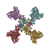
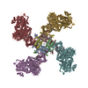
 Citation Citation |  Journal: Nat Commun / Year: 2024 Journal: Nat Commun / Year: 2024Title: Structure of an open K channel reveals tandem PIP binding sites mediating the Kir6.2 and SUR1 regulatory interface. Authors: Camden M Driggers / Yi-Ying Kuo / Phillip Zhu / Assmaa ElSheikh / Show-Ling Shyng /   Abstract: ATP-sensitive potassium (K) channels, composed of four pore-lining Kir6.2 subunits and four regulatory sulfonylurea receptor 1 (SUR1) subunits, control insulin secretion in pancreatic β-cells. K ...ATP-sensitive potassium (K) channels, composed of four pore-lining Kir6.2 subunits and four regulatory sulfonylurea receptor 1 (SUR1) subunits, control insulin secretion in pancreatic β-cells. K channel opening is stimulated by PIP and inhibited by ATP. Mutations that increase channel opening by PIP reduce ATP inhibition and cause neonatal diabetes. Although considerable evidence has implicated a role for PIP in K channel function, previously solved open-channel structures have lacked bound PIP, and mechanisms by which PIP regulates K channels remain unresolved. Here, we report the cryoEM structure of a K channel harboring the neonatal diabetes mutation Kir6.2-Q52R, in the open conformation, bound to amphipathic molecules consistent with natural C18:0/C20:4 long-chain PI(4,5)P at two adjacent binding sites between SUR1 and Kir6.2. The canonical PIP binding site is conserved among PIP-gated Kir channels. The non-canonical PIP binding site forms at the interface of Kir6.2 and SUR1. Functional studies demonstrate both binding sites determine channel activity. Kir6.2 pore opening is associated with a twist of the Kir6.2 cytoplasmic domain and a rotation of the N-terminal transmembrane domain of SUR1, which widens the inhibitory ATP binding pocket to disfavor ATP binding. The open conformation is particularly stabilized by the Kir6.2-Q52R residue through cation-π bonding with SUR1-W51. Together, these results uncover the cooperation between SUR1 and Kir6.2 in PIP binding and gating, explain the antagonistic regulation of K channels by PIP and ATP, and provide a putative mechanism by which Kir6.2-Q52R stabilizes an open channel to cause neonatal diabetes. | |||||||||
| History |
|
- Structure visualization
| Structure viewer | Molecule:  Molmil Molmil Jmol/JSmol Jmol/JSmol |
|---|
- Downloads & links
- Download
| PDBx/mmCIF format |  8ti2.cif.gz 8ti2.cif.gz | 1.5 MB | Display |  PDBx/mmCIF format PDBx/mmCIF format |
|---|---|---|---|---|
| PDB format |  pdb8ti2.ent.gz pdb8ti2.ent.gz | 1014.6 KB | Display |  PDB format PDB format |
| PDBx/mmJSON format |  8ti2.json.gz 8ti2.json.gz | Tree view |  PDBx/mmJSON format PDBx/mmJSON format | |
| Others |  Other downloads Other downloads |
-Validation report
| Arichive directory |  https://data.pdbj.org/pub/pdb/validation_reports/ti/8ti2 https://data.pdbj.org/pub/pdb/validation_reports/ti/8ti2 ftp://data.pdbj.org/pub/pdb/validation_reports/ti/8ti2 ftp://data.pdbj.org/pub/pdb/validation_reports/ti/8ti2 | HTTPS FTP |
|---|
-Related structure data
| Related structure data |  41278MC  8ti1C M: map data used to model this data C: citing same article ( |
|---|---|
| Similar structure data | Similarity search - Function & homology  F&H Search F&H Search |
- Links
Links
- Assembly
| Deposited unit | 
|
|---|---|
| 1 |
|
- Components
-Protein , 2 types, 8 molecules ABCDEHGF
| #1: Protein | Mass: 43690.828 Da / Num. of mol.: 4 / Mutation: Q52R Source method: isolated from a genetically manipulated source Source: (gene. exp.)   Rattus norvegicus (Norway rat) / Cell: Beta cell / Gene: Kcnj11 / Organ: Pancreas Rattus norvegicus (Norway rat) / Cell: Beta cell / Gene: Kcnj11 / Organ: Pancreas / Cell line (production host): COS-M6 (RRID:CVCL_8561) / Production host: / Cell line (production host): COS-M6 (RRID:CVCL_8561) / Production host:   Chlorocebus aethiops (grivet) / References: UniProt: P70673 Chlorocebus aethiops (grivet) / References: UniProt: P70673#2: Protein |  ABCC8 ABCC8Mass: 177296.578 Da / Num. of mol.: 4 Source method: isolated from a genetically manipulated source Source: (gene. exp.)   Mesocricetus auratus (golden hamster) / Cell line (production host): COS-M6 (RRID:CVCL_8561) / Production host: Mesocricetus auratus (golden hamster) / Cell line (production host): COS-M6 (RRID:CVCL_8561) / Production host:   Chlorocebus aethiops (grivet) / References: UniProt: A0A1S4NYG1 Chlorocebus aethiops (grivet) / References: UniProt: A0A1S4NYG1 |
|---|
-Sugars , 1 types, 4 molecules 
| #7: Sugar | ChemComp-NAG /  N-Acetylglucosamine N-Acetylglucosamine |
|---|
-Non-polymers , 5 types, 38 molecules 


| #3: Chemical | ChemComp-PT5 / [(  Phosphatidylinositol 4,5-bisphosphate Phosphatidylinositol 4,5-bisphosphate#4: Chemical | ChemComp-K / #5: Chemical | ChemComp-PEF /  Phosphatidylethanolamine Phosphatidylethanolamine#6: Chemical | ChemComp-P5S /  Phosphatidylserine Phosphatidylserine#8: Water | ChemComp-HOH / |  Water Water |
|---|
-Details
| Has ligand of interest | Y |
|---|
-Experimental details
-Experiment
| Experiment | Method:  ELECTRON MICROSCOPY ELECTRON MICROSCOPY |
|---|---|
| EM experiment | Aggregation state: PARTICLE / 3D reconstruction method:  single particle reconstruction single particle reconstruction |
- Sample preparation
| Component | Name: Kir6.2-Q52R/SUR1 open channel / Type: COMPLEX Details: Kir6.2-Q52R/SUR1 open KATP channel in the open conformation in complex with PIP2 and other phospholipids Entity ID: #1-#2 / Source: MULTIPLE SOURCES | ||||||||||||||||||||||||||||||
|---|---|---|---|---|---|---|---|---|---|---|---|---|---|---|---|---|---|---|---|---|---|---|---|---|---|---|---|---|---|---|---|
| Molecular weight | Value: 0.880 MDa / Experimental value: NO | ||||||||||||||||||||||||||||||
| Source (natural) | Organism:   Rattus norvegicus (Norway rat) Rattus norvegicus (Norway rat) | ||||||||||||||||||||||||||||||
| Source (recombinant) | Organism:   Chlorocebus aethiops (grivet) / Cell: COS-M6 cells / Plasmid Chlorocebus aethiops (grivet) / Cell: COS-M6 cells / Plasmid : recombinant adenovirus : recombinant adenovirus | ||||||||||||||||||||||||||||||
| Buffer solution | pH: 7.4 / Details: MSB with digitonin and PIP2 | ||||||||||||||||||||||||||||||
| Buffer component |
| ||||||||||||||||||||||||||||||
| Specimen | Conc.: 0.15 mg/ml / Embedding applied: NO / Shadowing applied: NO / Staining applied : NO / Vitrification applied : NO / Vitrification applied : YES : YESDetails: 3 microliters of purified Kir6.2-Q52R/FLAG-SUR1 was loaded onto Quantifoil R 1.2/1.3 Au 300 grids prepared with a fresh Graphene Oxide surface | ||||||||||||||||||||||||||||||
| Specimen support | Details: The grid was prepared with a Graphene Oxide coating before use. Grid material: GOLD / Grid mesh size: 300 divisions/in. / Grid type: Quantifoil R1.2/1.3 | ||||||||||||||||||||||||||||||
Vitrification | Instrument: FEI VITROBOT MARK III / Cryogen name: ETHANE / Humidity: 100 % / Chamber temperature: 279 K |
- Electron microscopy imaging
| Experimental equipment |  Model: Titan Krios / Image courtesy: FEI Company |
|---|---|
| Microscopy | Model: FEI TITAN KRIOS Details: Titan Krios #3 at the Pacific Northwest National Lab |
| Electron gun | Electron source : :  FIELD EMISSION GUN / Accelerating voltage: 300 kV / Illumination mode: SPOT SCAN FIELD EMISSION GUN / Accelerating voltage: 300 kV / Illumination mode: SPOT SCAN |
| Electron lens | Mode: BRIGHT FIELD Bright-field microscopy / Nominal magnification: 105000 X / Nominal defocus max: 2500 nm / Nominal defocus min: 1000 nm / Cs Bright-field microscopy / Nominal magnification: 105000 X / Nominal defocus max: 2500 nm / Nominal defocus min: 1000 nm / Cs : 2.7 mm / C2 aperture diameter: 70 µm / Alignment procedure: COMA FREE : 2.7 mm / C2 aperture diameter: 70 µm / Alignment procedure: COMA FREE |
| Specimen holder | Cryogen: NITROGEN / Specimen holder model: FEI TITAN KRIOS AUTOGRID HOLDER |
| Image recording | Average exposure time: 2.2 sec. / Electron dose: 55 e/Å2 / Film or detector model: GATAN K3 (6k x 4k) / Num. of grids imaged: 3 / Num. of real images: 5241 |
- Processing
| EM software |
| ||||||||||||||||||||||||||||||||||||
|---|---|---|---|---|---|---|---|---|---|---|---|---|---|---|---|---|---|---|---|---|---|---|---|---|---|---|---|---|---|---|---|---|---|---|---|---|---|
CTF correction | Type: PHASE FLIPPING AND AMPLITUDE CORRECTION | ||||||||||||||||||||||||||||||||||||
| Particle selection | Num. of particles selected: 14115 | ||||||||||||||||||||||||||||||||||||
| Symmetry | Point symmetry : C4 (4 fold cyclic : C4 (4 fold cyclic ) ) | ||||||||||||||||||||||||||||||||||||
3D reconstruction | Resolution: 3.28 Å / Resolution method: FSC 0.143 CUT-OFF / Num. of particles: 14115 / Algorithm: FOURIER SPACE / Num. of class averages: 1 / Symmetry type: POINT | ||||||||||||||||||||||||||||||||||||
| Atomic model building | Protocol: FLEXIBLE FIT | ||||||||||||||||||||||||||||||||||||
| Atomic model building | PDB-ID: 6BAA Accession code: 6BAA / Source name: PDB / Type: experimental model | ||||||||||||||||||||||||||||||||||||
| Refinement | Cross valid method: NONE / Stereochemistry target values: GeoStd + Monomer Library | ||||||||||||||||||||||||||||||||||||
| Displacement parameters | Biso mean: 165.54 Å2 | ||||||||||||||||||||||||||||||||||||
| Refine LS restraints |
|
 Movie
Movie Controller


 PDBj
PDBj


