 Movie
Movie Controller


+ Open data

- Basic information
| Entry | Database: PDB / ID: 8poc | ||||||
|---|---|---|---|---|---|---|---|
| Title | Cryo-EM structure of Dickeya dadantii BcsD | ||||||
 Components Components | Cellulose synthase operon protein D | ||||||
 Keywords Keywords |  CYTOSOLIC PROTEIN / Cellulose secretion / bacterial biofilms / cytoskeleton CYTOSOLIC PROTEIN / Cellulose secretion / bacterial biofilms / cytoskeleton | ||||||
| Function / homology | Cellulose synthase operon protein D, bacterial / Cellulose synthase subunit D superfamily / Cellulose synthase subunit D / cellulose biosynthetic process / Cellulose synthase operon protein D Function and homology information Function and homology information | ||||||
| Biological species |  Dickeya dadantii 3937 (bacteria) Dickeya dadantii 3937 (bacteria) | ||||||
| Method | ELECTRON MICROSCOPY / single particle reconstruction / cryo EM / Resolution: 4 Å | ||||||
 Authors Authors | Notopoulou, A. / Krasteva, P.V. | ||||||
| Funding support | European Union, 1items
| ||||||
 Citation Citation | Journal: Curr Biol / Year: 2024 Title: Structures and roles of BcsD and partner scaffold proteins in proteobacterial cellulose secretion. Authors: Thibault G Sana / Areti Notopoulou / Lucie Puygrenier / Marion Decossas / Sandra Moreau / Aurélien Carlier / Petya V Krasteva /   Abstract: Cellulose is the world's most abundant biopolymer, and similar to its role as a cell wall component in plants, it is a prevalent constituent of the extracellular matrix in bacterial biofilms. ...Cellulose is the world's most abundant biopolymer, and similar to its role as a cell wall component in plants, it is a prevalent constituent of the extracellular matrix in bacterial biofilms. Although bacterial cellulose (BC) was first described in the 19 century, it was only recently revealed that it is produced by several distinct types of Bcs secretion systems that feature multiple accessory subunits in addition to a catalytic BcsAB synthase tandem. We recently showed that crystalline cellulose secretion in the Gluconacetobacter genus (α-Proteobacteria) is driven by a supramolecular BcsH-BcsD scaffold-the "cortical belt"-which stabilizes the synthase nanoarrays through an unexpected inside-out mechanism for secretion system assembly. Interestingly, while bcsH is specific for Gluconacetobacter, bcsD homologs are widespread in Proteobacteria. Here, we examine BcsD homologs and their gene neighborhoods from several plant-colonizing β- and γ-Proteobacteria proposed to secrete a variety of non-crystalline and/or chemically modified cellulosic polymers. We provide structural and mechanistic evidence that through different quaternary structure assemblies BcsD acts with proline-rich BcsH, BcsP, or BcsO partners across the proteobacterial clade to form synthase-interacting intracellular scaffolds that, in turn, determine the biofilm strength and architecture in species with strikingly different physiology and secreted biopolymers. | ||||||
| History |
|
- Structure visualization
| Structure viewer | Molecule: MolmilJmol/JSmol |
|---|
- Downloads & links
-Download
| PDBx/mmCIF format | 8poc.cif.gz | 115.3 KB | Display | PDBx/mmCIF format |
|---|---|---|---|---|
| PDB format | pdb8poc.ent.gz | 90.1 KB | Display | PDB format |
| PDBx/mmJSON format | 8poc.json.gz | Tree view | PDBx/mmJSON format | |
| Others |  Other downloads Other downloads |
-Validation report
| Arichive directory | https://data.pdbj.org/pub/pdb/validation_reports/po/8pocftp://data.pdbj.org/pub/pdb/validation_reports/po/8poc | HTTPS FTP |
|---|
-Related structure data
| Related structure data |  17788MC  8pkdC  8pogC C: citing same article ( M: map data used to model this data |
|---|---|
| Similar structure data |


-Links
 PDBj
PDBj- Assembly
| Deposited unit | 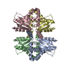
|
|---|---|
| 1 |
|
-Components
| #1: Protein | Mass: 17930.289 Da / Num. of mol.: 4 Source method: isolated from a genetically manipulated source Source: (gene. exp.) Dickeya dadantii 3937 (bacteria) / Gene: bcsD / Production host: Escherichia coli BL21(DE3) (bacteria) / References: UniProt: E0SES7 |
|---|
-Experimental details
-Experiment
| Experiment | Method: ELECTRON MICROSCOPY |
|---|---|
| EM experiment | Aggregation state: PARTICLE / 3D reconstruction method: single particle reconstruction |
- Sample preparation
| Component | Name: Tetrameric BcsD of Dickeya dadantii / Type: COMPLEX / Entity ID: all / Source: RECOMBINANT |
|---|---|
| Molecular weight | Value: 0.07 MDa |
| Source (natural) | Organism: Dickeya dadantii 3937 (bacteria) |
| Source (recombinant) | Organism: Escherichia coli BL21(DE3) (bacteria) |
| Buffer solution | pH: 8 / Details: 20 mM HEPES pH 8.0, 120 mM NaCl |
| Specimen | Conc.: 2 mg/ml / Embedding applied: NO / Shadowing applied: NO / Staining applied: NO / Vitrification applied: YES |
| Specimen support | Grid material: GOLD / Grid mesh size: 300 divisions/in. / Grid type: Quantifoil R1.2/1.3 |
| Vitrification | Instrument: FEI VITROBOT MARK IV / Cryogen name: ETHANE / Humidity: 100 % / Chamber temperature: 277 K |
- Electron microscopy imaging
| Experimental equipment |  Model: Talos Arctica / Image courtesy: FEI Company |
|---|---|
| Microscopy | Model: FEI TALOS ARCTICA |
| Electron gun | Electron source: FIELD EMISSION GUN / Accelerating voltage: 200 kV / Illumination mode: FLOOD BEAM |
| Electron lens | Mode: BRIGHT FIELDBright-field microscopy / Nominal defocus max: 2600 nm / Nominal defocus min: 400 nm / Cs: 2.7 mm |
| Image recording | Electron dose: 52.9 e/Å2 / Film or detector model: GATAN K2 SUMMIT (4k x 4k) |
| Image scans | Width: 3838 / Height: 3710 / Movie frames/image: 50 / Used frames/image: 0-50 |
- Processing
| EM software |
| |||||||||||||||||||||||||||
|---|---|---|---|---|---|---|---|---|---|---|---|---|---|---|---|---|---|---|---|---|---|---|---|---|---|---|---|---|
| Image processing | Details: 3168 out of 3379 movies retained for processing | |||||||||||||||||||||||||||
| CTF correction | Details: Gctf / Type: PHASE FLIPPING AND AMPLITUDE CORRECTION | |||||||||||||||||||||||||||
| Particle selection | Num. of particles selected: 1488279 Details: After first round of 2D classification, 406652 particles retained for Ab-initio model generation and downstream processing | |||||||||||||||||||||||||||
| Symmetry | Point symmetry: D2 (2x2 fold dihedral) | |||||||||||||||||||||||||||
| 3D reconstruction | Resolution: 4 Å / Resolution method: FSC 0.143 CUT-OFF / Num. of particles: 295199 / Symmetry type: POINT | |||||||||||||||||||||||||||
| Atomic model building | Protocol: OTHER / Space: REAL Details: Reiterative refinement in Phenix, Coot and Namdinator | |||||||||||||||||||||||||||
| Atomic model building | Source name: AlphaFold / Type: in silico model | |||||||||||||||||||||||||||
| Refine LS restraints |
|
