 Movie
Movie Controller


+ Open data

- Basic information
| Entry | Database: PDB / ID: 8pg0 | |||||||||
|---|---|---|---|---|---|---|---|---|---|---|
| Title | Human OATP1B3 | |||||||||
 Components Components |
| |||||||||
 Keywords Keywords |  TRANSPORT PROTEIN / organic anion / bicarbonate / SLCO1B3 / uptake / drug / transporter / polypeptide / liver TRANSPORT PROTEIN / organic anion / bicarbonate / SLCO1B3 / uptake / drug / transporter / polypeptide / liver | |||||||||
| Function / homology |  Function and homology information Function and homology informationDefective SLCO1B3 causes hyperbilirubinemia, Rotor type (HBLRR) / Transport of organic anions / sodium-independent organic anion transmembrane transporter activity / sodium-independent organic anion transport / negative regulation of peptidase activity / bile acid transmembrane transporter activity / organic anion transport / heme catabolic process / Atorvastatin ADME / organic anion transmembrane transporter activity ...Defective SLCO1B3 causes hyperbilirubinemia, Rotor type (HBLRR) / Transport of organic anions / sodium-independent organic anion transmembrane transporter activity / sodium-independent organic anion transport / negative regulation of peptidase activity / bile acid transmembrane transporter activity / organic anion transport / heme catabolic process / Atorvastatin ADME / organic anion transmembrane transporter activity / bile acid and bile salt transport / Heme degradation / monoatomic ion transport / Recycling of bile acids and salts / xenobiotic metabolic process / basal plasma membrane / serine-type endopeptidase inhibitor activity / basolateral plasma membrane / plasma membraneSimilarity search - Function | |||||||||
| Biological species |  Homo sapiens (human) Homo sapiens (human) | |||||||||
| Method | ELECTRON MICROSCOPY / single particle reconstruction / cryo EM / Resolution: 2.97 Å | |||||||||
 Authors Authors | Ciuta, A.-D. / Nosol, K. / Kowal, J. / Mukherjee, S. / Ramirez, A.S. / Stieger, B. / Kossiakoff, A.A. / Locher, K.P. | |||||||||
| Funding support |  Switzerland, Switzerland,  United States, 2items United States, 2items
| |||||||||
 Citation Citation | Journal: Nat Commun / Year: 2023 Title: Structure of human drug transporters OATP1B1 and OATP1B3. Authors: Anca-Denise Ciută / Kamil Nosol / Julia Kowal / Somnath Mukherjee / Ana S Ramírez / Bruno Stieger / Anthony A Kossiakoff / Kaspar P Locher / Abstract: The organic anion transporting polypeptides OATP1B1 and OATP1B3 are membrane proteins that mediate uptake of drugs into the liver for subsequent conjugation and biliary excretion, a key step in drug ...The organic anion transporting polypeptides OATP1B1 and OATP1B3 are membrane proteins that mediate uptake of drugs into the liver for subsequent conjugation and biliary excretion, a key step in drug elimination from the human body. Polymorphic variants of these transporters can cause reduced drug clearance and adverse drug effects such as statin-induced rhabdomyolysis, and co-administration of OATP substrates can lead to damaging drug-drug interaction. Despite their clinical relevance in drug disposition and pharmacokinetics, the structure and mechanism of OATPs are unknown. Here we present cryo-EM structures of human OATP1B1 and OATP1B3 bound to synthetic Fab fragments and in functionally distinct states. A single estrone-3-sulfate molecule is bound in a pocket located in the C-terminal half of OATP1B1. The shape and chemical nature of the pocket rationalize the preference for diverse organic anions and allow in silico docking of statins. The structure of OATP1B3 is determined in a drug-free state but reveals a bicarbonate molecule bound to the conserved signature motif and a histidine residue that is prevalent in OATPs exhibiting pH-dependent activity. | |||||||||
| History |
|
- Structure visualization
| Structure viewer | Molecule: MolmilJmol/JSmol |
|---|
- Downloads & links
-Download
| PDBx/mmCIF format | 8pg0.cif.gz | 165.2 KB | Display | PDBx/mmCIF format |
|---|---|---|---|---|
| PDB format | pdb8pg0.ent.gz | 123.3 KB | Display | PDB format |
| PDBx/mmJSON format | 8pg0.json.gz | Tree view | PDBx/mmJSON format | |
| Others |  Other downloads Other downloads |
-Validation report
| Arichive directory | https://data.pdbj.org/pub/pdb/validation_reports/pg/8pg0ftp://data.pdbj.org/pub/pdb/validation_reports/pg/8pg0 | HTTPS FTP |
|---|
-Related structure data
| Related structure data |  17655MC 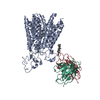 8phwC M: map data used to model this data C: citing same article ( |
|---|---|
| Similar structure data |

-Links
 PDBj
PDBj


- Assembly
| Deposited unit | 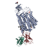
|
|---|---|
| 1 |
|
-Components
-Antibody , 2 types, 2 molecules HL
| #2: Antibody | Mass: 25599.549 Da / Num. of mol.: 1 Source method: isolated from a genetically manipulated source Source: (gene. exp.) Homo sapiens (human) / Production host:  Escherichia coli (E. coli) Escherichia coli (E. coli) |
|---|---|
| #3: Antibody | Mass: 23258.783 Da / Num. of mol.: 1 Source method: isolated from a genetically manipulated source Source: (gene. exp.) Homo sapiens (human) / Production host: Escherichia coli (E. coli) |
-Protein / Sugars , 2 types, 2 molecules A
| #1: Protein | Mass: 77478.586 Da / Num. of mol.: 1 Source method: isolated from a genetically manipulated source Source: (gene. exp.) Homo sapiens (human) / Gene: SLCO1B3, LST2, OATP1B3, OATP8, SLC21A8 / Production host: Homo sapiens (human) / References: UniProt: Q9NPD5 |
|---|---|
| #4: Sugar | ChemComp-NAG / N-Acetylglucosamine Type: D-saccharide, beta linking / Mass: 221.208 Da / Num. of mol.: 1 / Source method: obtained synthetically / Formula: C8H15NO6 Type: D-saccharide, beta linking / Mass: 221.208 Da / Num. of mol.: 1 / Source method: obtained synthetically / Formula: C8H15NO6 |
-Non-polymers , 2 types, 4 molecules 

| #5: Chemical | ChemComp-BCT / Bicarbonate Mass: 61.017 Da / Num. of mol.: 1 / Source method: obtained synthetically / Formula: CHO3 / Feature type: SUBJECT OF INVESTIGATION / Comment: pH buffer*YM Mass: 61.017 Da / Num. of mol.: 1 / Source method: obtained synthetically / Formula: CHO3 / Feature type: SUBJECT OF INVESTIGATION / Comment: pH buffer*YM |
|---|---|
| #6: Chemical | Cholesterol Mass: 386.654 Da / Num. of mol.: 3 / Source method: obtained synthetically / Formula: C27H46O Mass: 386.654 Da / Num. of mol.: 3 / Source method: obtained synthetically / Formula: C27H46O |
-Details
| Has ligand of interest | Y |
|---|
-Experimental details
-Experiment
| Experiment | Method: ELECTRON MICROSCOPY |
|---|---|
| EM experiment | Aggregation state: PARTICLE / 3D reconstruction method: single particle reconstruction |
- Sample preparation
| Component | Name: Human OATP1B3 / Type: COMPLEX / Entity ID: #1-#3 / Source: RECOMBINANT |
|---|---|
| Molecular weight | Value: 0.18 MDa / Experimental value: YES |
| Source (natural) | Organism: Homo sapiens (human) |
| Source (recombinant) | Organism: Homo sapiens (human) |
| Buffer solution | pH: 7.4 |
| Specimen | Conc.: 0.59 mg/ml / Embedding applied: NO / Shadowing applied: NO / Staining applied: NO / Vitrification applied: YES |
| Vitrification | Cryogen name: ETHANE-PROPANE |
- Electron microscopy imaging
| Experimental equipment |  Model: Titan Krios / Image courtesy: FEI Company |
|---|---|
| Microscopy | Model: FEI TITAN KRIOS |
| Electron gun | Electron source: FIELD EMISSION GUN / Accelerating voltage: 300 kV / Illumination mode: FLOOD BEAM |
| Electron lens | Mode: BRIGHT FIELDBright-field microscopy / Nominal defocus max: 2200 nm / Nominal defocus min: 600 nm / Cs: 2.7 mm / C2 aperture diameter: 100 µm |
| Specimen holder | Cryogen: NITROGEN / Specimen holder model: FEI TITAN KRIOS AUTOGRID HOLDER |
| Image recording | Electron dose: 60.5 e/Å2 / Film or detector model: GATAN K3 BIOQUANTUM (6k x 4k) |
- Processing
| CTF correction | Type: PHASE FLIPPING AND AMPLITUDE CORRECTION | ||||||||||||||||||||||||
|---|---|---|---|---|---|---|---|---|---|---|---|---|---|---|---|---|---|---|---|---|---|---|---|---|---|
| 3D reconstruction | Resolution: 2.97 Å / Resolution method: FSC 0.143 CUT-OFF / Num. of particles: 75610 / Symmetry type: POINT | ||||||||||||||||||||||||
| Refine LS restraints |
|
